See discussions, stats, and author profiles for this publication at: https://www.researchgate.net/publication/13489168
p53 mutation with frequent novel codons but
not a mutator phenotype in BRCA1- and
BRCA2-associated breast tumours
Article in Oncogene · November 1998 DOI: 10.1038/sj.onc.1202106 · Source: PubMed CITATIONS158
READS187
14 authors, including: Some of the authors of this publication are also working on these related projects: p53 and Cervical CancerView project Drug Resistance Studies in Ovarian Cancer
View project Tim Crook University of Dundee 187 PUBLICATIONS 13,043 CITATIONS SEE PROFILE Isik Yulug Bilkent University 65 PUBLICATIONS 1,881 CITATIONS SEE PROFILE
All content following this page was uploaded by Isik Yulug on 26 March 2014.
p53 mutation with frequent novel codons but not a mutator phenotype in
BRCA1- and BRCA2-associated breast tumours
Tim Crook
1, Louise A Brooks
2, Susan Crossland
1, P Osin
1, Karen T Barker
1, Joanne Waller
1,
Elizabeth Philp
1, Paul D Smith
1, Isik Yulug
1,4, Julian Peto
1, Gillian Parker
3, Martin J Allday
3,
Mark R Crompton
1and Barry A Gusterson
11Institute of Cancer Research, Haddow Laboratories, 15 Cotswold Road, Sutton, Surrey, SM2 5NG;2Division of Clinical Sciences,
London School of Hygiene and Tropical Medicine, Keppel Street, London W1;3Ludwig Institute of Cancer Research and Section
of Virology and Cell Biology, Imperial College School of Medicine, St. Mary's Campus, Norfolk Place, London W2 1PG, UK
The status of p53 was investigated in breast tumours arising in germ-line carriers of mutant alleles of BRCA1 and BRCA2 and in a control series of sporadic breast tumours. p53 expression was detected in 20/26 (77%) BRCA1-, 10/22 (45%) BRCA2-associated and 25/72 (35%) grade-matched sporadic tumours. Analysis of p53 sequence revealed that the gene was mutant in 33/50 (66%) BRCA-associated tumours, whereas 7/20 (35%) sporadic grade-matched tumours contained p53 mutation (P50.05). A number of the mutations detected in the BRCA-associated tumours have not been previously described in human cancer databases, whilst others occur
extremely rarely. Analysis of additional genes, p16INK4,
Ki-ras and b-globin revealed absence or very low incidence of mutations, suggesting that the higher frequency of p53 mutation in the BRCA-associated tumours does not re¯ect a generalized increase in susceptibility to the acquisition of somatic mutation. Furthermore, absence of frameshift mutations in the polypurine tracts present in the coding sequence of the TGF b type II receptor (TGF b IIR) and Bax implies that loss of function of BRCA1 or BRCA2 does not confer a mutator phenotype such as that found in
tumours with microsatellite instability (MSI). p21Waf1
was expressed in BRCA-associated tumours regardless of p53 status and, furthermore, some tumours expressing
wild-type p53 did not express detectable p21Waf1. These
data do not support, therefore, the simple model based on studies of BRCA7/7 embryos, in which mutation of p53 in BRCA-associated tumours results in loss of
p21Waf1 expression and deregulated proliferation. Rather,
they imply that proliferation of such tumours will be subject to multiple mechanisms of growth regulation. Keywords: BRCA1; BRCA2; p53; mutation(s); breast; tumour; familial
Introduction
Inheritance of mutant alleles of the breast cancer susceptibility genes BRCA1 and BRCA2 confers a substantial risk of developing breast, ovarian and some other cancers (Mikki et al., 1994; Wooster et al., 1995).
BRCA1 and BRCA2 have numerous similarities (Zhang et al., 1998). Tumorigenesis in carriers of germ-line mutations in BRCA1 and BRCA2 is invariably accompanied by loss of the wild-type allele (Smith et al., 1992; Neuhausen and Marshall, 1994; Collins et al., 1995; Gudmundsson et al., 1995; Kelsell et al., 1996) suggesting that the proteins encoded by the two genes operate as tumour suppressors. An increasing-body of evidence favours a role for both BRCA1 and BRCA2 in cellular response to DNA damage (Connor et al., 1997; Scully et al., 1997; Patel et al., 1998). It is postulated that the BRCA genes act as caretakers, functioning to maintain genomic stability, rather than as gatekeepers which regulate cellular proliferation (Brugarolas and Jacks, 1997; Kinzler and Vogelstein, 1997). Embryos homozy-gously deleted for BRCA1 or 2 are inviable, cells
undergoing a p21Waf1-mediated growth arrest at around
day 5 ± 6 (Hakem et al., 1996; Suzuki et al., 1997). This lethality is partially reversed by breeding into a p53 or
p21Waf1null background (Hakem et al., 1997; Ludwig et
al., 1997). Paradoxically, tumours arising in carriers of BRCA1 mutations are frequently highly proliferative, despite being functionally null for BRCA1 (Eisenger et al., 1996; Marcus et al., 1996; Johannsson et al., 1997). The molecular basis for this is not known. Analysis of ®broblasts derived from mice homozygous for a truncating mutation in BRCA2 has revealed a proliferation defect associated with p53-dependent
induction of p21Waf1 expression, and a DNA repair
defect (Connor et al., 1997). In another study, mouse embryo ®broblasts carrying BRCA2 truncations, were shown to be signi®cantly more sensitive to UV irradiation and the alkylating agent methylmethanesul-phonate than wild-type controls, leading to the suggestion that BRCA2 may be associated with a nucleotide excision repair pathway (Patel et al., 1998). However, it is not known whether these defects occur in tumours arising in carriers of germline mutant alleles of BRCA1 and BRCA 2.
In sporadic breast cancer the frequency of p53 mutation is 20 ± 40%, the proportion of mutant tumours increasing with higher grade. In a previous study using DNA extracted from paran sections, p53 mutations were reported to occur at high frequency in familial tumours (of unknown BRCA status) but not sporadic breast tumours (Glebov et al., 1994). Furthermore, analysis of a small series of breast and ovarian tumours arising in carriers of mutant BRCA1 alleles revealed a high incidence of p53 mutation (Crook et al., 1997). In the present study we have Correspondence: T Crook
4Current address: Bilkent University, Ankara, Turkey
TC and LAB contributed equally to these studies
Received 2 January 1998; revised 1 May 1998; accepted 5 May 1998
investigated the status of p53 in a larger series of BRCA1- and BRCA2-associated breast carcinomas and in a group of sporadic breast tumours matched for grade, and have compared both the frequency and nature of the p53 mutations detected in the two groups.
Results
The expression and structure of p53 was examined in 50 tumours arising in carriers of germ-line mutations in BRCA1 (28 tumours) and BRCA2 (22 tumours). The germ-line mutations in these families have been described previously (Gayther et al., 1995, 1997). To control for a potentially biasing eect of grade, a series of sporadic grade 3 breast tumours, previously characterized by the Breast Cancer Linkage Consor-tium (1997), was analysed in parallel.
Expression of p53 in BRCA1- and BRCA2-associated breast tumours
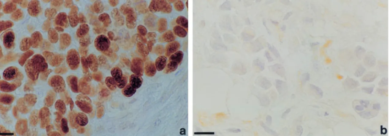
Expression of p53 in sporadic and familial tumours was analysed by immunocytochemistry (IC) in paran sections of formalin-®xed tissues, using the DO-1 antibody. 20/26 (77%) of BRCA1-, 10/22 (45%) of BRCA2-associated tumours and 25/72 (35%) of grade-matched sporadic tumours were positive (Figure 1 and Table 1). In all cases of BRCA1- and BRCA2-associated tumours where ductal carcinoma in situ (DCIS) was also present in the tissue sections examined, the p53 immunostaining detected in invasive tumour tissue was also present in the DCIS. In no section examined was p53 staining observed in normal tissue. Detection of p53 by IC in breast tumours frequently re¯ects stabilization due to muta-tion (Visscher et al., 1996). The absence of immuno-cytochemically detectable p53 in normal breast tissue in our study therefore implies somatic acquisition, rather than germline inheritance, of p53 mutation. It is estimated, however, that 10 ± 15% of mutants do not give rise to stabilized protein and that p53 becomes stabilized (and thereby detectable) in some breast tumours despite being wild-type (Visscher et al., 1996). We therefore analysed the sequence of p53 in the sporadic and BRCA-associated tumours.
p53 is mutant at high frequency in BRCA-associated tumours
Genomic DNA was extracted from formalin-®xed, paran-embedded tumour sections from 28 BRCA1-, 22 BRCA2-associated and 20 grade 3 sporadic tumours and used for analysis of the sequence of various genes. For p53 analysis, DNA was subjected to single strand conformation polymorphism analysis (SSCP), cloning and sequencing of each p53 exon (Visscher et al., 1996). However, when it was observed in preliminary studies that p53 mutation was common in the BRCA1-and BRCA2-associated tumours, the coding sequence of p53 was then analysed in its entirety in each BRCA-associated and sporadic tumour by sequencing at least 12 independent plasmid clones for each exon for each tumour, irrespective of the presence or not of SSCP mobility shifting. In the BRCA1-associated tumours, p53 mutations were detected in 19/28 tumours analysed, two of which were negative by IC (Table 1). In the case of the BRCA2-associated tumours, 14/ 22 tumours were mutant for p53 these being all nine tumours positive by IC and ®ve negative by IC (Table 1, Figure 2). Similar sequencing analysis of DNA extracted from paran sections of 20 sporadic grade 3 tumours revealed single missense mutations in 7/20 tumours (Table 1). The dierence in p53 mutation frequency between the sporadic and BRCA-associated tumours was statistically signi®cant (P50.05).
Multiple p53 mutations occur in some BRCA-associated but not sporadic breast tumours
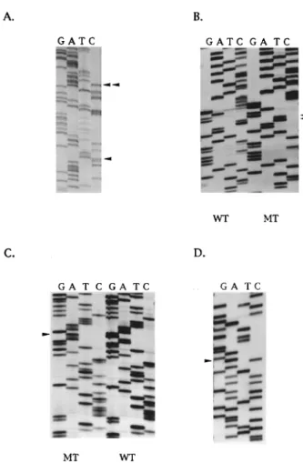
Sequencing analysis revealed that ®ve BRCA1-asso-ciated and three BRCA2-assoBRCA1-asso-ciated tumours contained two p53 mutations. One BRCA1-associated tumour and one BRCA2-associated tumour contained three independent mutations (Table 1). In four BRCA1- and three BRCA2-associated tumours with two mutations, a dierent coding change occurred with the identical silent, second mutation (codon 211 ACT4ACC). In the informative tumours, sequencing of multiple plasmid clones revealed that the mutations usually occurred on the same allele (Figure 2a). Thus in the BRCA2 tumour with three mutations (202R4S, 214 H4R and 219 P4H), all three occurred on the same allele. Similarly, in the two informative BRCA2
Figure 1 Immunocytochemical analysis of p53 expression in BRCA1-associated breast cancer. (a) positive tumour: (b) negative tumour. The scale bars are equivalent to 10 mm
p53 mutations in familial breast cancer T Crook et al
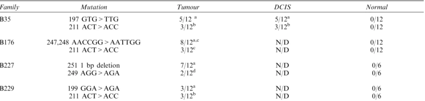
tumours with two mutations (202R4S, 211 Sil.) and (220Y4H, 211 Sil.), both mutations were on the same allele. Further sequence analysis of multiple indepen-dent plasmid clones from these two tumours iindepen-denti®ed numerous cases in which only the mutation resulting in the coding change (202R4S or 220Y4H) was present. However, no clone was observed in which the silent mutation at codon 211 alone was detected, implying that it arose subsequent to the coding mutation (Table 1). Previous studies using DNA extracted from paran sections have reported multiple p53 mutations in familial tumours (of unknown BRCA status) but not sporadic breast tumours. To further verify that the mutations in the present study were not artefacts attributable to tissue ®xation or contamination, and to con®rm that they were somatically acquired and not germ-line or polymorphisms, microdissected tumour, DCIS and normal from selected tissue sections were analysed for the presence of mutations. In each of the analysed cases, the coding mutations originally detected in DNA from undissected tissue sections were also identi®ed in the microdissected tumour and the single case of DCIS (Table 2). In the two cases analysed where the tumour contained a non-coding
change at codon 211, in addition to coding changes, sequencing analysis did not detect the codon 211 change in the microdissected DCIS. These observations are consistent with (i) absence of p53 expression in normal tissue present in any of the tissue sections analysed (ii) the presence of immunocytochemically detectable p53 protein in cases of DCIS where the associated tumours were also positive for p53 expression (iii) somatic acquisition rather than germ-line inheritance of the p53 sequence changes.
Absence of mutator phenotype in BRCA-associated tumours
The frequency and occasional multiplicity of p53 mutation suggested that loss of BRCA1 and BRCA2 function might confer a generalized increase in sensitivity to somatic mutation of cellular genes. To investigate this possibility, we performed analysis of the structure of additional genes which are rarely
mutant (Ki-ras) or almost never mutant (p16INK4) in
sporadic breast cancer. We also analysed a gene in which mutation could not provide a selective advantage (b-globin). Consistent with previous
stu-dies, no mutations were detected in p16INK4 in
paran-extracted DNA from the sporadic grade 3 tumours, nor in any of the BRCA-associated tumours. The
proposed absence of p16INK4 mutations in BRCA
tumours was further con®rmed by direct sequencing in 12 of the BRCA1-associated and 12 of the BRCA2-associated tumours. In the case of the Ki-ras oncogene which is often mutant in pancreatic carcinomas (Bos, 1987), but only rarely in sporadic breast tumours, a codon 12 mutation (GGT4GAT, Gly4Asp) was detected in one BRCA2-associated tumour, whereas 13 further tumours analysed (eight BRCA1 and ®ve BRCA2) did not contain activating mutations at these sites. Mutation at this codon has been previously detected in sporadic breast cancer and in a breast carcinoma cell line (Prosperi et al., 1990). Furthermore, this frequency of Ki-ras mutation is comparable to that reported in sporadic breast tumours (Rochlitz et al., 1989; Clark et al., 1995). Finally a 268 bp fragment of the b-globin gene was analysed by SSCP in each of the BRCA1 and BRCA2 tumours and the sporadic grade 3 breast tumours. No mobility shifts indicative of mutation were detected. Because the incidence of p53 mutations suggested the possibility of a mutator
phenotype, we analysed the poly(A)10 tract of
TGF-bIIR and the poly(G)8 tract of Bax in the
BRCA-associated tumours, since studies of human tumours with microsatellite instability (MSI) arising in a classical mutator background have revealed frameshift mutations within these regions of each gene. No aberrant mobility bands suggestive of frameshift mutation were observed in any of 28 tumours analysed (16 BRCA1, 12 BRCA2).
Expression of p21WAF1 occurs in BRCA1- and
BRCA2-associated tumours irrespective of p53 status
Studies of embryos deleted for both BRCA2 and p53,
or BRCA2 and p21Waf1, have revealed that development
progresses to a signi®cantly later stage (in such double knockouts) than in BRCA27/7 embryos which undergo cell cycle arrest at day 5 ± 6. This suggests Figure 2 Sequence analysis of p53 in BRCA-associated breast
tumours. (a) Presence of two p53 mutations in the same allele of p53 in BRCA1-associated tumour. The sequence changes at codon 224 gAg4gAC and codon 211 ACT4ACC are arrowed; (b) Tandem CC4TT transition at codons 247/248 (arrowed in lane MT) in BRCA2-associated tumour. The wild-type sequence (WT) is also shown; (c) Codon 199 mutation (ggA4AgA) arrowed in BRCA1-associated tumour; (d) Silent sequence change at codon 170 ACg4ACA in BRCA1-associated tumour
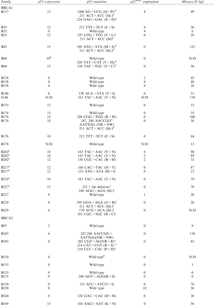
Table 1 p53 mutations in tumours arising in carriers of mutant BRCA1/2 alleles and grade-matched sporadic breast tumours
Family p53 expression p53 mutation p21Waf1expression Mitoses/10 hpf
BRCA1 B117 B35 B35 B35 B43 B64 B64 B138 B138 B138 B146 b146 B155 B176 B176 B176 B176 B178 B202e B202e B202e B217e B217e B218e B227e B227 B229 B229 12 12 0 12 15 10b 15 0 0 4 0 N/D 12 15 15 18 10 N/D 18 18 12 12 12 18 15 0 0 6 144CAG>CCG (Q>P);d 211 ACT>ACC (Sil.)a 224 GAG>GAC (E>D)a 212 TTT>TCT (F>S) Wild-type 197 GTG>TTG (V>L)a 211 ACT>ACC (Sil)a 169 ATG>ATA (M>I);d 211 ACT>ACC (Sil.)d Wild-type 220 TAT>CAT (Y>H);d 234 TAC>TGC (Y>C)d Wild-type Wild-type Wild type 150 ACA>ATA (T>I) 163 TAC>AAC (Y>N) Wild-type Wild-type 248 CGG>TGG (R>W) 247, 248 AACCGGd AATTGG (NR>NW) 211 ACT>ACC (Sil.)d 212 TTT>TCT (F>S) Wild-type 163 TAC>AAC (Y>N) 163 TAC>AAC (Y>N) 158 CGC>CAC (R>H) 168 CAC>TAC (H>Y) 133 ATG>ATA (M>I) 163 TAC>AAC (Y>N) 251 1 bp deletion;c 249 AGG>AGA (Sil.)c Wild-type 199 GGA>AGA (G>R)a 211 ACT>ACC (Sil.)a 170 ACG>ACA (Sil.)c 181 CGC>TGC (R>C)c 4 4 4 0 0 0 0 2 4 0 0 N/D 0 0 0 0 0 N/D 6 2 2 0 0 0 0 1 0 0 49 36 6 29 122 N/D 56 42 46 26 51 110 53 35 100 56 64 13 90 89 32 87 12 79 70 30 28 N/D BRCA2 B97 B105 B105 B124 B135 B135 B135 B150 B150 B164 B169 2 6 0 0 0 0 0 0 0 0 15 Wild-type 247/248 AACCGG> AATTGG(NR>NW) 202 CGT>AGT(R>R)a 214 CAT>CGT (R>S)a 219 CCC>CAC (P>H)a Wild-typeb Wild-type Wild-type 240 AGT>AGG(R>S) 155 ACC>ATC(T>I) Wild type 228 GAC>CAC (D>H) 164 AAG>AAT (K>N) 0 0 0 0 0 0 0 0 10 0 0 9 110 65 N/D 1 0 0 76 36 10 56
p53 mutations in familial breast cancer T Crook et al
that loss of the p53-dependent, p21Waf1 mediated G 1-S
checkpoint confers partial rescue of the cell cycle arrest consequent to abrogation of BRCA1 or BRCA2
function. To determine whether expression of p21Waf1
is aected by p53 status in BRCA1- and BRCA2-associated tumorigenesis, expression was analysed in tumours by IC. In the 20 sporadic grade 3 tumours, expression was absent or at extremely low levels irrespective of p53 status or expression, even in the three tumours which expressed signi®cant wild-type p53 (Table 1). In the case of the BRCA1-associated
tumours, expression of p21Waf1 was detected in 9/26
tumours analysed, these comprising four wild-type and
®ve mutant for p53. Three tumours wild-type for p53
did not express p21Waf1despite having high levels of p53
protein (Table 1). Analysis of BRCA2-associated
tumours revealed expression of p21Waf1 in 5/22
tumours including three tumours mutant for p53
(Table 1). The highest expression of p21Waf1 was
observed in two of the eight BRCA2-associated tumours which retained wild-type p53 (Table 1).
However, in these tumours p21Waf1 expression was not
detected in every tumour cell. Rather, intense expression was observed in some cells, whereas expression was either much reduced or absent in other cells. Of the remaining six BRCA2 tumours wild-type Table 2 p53 status of microdissected BRCA1-associated tissues
Family Mutation Tumour DCIS Normal
B35 B176 B227 B229 197 GTG>TTG 211 ACT>ACC 247,248 AACCGG>AATTGG 211 ACT>ACC 251 1 bp deletion 249 AGG>AGA 199 GGA>AGA 211 ACT>ACC 5/12a 3/12b 8/12a,c 3/12c 7/12a 2/12d 3/12a 3/12b 5/12a 3/12b N/D N/D N/D N/D N/D N/D 0/12 0/12 0/12 0/12 0/6 0/6 0/6 0/6
aTotal number of clones in which coding change was detected.bNumber of clones in which non-coding change was detected with coding change. cCannot be determined whether these changes occur together on same allele.dNumber of clones in which silent mutation was detected. N/D:
Not done (no DCIS in section)
Table 1 continued
Family p53 expression p53 mutation p21Waf1expression Mitoses/10 hpf
B186 B186 B186 B186 B186 B186 B186 B186 B196 B196 B211 Sporadic Sporadic Sporadic Sporadic Sporadic Sporadic Sporadic Sporadic Sporadic Sporadic Sporadic Sporadic Sporadic Sporadic Sporadic Sporadic Sporadic Sporadic Sporadic Sporadic 18 8 0 0 2 0 15 15 6 0 18 15 15 0 0 0 0 18 0 0 0 0 0 12 0 10 10 8 10 10 15 220 TAT>CAT (Y>H)a 211 ACT>ACC (Sil.)a 207 GAT>GGT (D>G) 202 CGT>AGT (R>S)a 211 ACT>ACC (Sil.)a Wild-type Wild-type Wild-type 163 TAC>AAC (Y>N) 156 CGC>CCC (R>P) 202 CGT>AGT (R>S) 181 CGC>TGC (R>C) 163 TAC>AAC (Y>N)c 211 ACT>ACC (Sil.)c 175 CGC>CAC (R>H) 245 GGC>GTC (G>V) Wild-type Wild-type Wild-type Wild-type 248 CGG>CAG (R>Q) Wild-type Wild-type Wild-type Wild-type Wild-type 258 GAA>AAA (E>K) Wild-type 171 GAG>AAG (E>K) Wild-type Wild type Wild type 163 TAC>TCC(Y>S) 272 GTG>GCG (V>A) 0 1 6 0 9 0 0 0 0 6 0 0 0 2 0 1 0 0 0 0 0 0 0 0 0 0 0 0 0 0 0 36 30 56 12 23 1 98 72 36 36 136 120 150 61 33 32 7 100 110 160 45 19 4 66 46 N/D 91 44 28 16 46
aMutations occurring on same allele; bDuctal Carcinoma in situ; cMutations occurring on dierent alleles;dCannot be determined whether
mutations are on the same allele;eMutation previously reported (Crook et al., 1997); N/D: not determined
for p53, only a single tumour expressed detectable p53
protein and none expressed p21Waf1 (Table 1). Taken
together, these data imply that, as in sporadic breast
and other tumours, p21Waf1 expression is induced in
some cases by p53-independent mechanisms. Eect of p53 mutation on mitosis in BRCA1- and BRCA2-associated tumours
The higher frequency of p53 mutation in the BRCA1 and BRCA2 tumours than in sporadics implies some form of selective pressure. Although abrogation of
p21Waf1 expression does not appear to be the basis for
this, we sought to establish whether proliferation was aected by p53 status by determining mitotic fraction in BRCA-associated tumours mutant or wild-type for p53. These analyses suggested that no clear relationship exists between p53 mutation and proliferation (Table 1). For example, although mitosis is higher in some tumours with mutant p53 in certain families (see for example family B186), in other families mitotic fraction was not elevated despite the presence of mutation. For example in family B135, two of the three analysed tumours retained wild-type p53 and exhibited an extremely low mitotic fraction. In the third tumour this extremely low proliferation was unaected by p53 mutation (codon 240R4S). Taken together, with the
analysis of p21Waf1 expression, these results are not
consistent with a model in which p53 mutation simply
re¯ects a requirement to abrogate a p21Waf1-dependent
proliferation block.
Discussion
In this study we demonstrate that p53 is mutant in a higher proportion of BRCA1- and BRCA2-associated breast tumours than in a series of sporadic breast tumours matched for grade. The higher frequency implies that mutation in p53 may have a role in tumorigenesis in a proportion of the BRCA1- and BRCA2-associated tumours in the present series. However, the presence of a signi®cant number of tumours which retain wild-type p53 and of a number
of tumours which express p21Waf1, suggests that neither
mutation of p53 nor loss of p21Waf1 expression is
required for BRCA-associated tumorigenesis.
The identity of the p53 mutations detected in both BRCA1- and BRCA2-associated breast tumours is most unusual and of interest in comparison with numerous previous analyses of p53 in human cancer in general, and in breast cancer speci®cally. Although some of the mutants, such as 163Y4N, have been previously documented in human breast cancer, a signi®cant number have not been described. For example, analysis of the IARC human cancer p53 mutation database revealed codon 211 mutations in occasional tumours, but the ACT4ACC mutation detected in the BRCA-associated tumours herein is not present. Similarly, mutation at codon 150 is extremely rare, being reported in only three cases, two of which involve deletion. The ACA4ATA mutation we have observed is the ®rst report of this sequence change, and only the second observation of any missense mutation at this codon in human cancer. A third example of an
apparently previously unobserved mutation is the codon 202 CGT4AGT change we have detected in three BRCA2-associated tumours. Although mutations at codon 202 have been occasionally described, the CGT4AGT mutation has not. Yet another example is at codon 155. Ten point mutations at this codon are in the IARC database, of which ®ve are ACC4CCC, two
are ACC4GCC, two ACC4AAC and one
ACC4ACT. The mutation detected in the BRCA2-associated tumour in our study, ACC4ATC, has not been previously described. Similarly, 11 point muta-tions at codon 197 are present in the database, yet the GTG4TTG mutation observed in a BRCA1-asso-ciated tumour in the present study has not been described. Additionally, in some tumours, mutations were detected which are exceedingly rare in human cancer. One such is the codon 199 change GGA4AGA. This mutation occurs only once, this being in a Wilm's tumour, in the IARC database. A second, and related, observation of interest from our work is the occurrence of p53 mutations in BRCA-associated breast tumours which have previously only been reported in cutaneous tumours. The most striking example of this is the presence of a CC-TT tandem transition at codons 247, 248 in one BRCA1- and one BRCA2-associated tumour. Previously, such tandem mutations at pyrimidine dimers have been described exclusively, with a single exception, in cutaneous neoplasia, their presence being attributed to produc-tion of photolesions between adjacent pyrimidine bases by UVB and subsequent impaired repair of the lesion (Hutchinson, 1994). The observation of such sequence changes in familial breast cancer, although most unexpected, may result from exposure to oxidising or other mutagenic agents which act with increased eciency in cells with impaired DNA repair function (Reid and Loeb, 1993). This hypothesis is strengthened by the observation of the mutation TTT4TCT at codon 212 in 2 BRCA1-associated tumours. Point mutation at this codon is rare in human tumours, the change TTT4TTA being reported in just three tumours in the IARC database. Only a single other codon 212 mutation is represented on the IARC database, this being TTT4TCT in a skin tumour. The recent demonstration that mouse ®broblasts with truncated BRCA2 are more sensitive to UV irradiation than controls (perhaps due to defective nucleotide or base excision repair) is clearly of interest in the context of this observation (Patel et al., 1998). Previous studies of sporadic breast cancer have revealed three codons at which mutation most commonly occurs: 175, 248 and 273 (Levine et al., 1994; Hollstein et al., 1994). Two BRCA1-associated and one BRCA2-associated tumour contained mutation at these `hot spot' codons, these being the tandem 247, 248 CC4TT mutation alluded to above in two cases and a single CGG4TGG transition at codon 248 in one BRCA1 tumour. The biological signi®cance of the unusual spectrum of p53 changes, and the mechanism underlying their presence in BRCA-associated but not sporadic breast cancer requires additional study. The presence of multiple p53 mutations, some of which were non-coding, was described in by Glebov et al. (1994) in a study of familial breast tumours of unknown BRCA status and we now demonstrate multiple mutations in eight of the BRCA1 and four of the BRCA2-associated tumours.
p53 mutations in familial breast cancer T Crook et al
The explanation for the multiple mutations is not clear. However, it is noteworthy that in tumours with both coding and silent mutations, the coding mutation is clearly acquired before the silent change. It is not possible from the present data to determine whether loss of the wild-type BRCA allele precedes somatic acquisition of p53 mutation or vice versa. The relatively high frequency of mutation and presence of multiple p53 mutations in some tumours is consistent with a generalised increase in sensitivity to acquisition of somatic gene mutations in BRCA-associated tumour-igenesis. To address this possibility, we performed sequence analysis of ®ve additional genes. We analysed two genes in which mutation confers growth advantage
(Ki-ras by gain of function, p16INK4 by loss of
function), but which are infrequently mutant in sporadic breast cancer, and it was reasoned that mutation in either would be likely to confer a selective advantage to cells in which they occur and thereby favour their retention in tumour progression. However, our analysis did not reveal a frequency of mutation in either BRCA1- or BRCA2-associated tumours above that observed in sporadic breast tumours of similar
grade. Thus, no mutations were detected in p16INK4 in
any of the familial tumours analysed, as has previously been demonstrated in sporadic breast tumours (Xu et al., 1994; Quesnel et al., 1995) and veri®ed in this study. Similarly, analysis of Ki-ras revealed only a single codon 12 mutation in 14 analysed tumours. Both the identity and frequency of this change is consistent with previous studies of ras genes in breast cancer (Clark and Der, 1995). The absence of mutation in globin further argues against a global increase in mutation frequency in BRCA1- and BRCA2-associated tumours. The presence of a mutator phenotype, similar to that observed in Hereditary Non-Polyposis Colon Cancer (HNPCC) and gastric carcinomas was investi-gated by searching for frameshift mutations in TGF-b type IIR and Bax. Mutations in polypurine tracts within these genes have been reported in human tumours, such as gastric carcinoma, with MSI (Markowitz et al., 1995; Parsons et al., 1995; Chung et al., 1997). However, the absence of any such changes in the BRCA-associated tumours in the present series implies that a classical mutator phenotype is not present in BRCA-associated tumours. Consistent with this observation, we failed to observe instability in several microsatellites in the same tumours (data not shown). Taken together, these results suggest that the high frequency of p53 mutation is not the result of a mutator phenotype.
Given the apparent absence of a global mutator phenotype in BRCA1- and BRCA2-associated tumours, the implication of our data is, therefore, that selective pressure to abrogate one or more of the wild-type functions of p53, or to acquire some gain of function phenotype, (at least in some tumours) underlies the increased mutation frequency. Neither the expression of
p21Waf1 in a signi®cant number of tumours mutant for
p53 nor the absence of a clear relationship between p53 status and proliferation is consistent with the hypothesis that selection for p53 mutation in such tumours simply
re¯ects a requirement to abrogate a p21Waf1-dependent
G1 block. The presence of a signi®cant proportion of tumours which retain (and in some cases express) wild-type p53 further refutes this hypothesis. Taken together,
therefore, our data imply that proliferation of BRCA tumours, like sporadic breast cancers, is subject to regulation by multiple factors, genetic, epigenetic and hormonal, rather than by activation of a single cell cycle checkpoint. The role of the p53 mutations will require further study.
Materials and methods Tumours
The tumours analysed in this study arose in con®rmed carriers of germ-line mutant alleles of BRCA1 and BRCA2 and were identi®ed from databases compiled by Professor M Stratton. The cases were unselected and were all of those available to us where paran blocks of the tumours had been collected into the department and sections had been taken for immunohistochemistry and thick sections taken for DNA isolation. On pathology review by BG and PO, there were 27 grade 3 BRCA1 tumours and one case of pure DCIS. The BRCA2 tumours comprised 16 cases that were grade 3 and six cases that were grade 2. Although this re¯ects the previous high percentage of BRCA1 and BRCA2 tumours of high grade, it was clearly important to use as controls a group of tumours matched for grade. We therefore took 72 grade-matched tumours from the control group used by the Breast Cancer Linkage Consortium (1997) for immunocytochemical analysis of p53. These comprised seven grade 2, 64 grade 3 and one pure DCIS of comedo type. In addition to the selection of these tumours on the basis of grade, it was possible to assess whether they were biologically representative of sporadic tumours of the designated grade on the basis of published data for expression of p27Kip1(Fredersdorf et al., 1997) and the oestrogen receptor. These analyses con®rmed that the control sporadic tumours were representative of their allocated grade. All tumour grading was carried out using the recommendations of the National Co-ordinating Group for Breast Cancer Screening Pathology. For sequencing, 20 of the control grade 3 tumours were selected for DNA isolation and subsequent SSCP and sequencing analysis. Proliferation was assessed by counting the number of mitotic ®gures per 10 high-powered ®elds, using the methodology advised by the National Co-ordinating Group for Breast Screening Pathology.
Immunocytochemistry
Formalin-®xed, paran-embedded 5 mm sections were mounted on glass slides then deparanized by passage through graded alcohols. Following microwave antigen retrieval, immunocytochemisty was performed as follows: For p53: antibody DO-1 (purchased from Oncogene Science) was diluted 1 : 1000 then applied to sections. For P21Waf1: Tissue culture supernatant containing antibody SX21 (Fredersdorf et al., 1996) was applied directly to sections. The detection system used a biotinylated rabbit anti-mouse polyclonal serum (Dako Cat. No. E0354) at 1 : 200 dilution, followed by a streptAB complex/HRP (Dako. Cat. No. K0377) according to the manufacturer's instructions.
p53 and p21Waf1scoring
Each tumour was evaluated by two breast pathologists (PO and BAG) without prior knowledge of the patient group. Sections were scored for immunocytochemical positivity using the `quick score' method (Detre et al., 1995). This combines both intensity of staining (scored 0 ± 3), multi-plied by the percentage of tumour cells positive (on a scale
1 ± 6) giving a range of 1 ± 18 (within which range tumours were designated positive).
Analysis of gene sequence
DNA was isolated from paran-embedded tissue following xylene dewaxing by incubation for 5 days in SDS/ proteinase K at 558C. DNA was recovered by phenol extraction and ethanol precipitation. For analysis of mutation in exons 2 ± 11 of p53, SSCP was performed as described (Visscher et al., 1996). Brie¯y, 50 ng of genomic DNA was subjected to PCR in a total volume of 50 ml containing 61 PCR buer (10 mM Tris.Cl, pH 8.3, 50 mM KCl, 1.5 mM MgCl2 and 0.01% gelatin), 200 mM each dNTP, 0.4 mM primers, 4 mCi a-32P dCTP and 2 units AmpliTaq DNA polymerase (Cetus). Ampli®ed products were denatured, then resolved on 5 or 6% native polyacrylamide gels with and without 10% glycerol. DNA samples with apparently abnormally migrating conformers were reampli®ed, then ligated into pGEM-T. Multiple plasmid clones were sequenced using T7 DNA polymerase. Proposed mutations were sequenced on both strands and from at least two paran sections. In view of the high frequency of p53 mutations in the BRCA-associated tumours suggested by initial SSCP analysis, the coding sequence of p53 was determined in its entirety for each tumour (irrespective of the presence of SSCP shifts). At least 12 clones from each exon for each tumour were sequenced to exclude the possible presence of p53 mutations which were not detected by SSCP. For analysis of p16INK4, SSCP was performed using the PCR primers and conditions described by Zhang et al. (1994). Following PCR, ampli®ed products were resolved on 6% native polyacrylamide gels. All reactions were run on gels with 5% glycerol (in 0.56 TBE) and 10% glycerol (in 16 TBE). Each gel run included positive control DNA samples known to contain point mutations in the region of p16 under analysis. To verify absence of mutations, the
sequence of exons 1 and 2 of p16 was determined in 24 tumours (12 BRCA1 and 12 BRCA2). Sequencing templates were generated by asymmetric PCR and sequenced in both directions. For analysis of Ki-ras, a 106 bp region of the gene spanning codons 12 and 13 was ampli®ed with the following primers: 5'-GACTGAATA-TAAACTTGTGG-3' (sense) and 5'-CTATTGTTGGAT-CATATTCG-3' (anti-sense). Reactions were performed in 61 reaction buer (60 mM KCl, 15 mM Tris pH 8.8, 2.2 mM MgCl2 200 mM of each dNTP, 20 pmol each primer and 1 U Taq DNA polymerase. Reactions were cycled 40 times at 948C (1 min), 558C (2 min) and 728C (1 min). Ampli®ed products were resolved on 1.5% agarose TAE gels, excised and puri®ed using a Qiaex II gel extraction system, then ligated into pGEM T-easy and sequenced. The presence of sequence changes in a 268 bp fragment of b-globin was tested by SSCP using PCR conditions previously described (Bauer et al., 1991). Analysis for the presence of MSI-associated frameshift mutations in the coding regions of TGF bRII [poly(A)10] tract and Bax [poly(G)8] tract was performed as described previously by Chung et al. (1997).
Acknowledgements
T Crook is a Leopold Muller Fellow. J Waller is funded by US Army grant number DAMD 17-94-4066, L Brooks by the Medical Research Council, P Osin by the Gilbert Fellowship and G Parker by the Wellcome Trust. IG Yulug received an award from the British Council in Turkey. Various parts of the study were supported by the Cancer Research Campaign and Breakthrough Breast Cancer. We thank X Lu for supplying the anti-p21Waf1 antibody. Professors Mike Stratton, Doug Easton and Bruce Ponder generously facilitated access to clinical material used in the study.
References
Bauer H, Ting Y, Greer CE, Chambers JC, Tashiro CJ, Chimera J, Reingold A and Manos MM. (1991). JAMA, 265, 472 ± 477.
Bos JL. (1987). Cancer Res. 49, 4682 ± 4689.
Breast Cancer Linkage Consortium. (1997). Lancet, 349, 1505 ± 1509.
Brugarolas J and Jacks T. (1997). Nature Medicine, 3, 721 ± 722.
Chung Y-J, Park W, Song J-M, Lee K-Y, Seo E-J, Choi S-W and Rhyu M-G. (1997). Oncogene 15, 1719 ± 1726. Clark GJ and Der CJ. (1995). Breast Cancer Res. Treat., 35,
133 ± 144.
Collins N, McManus R, Wooster R, Mangion J, Seal S, Lakhani SR, Ormiston W, Daly PA, Ford D, Easton DF and Stratton MR. (1995). Oncogene, 10, 1673 ± 1675. Connor F, Bertwistle D, Mee PJ, Ross GM, Swift S,
Grigorieva E, Tybulewicz VLJ and Ashworth A. (1997). Nature Genetics, 17, 423 ± 430.
Crook T, Crossland S, Crompton MR, Osin P and Gusterson BA. (1997). Lancet, 350, 638 ± 639.
Detre S, Saccani Jotti G and Dowsett M. (1995). J. Clin. Path., 48, 876 ± 878.
Eisinger F, Stoppa-Lyonnet D, Longy M, Kerangueven F, Noguchi T, Bailly C, Vincent-Salomon A, Jacquemier J, Birnbaum D and Sobol H. (1996). Cancer Res., 56, 471 ± 474.
Fredersdorf S, Milne AW, Hall PA and Lu X. (1996). Am. J. Path., 148, 825 ± 835.
Fredersdorf S, Burns J, Milne AM, Packham G, Fallis L, Gillett CE, Royds JA, Peston D, Hall PA, Hanby AM, Barnes DM, Shousha S, O' Hare MJ and Lu X. (1997). Proc. Natl. Acad. Sci. USA, 94, 6380 ± 6385.
Gayther SA, Warren W, Mazoyer S, Russell PA, Harrington PA, Chiano M, Seal S, Hamoudi R, van Rensburg EJ, Dunning AM, Love R, Evans G, Easton D, Clayton D, Stratton MR and Ponder BAJ. (1995). Nature Genetics, 11, 428 ± 433.
Gayther SA, Mangion J, Russell P, Seal S, Barfoot R, Ponder BAJ, Stratton MR and Easton D. (1997). Nature Genetics, 15, 103 ± 105.
Glebov OK, McKenzie KE, White CA and Sukumar S. (1994). Cancer Res., 54, 3703 ± 3709.
Gudmundsson J, Johannesdottir G, Bergthorsson JT, Arason A, Ingvarsson S, Egilsson V and Barkardottir RB. (1995). Cancer Res., 55, 4830 ± 4832.
Hakem R, de la Pompa JL, Sirard C, Mo R, Woo M, Hakem A, Wakeham A, Potter J, Reitmair A, Billia F, Firpo F, Hui CC, Roberts J, Rossant J, Mak TW. (1996). Cell, 11, 1009 ± 1023.
Hakem R, de la Pompa JL, Elia A, Potter J and Mak TW. (1997). Nature Genetics, 16, 298 ± 301.
Hollstein M, Rice K, Greenblatt MS, Soussi T, Fuchs R, Sorlie T, Hovig E, Smith-Sorensen B, Montesano R and Harris CC. (1994). Nucl. Acid Res., 22, 3551 ± 3555. Hutchinson F. (1994). Mutation Res., 309, 11 ± 15.
p53 mutations in familial breast cancer T Crook et al
Johannsson OT, Idvall I, Anderson C, Borg A, Barkardottir RB, Egilsson V and Olsson H. (1997). Eur. J. Cancer, 33, 362 ± 371.
Kelsell DP, Spurr KN, Barns DM, Gusterson B and Bishop DT. (1996). Lancet, 347, 1554 ± 1555.
Kinzler KW and Vogelstein B. (1997). Nature, 386, 761 ± 763. Levine AJ, Perry ME, Chaing A, Silver A, Dittmer D, Wu M
and Welsh D. (1994). Br. J. Cancer, 69, 409 ± 416. Ludwig T, Chapman DL, Papaioannou VE and Efstratiadis
A. (1997). Genes and Dev., 11, 1226 ± 1241.
Marcus J, Watson P, Page DL, Narod SA, Lenoir GM, Tonin P, Linder-Stephenson L, Salerno G, Conway TA and Lynch HT. (1996). Cancer, 77, 697 ± 709.
Markowitz S, Wang J, Myero L, Parsons R, Sun L, Lutterbaugh J, Fan RS, Zborowska E, Kinzler KW and Vogelstein B. (1995). Science, 268, 1336 ± 1338.
Miki Y, Swensen J, Shattuck-Eidens D, Futreal PA, Harshman K, Tavtigian S, Liu Q, Cochran C, Bennett IM, Ding W, Bell R, Rosenthal J, Hussey C, Tran T, McLure M, Frye C, Hattier T, Phelps R, Haugen-Strand A, Katcher H, Yakumo K, Gholami Z, Shaer D, Stone S, Bayer S, Wray C, Bogden R, Daynath P, Ward J, Tonin P, Narod S, Bristow PK, Norris FH, Helvering L, Morrison P, Rostek P, Lai M, Barrett JC, Lewis C and Skolnick MH. (1994). Science, 266, 66 ± 71.
Neuhausen SL and Marshall CJ. (1994). Cancer Res., 54, 6069 ± 6072.
Parsons R, Myero LL, Liu B, Willson JK, Markowitz SD, Kinzler KW and Vogelstein B. (1995). Science, 268, 738 ± 740.
Patel KJ, Yu VPCC, Lee H, Corcoran A, Thistlethwaite FC, Evans MJ, Colledge WH, Friedman S, Ponder BAJ and Venkitaraman AR. (1998). Mol. Cell., 1, 347 ± 357. Prosperi M-T, Dupre G, Lidereau R and Goubin G. (1990).
Cancer Lett., 51, 169 ± 174.
Quesnel B, Fernaux P, Philippe N, Fournier J, Bonneterre J, Preudhomme C and Peyrat JP. (1995). Br. J. Cancer, 72, 351 ± 353.
Reid TM and Loeb LA. (1993). Proc. Natl. Acad. Sci. USA, 84, 3904 ± 3907.
Rochlitz CF, Scott GK, Dodson JM, Liu E, Dollbaum C, Smith HS and Benz CC. (1989). Cancer Res., 49, 357 ± 360. Scully R, Chen J, Plug A, Xiao Y, Weaver D, Feunteun J, Ashley T and Livingston DM. (1997). Cell, 88, 265 ± 275. Smith SA, Easton DF, Evans DGR and Ponder BA. (1992).
Nature Genetics, 2, 128 ± 131.
Suzuki A, de la Pompa JL, Hakem R Elia A, Yoshida R, Mo R, Nishina H, Chuang T, Wakeham A, Itie A, Koo W, Billia P, Ho A, Fukumoto M, Hui CC and Mak TW. (1997). Genes and Dev., 11, 1242 ± 1252.
Visscher DW, Sarkar FH, Shimoyama RK and Crissman JD. (1996). Diagn. Mol. Path., 5, 187 ± 193.
Wooster R, Bignell G, Lancaster J, Swift S, Seal S, Mangion J, Collins N, Gregory S, Gumbs C, Micklem G, Barfoot R, Hamoudi R, Patel S, Rice C, Biggs P, Hashim Y, Smith A, Connor F, Arason A, Gudmundsson J, Ficenec D, Kelsell D, Ford D, Tonin P, Bishop TD, Spurr NK, Ponder BAJ, Eeles R, Peto J, Devilee P, Cornelisse C, Lynch H, Narod S, Lenoir G, Egilsson V, Barkadottir RB, Easton DF, Bentley DR, Futreal PA, Ashworth A and Stratton MR. (1995). Nature, 378, 789 ± 792.
Xu L, Sgroi D, Sterner CJ, Beauchamp RL, Pinney DM, Keel S, Ueki K, Rutter JL, Buckler AJ and Louis DN. (1994). Cancer Res., 54, 5262 ± 5264.
Zhang H, Tombline G and Weber BL. (1998). Cell, 92, 433 ± 436.
Zhang SY, Klein-Szanto AJP, Sauter ER, Shafarenko M, Mitsunaga S, Norbori T, Carson DA, Ridge JA and Goodrow TL. (1994). Cancer Res., 54, 5050 ± 5053.
1689
View publication stats View publication stats