ORI GI NAL ARTICLE
167
©Copyright 2020 by the Turkish Spine Society / The Journal of Turkish Spinal Surgery published by Galenos Publishing House. J Turk Spinal Surg 2020;31(3):167-9
Ad dress for Cor res pon den ce: T. Ali Zırh, İstanbul Medipol University Faculty of Medicine, Department of Neurosurgery, İstanbul, Turkey E-mail: [email protected] Received: 26.06.2020 Accepted: 28.06.2020
ORCID ID: orcid.org/0000-0002-8811-2263
INTRODUCTION
Development and recurrence of disc herniations have tight relationship with the size of annulus defect(1,2).
Intervertebral disc herniation occurs after the development of an annulus defect whose sizes differ. The large defects are more problematic in regard to the development of recurrent disc herniation. During the discectomy, the piece of fragments pressing on the nerve root is removed. When the patient is mobilised after the discectomy, the body weight overlaps the anulus defect and triggers the formation of recurrence.
There is currently no accepted technique to repair the annulus. We examined the annulus of patients with disc herniation who were stabilised with dynamic constructs after discectomy.
MATERIALS AND METHODS
Since this study is a retrospective study, ethics committee approval was not obtained. The patients with disc herniations whose posterior annulus defect was large (>6 mm) were included in the study. Large defects were identified in 20 patients of whom 11 were female and nine were male with mean age was 52.2 (ranges: 35-80). Foraminal and extraforaminal herniations were excluded from the study.
The size of the annulus defects of the patients was evaluated by magnetic resonance (MR). Defect types were categorised
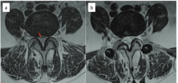
in two groups. The first group comprised of 12 patients with undisrupted annulus integrity, average thickness of the annulus taken in the axial section, and the defect between the point where the thickness began to decrease in the posterior wall and the point where the thickness returned to normal again was accepted as defective area (Figure 1a). The second group, made up of eight patients had the herniations developed through the totally ruptured annulus. The distance between the points where the annulus rupture began and ended was accepted as a defect and measured (Figure 1b).
Objective: Intervertebral disc herniations with wide annulus defects have been stabilised with the posterior dynamic stabilisation method following microlumbar discectomy.
Materials and Methods: The injured anulus is divided into two groups; a) the anulus weakens without losing its integrity and b) tearing all layers of the annulus.
Results: In a one year control, it was found that the annulus was adequately repaired in both groups. The repair process in the full-layer tear occurred with connective tissue. There was no recurrence.
Conclusion: Posterior transpedicular stabilisation is an effective treatment method for annulus repair. Keywords: Disc herniation, dynamic system, annulus repair
ABSTRACT
T. Ali ZIRH
İstanbul Medipol University Faculty of Medicine, Department of Neurosurgery, İstanbul, Turkey
POSTERIOR ANNULUS REPAIR AFTER DYNAMIC
STABILISATION
DOI: 10.4274/jtss.galenos.2020.247
Figure 1. a) weaken but preserved integrity of posterior annulus,
preop axial T2 image b) postop 1-year axial; T2 image shows
healing of annulus
T. Ali Zırh. Posterior Annulus Repair After Dynamic Stabilisation J Turk Spinal Surg 2020;31(3):167-9
168
In microlumbar discectomy procedure for the first group, they tried to preserve the integrity of the wall, and only an incision was made parallel to the annulus fibres and the intact part of the nucleus was not touched. The degenerative parts of the nucleus that moved into the annulus fibers were removed. For the second group, classic microlumbar discectomy procedure was performed. All the patients in two groups were stabilised under C-arm scopy. The Dynesys system (Zimmer Inc., Warsaw, IN, USA) was placed as posterior dynamic stabilisation (PDS) method.
After one year of follow-up, all patients were examined with MR protocol and the results were compared with preoperative MR findings.
RESULTS
After one year of follow-up, annulus repair was achieved in all patients in both groups. The patients in the first group with unimpaired annulus integrity observed the annulus healed close to the original, while those in the second group with full-layers rupture, observed the defects repaired with connective tissue (Figure 2 a,b).
Another remarkable observation was that, the annulus defect healed by making an inward fold in some patients (Figure 3). Recurrence was not detected in both groups.
DISCUSSION
Disc herniation develops as a result of weakening and tearing of the annulus. The greater the defect, the greater the risk of developing herniation. The relationship between the size of the defect and recurrence was systematically emphasised by Carragee et al.(2) for the first time. He noticed that there was a close relationship between annulus defect size and recurrence rate.
Subtotal discectomy is recommended to minimise recurrence rate in microlumbar discectomy patients. From our own experience, the general opinion is that subtotal discectomy reduces recurrence rates. However, it is also known that segmental instability is not tolerated by some patients where severe painful clinical picture appear(3).
The proper healing of the annulus is the most important point
after discectomy. However, if the patient does not have strong muscle compensation, it is almost impossible to repair the annulus properly. Nucleus fragments, which remained under load after discectomy, leak through the defect and prevent proper healing process.
Anatomically, in wide annulus defects, after discectomy, the only barrier between the residual disc tissue inside and the spinal canal is the posterior longitudinal ligament. This ligament is thick in the midline and weakens laterally. From this point of view, after the defect was developed in the annulus, there was no barrier to protect nerve tissue, whether discectomy was performed or not. Subtotal discectomy reduces the nucleus stock that will come out from the inside, but does not completely eliminate the risk.
In subtotal discectomy, it is a known fact that the disc, which has been severely damaged after surgery, spontaneously fused in the long term. Another fact is that, the level of fusion over the years cause disruption of adjacent segments, therefore, we stabilise the patients with large annulus defects using the posterior transpedicular dynamic system. If the disc is not severely damaged, the annulus heals and the disc recovers itself, and in some cases rehydration may occur. In this case, since the movement is preserved, stress in the adjacent segment is reduced and adjacent segment degeneration risk
Figure 3. Annulus defect healed by making an inward fold
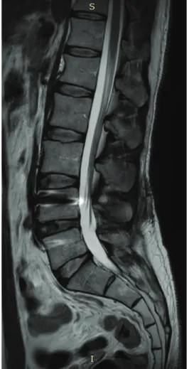
Figure 2. a) All layers of ruptured annulus and extrude herniation
T. Ali Zırh. Posterior Annulus Repair After Dynamic Stabilisation J Turk Spinal Surg 2020;31(3):167-9
169
is less(4). However, in cases where the disc is severely damaged, spontaneous fusion develops even if the level is stabilised with dynamic constructs. The patient would be painless; however, the possibility of adjacent segments problem cannot be eliminated.
CONCLUSION
The annulus integrity is important for recurrent disc herniation. One should bear in mind that dynamic stabilisation accomplishes this reality and this surgical method should be chosen in patients with significant annulus defects. Moreover, the PDS provides stability, therefore the risk of painful period following surgery is less.
Ethics
Ethics Committee Approval: Since this study is a retrospective
study, ethics committee approval was not obtained.
Informed Consent: Retrospective study.
Peer-review: Externally peer-reviewed.
Financial Disclosure: The author declared that this study
received no financial support.
REFERENCES
1. Ozer AF, Keskin F, Oktenoglu T, Suzer T, Ataker Y, Gomleksiz C, et al. A novel approach to the surgical treatment of lumbar disc herniations: indications of simple discectomy and posterior transpedicular dynamic stabilization based on carragee classification. Adv Orthop. 2013;2013:270565. doi: 10.1155/2013/270565 [Epub 2013 Apr 9]. 2. Carragee EJ, Han MY, Suen PW, Kim D. Clinical outcomes after lumbar
discectomy for sciatica: the effects of fragment type and anular competence. J Bone Joint Surg Am. 2003;85:102-8.
3. Schaller B. Failed back surgery syndrome: the role of symptomatic segmental single-level instability after lumbar microdiscectomy. Eur Spine J. 2004;13:193-8.
4. Yilmaz A, Senturk S, Sassanid M, Oktenoglu T, Yaman O, Yildirim H, et al. Disc rehydration after Dynamic Stabilization: A Report of 59 Cases. Asian Spine J. 2017;11:348-55.