DOI:10.25092/baunfbed.340575 J. BAUN Inst. Sci. Technol., 19(2), 123-136, (2017)
The effect of molecular interference on coherent
scattering
Aysun BÖKE
Balıkesir University, Faculty of Arts and Sciences, Department of Physics
Geliş Tarihi (Recived Date): 04.07.2017 Kabul Tarihi (Accepted Date): 09.08.2017
Abstract
The effect of molecular interference on coherent scattering is studied for human tissues (liver, kidney, muscle and fat). The coherent scattering cross sections are computed by numerical integration. The calculation of molecular form factors is performed as function of the momentum transfer variable formulated with x=sin ( /2)/ in the presence of small concentrations of different elements constituting tissue. It is seen that theoretical values of molecular form factors can be used in the region (x ≥ 1 Å-1) where there is no experimental data. Because of the lack of data on calculation of coherent scattering of tissues, the results of this study will provide valuable knowledge in literature.
Keywords: Coherent (Rayleigh) scattering, interference effect, tissue.
Koherent saçılma üzerine moleküler girişimin etkisi
Özet
Koherent saçılma üzerine moleküler girişimin etkisi, insan dokuları (karaciğer, böbrek, kas ve yağ) için çalışılmıştır. Koherent saçılma tesir kesitleri nümerik integrasyonla hesaplanmıştır. Moleküler form faktörlerin hesabı, x=sin ( /2)/ ile formüle edilen momentum transfer değişkeninin bir fonksiyonu olarak, dokuyu oluşturan farklı elementlerin küçük konsantrasyonlarının varlığında gerçekleştirilmiştir. Moleküler form faktörlerin teorik değerlerinin, deneysel verilerin olmadığı bölgede (x ≥ 1 Å-1
) kullanılabileceği görülmüştür. Dokuların koherent saçılmasının hesabı üzerine verilerin eksikliğinden dolayı, bu çalışmanın sonuçları literatürde değerli bilgi temin edecektir.
Anahtar kelimeler: Koherent (Rayleigh) saçılma, girişim etkisi, doku.
1. Introduction
Coherent scattering which gives rise to interference effects is found to be the most significant scattering mechanism. These interference effects are most important in the forward direction, where the path length differences are small. Interference effects are commonly used in low-energy x-ray diffractometry for material characterization to get information about the atomic and molecular structure. It is concluded by Leliveld et al [1] that Monte Carlo calculations are significantly in error when interference effects are ignored in the model for coherent scattering. The predominance of coherent scattering in photon transport occurrences at low x allows the exploitation of its dependence on molecular interference effects which results in the diffraction pattern itself in terms of tissue characterization. However, practically every molecular compound has its own diffraction pattern, which is not easy to compute or measure. It is, therefore, advisable that the usual tabulations of coherent scattering data performed in the frame of the independent atomic modeling (IAM) are updated or replaced with an appropriate customization [2].
Reliable photon cross section data are required in several fields that include analytical techniques (X-ray fluorescence, electron probe microanalysis, particle included x-ray emission), medical applications (radiology, radiotherapy, nuclear medicine), detector design and quantification, shielding design and industrial quality control [3]. A new method for the determination of molecular scattering differential cross sections for compounds has been early worked [4, 5]. The renewed interest in radiological diagnosis based on x-ray coherent scattering demands careful modeling of the proposed techniques to be optimized for clinical practice [6-8]. A library of the scattering properties of tissues will be an important tool for the developing field of x-ray scatter imaging [8]. Coherent scattering cross sections and molecular form factors of tissues are required especially in a low-energy application, such as mammographic imaging. Their knowledge would help to understand artifacts due to single and multiple scattering in computerized tomography (CT) scanners.
The interference effects are well known in crystallography. A collimated beam of x-rays incident on a crystal produce a scattered beam, very intense in certain directions, corresponding to constructive interference from waves reflected from layers of atoms in the crystal. Interference is possible because the wavelength of the x-rays is about the atomic spacing in a solid (≈1 Å-1
). In an amorphous solid, the arrangement of the molecules is not completely featureless. There is still a weak ordering. Therefore, interference is also possible for coherent x-ray scattering from amorphous materials. In contrast to the sharp diffraction patterns of crystals, amorphous materials generate only one or more broad halos [1].
Some researchers exerted an effort to provide molecular form factors including molecular interference effects from experimental data. Morin [9] have tabulated molecular form factor for liquid water. Tartari et al [2] developed molecular form factor tabulations for coherent scattering of photons in tissues. Peplow and Verghese [10] extracted molecular form factors from experimental measurements. Recently, King et al [11] have reported the experimental molecular form factors of tissues. Rao et al [12] have calculated x-ray scattering cross sections for molecules, plastics, tissues, and few biological materials. Hubbell [13] has tabulated the cross sections for elements and compounds which include muscle and compact bone tissue.
As far as it is known by now, there has not been any theoretical prediction on the molecular form factors obtained by using the relativistic modified atomic form factor (RMFF) approximation. The results are presented in Table 1 for use by others modeling photon transport problems. The calculation of molecular form factors is made according to tissue compositions, taken from ICRP 23 [14] for kidney, muscle, fat and from Kosanetzky et al [15] for liver. The number of elements involved in tissue compositions is 5 for liver, 47 for kidney, 44 for muscle and 3 for fat. The molecular coherent scattering cross sections are computed both with inclusion and without inclusion of the molecular interference effects. The new data are listed in Table 2 for this time by this study. Comparison on coherent cross section is presented in Table 3 for muscle tissue.
2. Theoretical background
2.1. The molecular form factors
Mainly there are two approaches, to obtain a detailed description of coherent scattering process: the numerical partial wave calculations of elastic scattering amplitudes using second-order S-matrix-theory and the form factor (FF) approximation. The FF approximation is often used to predict the Rayleigh scattering amplitudes. A detailed explanation on the FF approximations is given by Böke [16].
Theoretical predictions of the FF are based on nonrelativistic and relativistic individual electron and total atom wave functions. There is also the RMFF approximation, which accounts for the correction for binding of the atomic electrons. Hubbell et al [17], Hubbell and Øverbø [18] and Schaupp et al [19] have tabulated the nonrelativistic, relativistic and relativistic modified FF for a wide range of momentum transfer and for all elements, respectively. Detailed tabulation of the atomic FF is also presented by Chantler [20].
Clearly, experimental FF results are in better agreement with the RMFF approximation. The findings suggesting the superiority of the RMFF theory have been reported earlier [16, 19, 21-35]. It has been established both from comparison of theoretical results and from experiments that the RMFF, in general, produces better results than other choices of the FF [34].
The FF is usually calculated assuming a free atom with a spherically symmetric electron charge distribution. Furthermore, when the FF of a mixture of elements is calculated, it is assumed that the FF combines independently. As a result of these assumptions the scattering cross section model for coherent scattering only accounts for the interference between scatterings from electrons in the same atom.
When considering a molecule, the FF is often calculated [36] by adding the squares of the individual atomic FF, weighted by their respective atomic abundances ni.
2 2
( ) ( , )
mol i i i
f x n f x Z (1) For most composite materials, the atomic abundances are not known so the molecular coherent scatter FF can be expressed [2, 6, 10, 37-39] without knowing the molecular formula by using the free atom model:
2 2 ( ) ( , ) mol i i i i f x w f x Z W M (2)
where w,, Mi, Zi and f(x,Zi) are the mass fraction, the atomic mass, the atomic number and the atomic FF of element i, respectively. W is the molecular weight. The fmol(x), function of the momentum transfer variable x, is the molecular FF.
This approach does not consider the interference effects between the electrons of the various atoms when they are all assembled into the molecule. The approach also leaves out intermolecular interference effects, which depend on the spatial distribution of molecules in the material. Since the sum rule ignores these effects, it is called the free-gas model.
2.2. The coherent scattering cross section
The coherent (Rayleigh) scattering cross section per molecule is given as
2 0 ) ( ) ( f x d T mol mol (3)
Total coherent scattering cross section is computed by using numerical integration of the Thomson [40] formula weighted by the f 2mol(x) defined in Eq. (3). The differential Thomson scattering cross section per electron, is given as
) cos 1 ( 2 2 2 e T r d d (4)
where the classical electron radius is re=e2/mec2.
3. Results and discussion
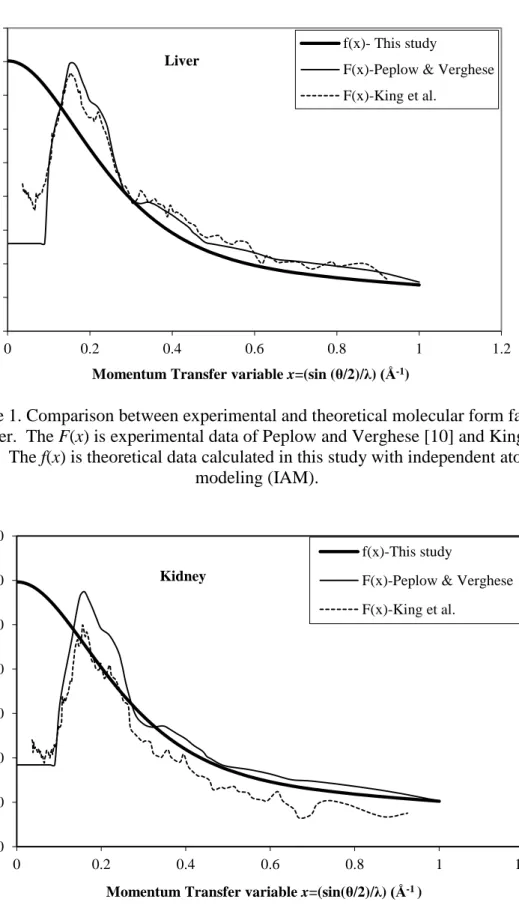
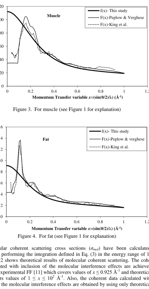
The theoretical FF values of tissues are calculated by using formula defined in Eq. (2) from x=0 Å-1 to 102 Å-1 and listed in Table 1. In calculations, the RMFF approximation of Schaupp et al [19] is used. The theoretical FF values are shown in Figures 1-4 together with those of measured by King et al [11] and Peplow and Verghese [10]. The theoretical FF values provide an approximation in the region (x ≥ 1 Å-1) where there is no experimental data. As discussed by some researchers [9, 10, 13, 38, 41, 42, 43], the measured values approach the IAM values at large values of x. As can be seen in figures 1-4, the theoretical and experimental FF values are very close to each other around x=1 Å-1.
The differences between the experimental and theoretical FF are most important for low x values. These differences are due to intramolecular and intermolecular interferences effects. It is clear from figures 1-4 that the experimental FF values differ with theoretical predictions. The FF values show a strong increase for small x values where interference effects are big. As a result of interference, the angular distribution does not
peak around zero scatter angle but at a specific angle. This peak angle depends on the primary photon energy and the type and structure of the scatter material.
Figure 1. Comparison between experimental and theoretical molecular form factors for liver. The F(x) is experimental data of Peplow and Verghese [10] and King et al
[11]. The f(x) is theoretical data calculated in this study with independent atomic modeling (IAM).
Figure 2. For kidney (see Figure 1 for explanation)
0 2 4 6 8 10 12 14 16 18 0 0.2 0.4 0.6 0.8 1 1.2 M o lecula r F o rm F a ct o r
Momentum Transfer variable x=(sin (θ/2)/λ) (Å-1)
Liver
f(x)- This study
F(x)-Peplow & Verghese F(x)-King et al. 0 20 40 60 80 100 120 140 0 0.2 0.4 0.6 0.8 1 1.2 M o lecu la r Fo rm Fa ct o r
Momentum Transfer variable x=(sin(θ/2)/λ) (Å-1 )
Kidney
f(x)-This study
F(x)-Peplow & Verghese F(x)-King et al.
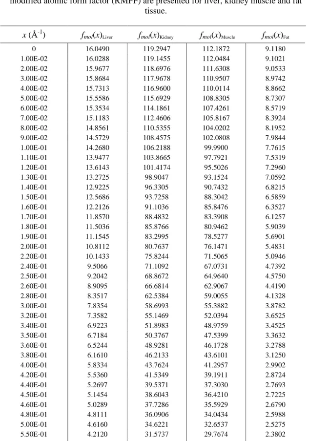
Figure 3. For muscle (see Figure 1 for explanation)
Figure 4. For fat (see Figure 1 for explanation)
The molecular coherent scattering cross sections (σmol) have been calculated by numerically performing the integration defined in Eq. (3) in the energy range of 1-150 keV. Table 2 shows theoretical results of molecular coherent scattering. The coherent data calculated with inclusion of the molecular interference effects are achieved by using both experimental FF [11] which covers values of x ≤ 0.925 Å-1 and theoretical FF which covers values of 1 ≤ x ≤ 102
Å-1. Also, the coherent data calculated without inclusion of the molecular interference effects are obtained by using only theoretical FF
0 20 40 60 80 100 120 0 0.2 0.4 0.6 0.8 1 1.2 M o lecula r F o rm F a ct o r
Momentum Transfer variable x=(sin(θ/2)/λ) (Å-1)
Muscle
f(x)- This study
F(x)-Peplow & Verghese F(x)-King et al. 0 2 4 6 8 10 12 14 16 0 0.2 0.4 0.6 0.8 1 1.2 M o lecula r F o rm F a ct o r
Momentum Transfer variable x=(sin(θ/2)/λ) (Å-1)
Fat
f(x)- This study
F(x)-Peplow & verghese F(x)-King et al.
which covers values of 0 ≤ x ≤ 102
Å-1.In Table 3, the molecular coherent coefficients (σmol) are compared for only muscle tissue because of the lack of data in the literature.
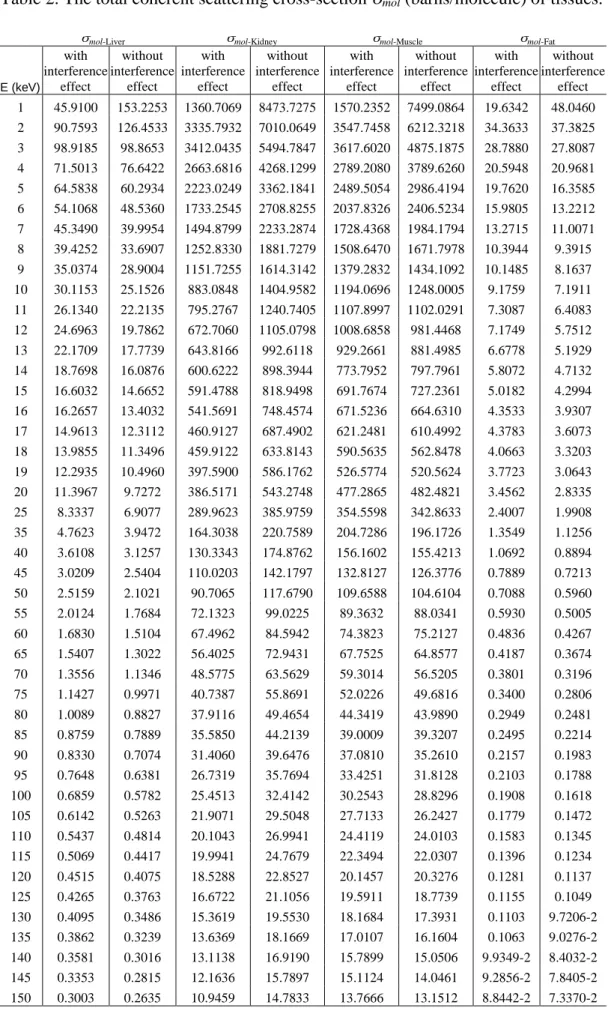
Table 1. The theoretical molecular form factors calculated by using the relativistic modified atomic form factor (RMFF) are presented for liver, kidney muscle and fat
tissue.
x (Å-1) fmol(x)Liver fmol(x)Kidney fmol(x)Muscle fmol(x)Fat
0 16.0490 119.2947 112.1872 9.1180 1.00E-02 16.0288 119.1455 112.0484 9.1021 2.00E-02 15.9677 118.6976 111.6308 9.0533 3.00E-02 15.8684 117.9678 110.9507 8.9742 4.00E-02 15.7313 116.9600 110.0114 8.8662 5.00E-02 15.5586 115.6929 108.8305 8.7307 6.00E-02 15.3534 114.1861 107.4261 8.5719 7.00E-02 15.1183 112.4606 105.8167 8.3924 8.00E-02 14.8561 110.5355 104.0202 8.1952 9.00E-02 14.5729 108.4575 102.0808 7.9844 1.00E-01 14.2680 106.2188 99.9900 7.7615 1.10E-01 13.9477 103.8665 97.7921 7.5319 1.20E-01 13.6143 101.4174 95.5026 7.2960 1.30E-01 13.2725 98.9047 93.1524 7.0592 1.40E-01 12.9225 96.3305 90.7432 6.8215 1.50E-01 12.5686 93.7258 88.3042 6.5859 1.60E-01 12.2126 91.1036 85.8476 6.3527 1.70E-01 11.8570 88.4832 83.3908 6.1257 1.80E-01 11.5036 85.8766 80.9462 5.9039 1.90E-01 11.1545 83.2995 78.5277 5.6901 2.00E-01 10.8112 80.7637 76.1471 5.4831 2.20E-01 10.1433 75.8244 71.5065 5.0946 2.40E-01 9.5066 71.1092 67.0731 4.7392 2.50E-01 9.2042 68.8672 64.9640 4.5750 2.60E-01 8.9095 66.6814 62.9067 4.4190 2.80E-01 8.3517 62.5384 59.0055 4.1328 3.00E-01 7.8354 58.6993 55.3882 3.8782 3.20E-01 7.3582 55.1469 52.0394 3.6525 3.40E-01 6.9223 51.8983 48.9759 3.4525 3.50E-01 6.7184 50.3767 47.5399 3.3632 3.60E-01 6.5244 48.9281 46.1728 3.2788 3.80E-01 6.1610 46.2133 43.6101 3.1250 4.00E-01 5.8334 43.7624 41.2957 2.9902 4.20E-01 5.5360 41.5349 39.1911 2.8724 4.40E-01 5.2697 39.5371 37.3030 2.7693 4.50E-01 5.1454 38.6043 36.4210 2.7225 4.60E-01 5.0289 37.7286 35.5929 2.6790 4.80E-01 4.8111 36.0906 34.0434 2.5988 5.00E-01 4.6160 34.6221 32.6537 2.5275 5.50E-01 4.2120 31.5737 29.7674 2.3802 6.00E-01 3.8998 29.2108 27.5280 2.2643
Table 1. (Continued.)
x (Å-1) fmol(x)Liver fmol(x)Kidney fmol(x)Muscle fmol(x)Fat
6.50E-01 3.6565 27.3660 25.7792 2.1659 7.00E-01 3.4620 25.8886 24.3786 2.0789 8.00E-01 3.1654 23.6388 22.2487 1.9193 9.00E-01 2.9379 21.9215 20.6286 1.7669 1.00E+00 2.7424 20.4551 19.2498 1.6189 1.10E+00 2.5635 19.1208 17.9983 1.4763 1.20E+00 2.3906 17.8354 16.7939 1.3398 1.30E+00 2.2243 16.6015 15.6380 1.2118 1.40E+00 2.0647 15.4193 14.5305 1.0933 1.50E+00 1.9110 14.2803 13.4628 0.9848 1.60E+00 1.7646 13.1962 12.4459 0.8860 1.70E+00 1.6263 12.1711 11.4838 0.7968 1.80E+00 1.4965 11.2091 10.5804 0.7164 1.90E+00 1.3755 10.3120 9.7376 0.6443 2.00E+00 1.2632 9.4792 8.9548 0.5799 2.20E+00 1.0638 8.0002 7.5639 0.4709 2.40E+00 8.9571E-01 6.7520 6.3893 0.3841 2.50E+00 8.2215E-01 6.2058 5.8752 0.3475 2.60E+00 7.5502E-01 5.7071 5.4057 0.3149 2.80E+00 6.3772E-01 4.8355 4.5851 0.2594 3.00E+00 5.4018E-01 4.1105 3.9022 0.2150 3.30E+00 4.2392E-01 3.2457 3.0875 0.1640 3.50E+00 3.6217E-01 2.7860 2.6541 0.1378 3.60E+00 3.3537E-01 2.5863 2.4658 0.1266
3.90E+00 2.6782E-01 2.0824 1.9905 9.8932E-02
4.00E+00 2.4878E-01 1.9401 1.8562 9.1321E-02
4.20E+00 2.1571E-01 1.6927 1.6224 7.8165E-02
4.60E+00 1.6379E-01 1.3029 1.2536 5.8000E-02
5.00E+00 1.2625E-01 1.0194 0.9845 4.3784E-02
5.40E+00 9.8635E-02 0.8093 0.7843 3.3565E-02
5.50E+00 9.2922E-02 0.7656 0.7425 3.1481E-02
5.80E+00 7.8043E-02 0.6513 0.6330 2.6102E-02
6.00E+00 6.9710E-02 0.5868 0.5711 2.3123E-02
6.20E+00 6.2453E-02 0.5304 0.5168 2.0550E-02
6.60E+00 5.0518E-02 0.4368 0.4263 1.6375E-02
7.00E+00 4.1262E-02 0.3633 0.3549 1.3188E-02
7.40E+00 3.3989E-02 0.3049 0.2979 1.0724E-02
8.00E+00 2.5792E-02 0.2380 0.2322 7.9932E-03
9.00E+00 1.6867E-02 0.1633 0.1582 5.0923E-03
1.00E+01 1.1438E-02 0.1164 0.1114 3.3784E-03
1.10E+01 7.9953E-03 8.5726E-02 8.0477E-02 2.3180E-03
1.20E+01 5.7319E-03 6.4901E-02 5.9464E-02 1.6355E-03
1.40E+01 3.1360E-03 3.9865E-02 3.4298E-02 8.7227E-04
1.60E+01 1.8311E-03 2.6329E-02 2.0972E-02 4.9941E-04
1.80E+01 1.1235E-03 1.8372E-02 1.3427E-02 3.0162E-04
2.00E+01 7.1662E-04 1.3369E-02 8.9237E-03 1.8977E-04
2.20E+01 4.7111E-04 1.0053E-02 6.1208E-03 1.2319E-04
2.50E+01 2.6183E-04 6.8575E-03 3.6489E-03 6.7218E-05
2.80E+01 1.5073E-04 4.8649E-03 2.2819E-03 3.7884E-05
Table 1. (Continued.)
x (Å-1) fmol(x)Liver fmol(x)Kidney fmol(x)Muscle fmol(x)Fat
3.50E+01 4.3309E-05 2.3968E-03 8.7668E-04 1.0020E-05
4.00E+01 1.6372E-05 1.5198E-03 4.8486E-04 3.1719E-06
4.50E+01 4.6102E-06 9.9039E-04 2.8667E-04 2.3799E-07
5.00E+01 3.4685E-06 6.5998E-04 1.8289E-04 1.3157E-06
6.00E+01 6.4303E-06 3.1065E-04 9.8708E-05 2.1072E-06
7.00E+01 6.5184E-06 1.5940E-04 7.2035E-05 2.0422E-06
8.00E+01 5.7895E-06 9.2446E-05 5.8703E-05 1.7863E-06
9.00E+01 4.9401E-06 6.2516E-05 4.9001E-05 1.5160E-06
1.00E+02 4.1660E-06 4.8090E-05 4.1116E-05 1.2773E-06
In the numerical integration, the integration variable was taken as 1-cosθ, from which the values of x=sin(θ/2)/λ(Å)=[(1-cosθ)/2]1/2/λ(Å) could be computed close to θ=0. The integration range used, was from 1-cos = 10-16 to 2.0 ( =0.0000008538 to 180 ), divided into intervals in the logarithm of 1-cos by using a modified formula [16]. However, in the studies of Hubbell et al [17] and Hubbell and Øverbø [18], the integration range was taken from 1-cos =10-12 to 2.0 ( =0.000081 to 180 ). Since the FF values change quickly for small x values, the integration mesh points can be increased in this range. Thus, with taking into account of the smaller scattering angles, the more accurate data are obtained.
The total coherent cross section is sensitive to the effects of interference at very low energies, where the integration covers values of small x, and it causes a significant decrease in the coherent cross section. It is observed in Table 2 at the photon energies below 3 keV for liver, fat and below 10 keV for kidney, muscle. For E<12 keV, the integration uses only values of x < 1 Å-1 and so total coherent coefficients (σmol) use experimental data. For E>12 keV, the integration extends to value of x ≥ 1 Å-1 and so coherent coefficients (σmol) use both experimental and theoretical data.
In Table 3, the differences between this study and Hubbell [13] are due to the inclusion of the molecular interference effects and smaller scattering angles. It is expected that the dissimilarity can be greater for values of E<10 keV. Besides, the reason for such a discrepancy can come from differences in the molecular weight and the elemental composition fractions. The number of elements involved in muscle tissue composition is 44 in this study and 10 in the study of Hubbell [13].
Table 2. The total coherent scattering cross-section mol (barns/molecule) of tissues.
mol-Liver mol-Kidney mol-Muscle mol-Fat
E (keV) with interference effect without interference effect with interference effect without interference effect with interference effect without interference effect with interference effect without interference effect 1 45.9100 153.2253 1360.7069 8473.7275 1570.2352 7499.0864 19.6342 48.0460 2 90.7593 126.4533 3335.7932 7010.0649 3547.7458 6212.3218 34.3633 37.3825 3 98.9185 98.8653 3412.0435 5494.7847 3617.6020 4875.1875 28.7880 27.8087 4 71.5013 76.6422 2663.6816 4268.1299 2789.2080 3789.6260 20.5948 20.9681 5 64.5838 60.2934 2223.0249 3362.1841 2489.5054 2986.4194 19.7620 16.3585 6 54.1068 48.5360 1733.2545 2708.8255 2037.8326 2406.5234 15.9805 13.2212 7 45.3490 39.9954 1494.8799 2233.2874 1728.4368 1984.1794 13.2715 11.0071 8 39.4252 33.6907 1252.8330 1881.7279 1508.6470 1671.7978 10.3944 9.3915 9 35.0374 28.9004 1151.7255 1614.3142 1379.2832 1434.1092 10.1485 8.1637 10 30.1153 25.1526 883.0848 1404.9582 1194.0696 1248.0005 9.1759 7.1911 11 26.1340 22.2135 795.2767 1240.7405 1107.8997 1102.0291 7.3087 6.4083 12 24.6963 19.7862 672.7060 1105.0798 1008.6858 981.4468 7.1749 5.7512 13 22.1709 17.7739 643.8166 992.6118 929.2661 881.4985 6.6778 5.1929 14 18.7698 16.0876 600.6222 898.3944 773.7952 797.7961 5.8072 4.7132 15 16.6032 14.6652 591.4788 818.9498 691.7674 727.2361 5.0182 4.2994 16 16.2657 13.4032 541.5691 748.4574 671.5236 664.6310 4.3533 3.9307 17 14.9613 12.3112 460.9127 687.4902 621.2481 610.4992 4.3783 3.6073 18 13.9855 11.3496 459.9122 633.8143 590.5635 562.8478 4.0663 3.3203 19 12.2935 10.4960 397.5900 586.1762 526.5774 520.5624 3.7723 3.0643 20 11.3967 9.7272 386.5171 543.2748 477.2865 482.4821 3.4562 2.8335 25 8.3337 6.9077 289.9623 385.9759 354.5598 342.8633 2.4007 1.9908 35 4.7623 3.9472 164.3038 220.7589 204.7286 196.1726 1.3549 1.1256 40 3.6108 3.1257 130.3343 174.8762 156.1602 155.4213 1.0692 0.8894 45 3.0209 2.5404 110.0203 142.1797 132.8127 126.3776 0.7889 0.7213 50 2.5159 2.1021 90.7065 117.6790 109.6588 104.6104 0.7088 0.5960 55 2.0124 1.7684 72.1323 99.0225 89.3632 88.0341 0.5930 0.5005 60 1.6830 1.5104 67.4962 84.5942 74.3823 75.2127 0.4836 0.4267 65 1.5407 1.3022 56.4025 72.9431 67.7525 64.8577 0.4187 0.3674 70 1.3556 1.1346 48.5775 63.5629 59.3014 56.5205 0.3801 0.3196 75 1.1427 0.9971 40.7387 55.8691 52.0226 49.6816 0.3400 0.2806 80 1.0089 0.8827 37.9116 49.4654 44.3419 43.9890 0.2949 0.2481 85 0.8759 0.7889 35.5850 44.2139 39.0009 39.3207 0.2495 0.2214 90 0.8330 0.7074 31.4060 39.6476 37.0810 35.2610 0.2157 0.1983 95 0.7648 0.6381 26.7319 35.7694 33.4251 31.8128 0.2103 0.1788 100 0.6859 0.5782 25.4513 32.4142 30.2543 28.8296 0.1908 0.1618 105 0.6142 0.5263 21.9071 29.5048 27.7133 26.2427 0.1779 0.1472 110 0.5437 0.4814 20.1043 26.9941 24.4119 24.0103 0.1583 0.1345 115 0.5069 0.4417 19.9941 24.7679 22.3494 22.0307 0.1396 0.1234 120 0.4515 0.4075 18.5288 22.8527 20.1457 20.3276 0.1281 0.1137 125 0.4265 0.3763 16.6722 21.1056 19.5911 18.7739 0.1155 0.1049 130 0.4095 0.3486 15.3619 19.5530 18.1684 17.3931 0.1103 9.7206-2 135 0.3862 0.3239 13.6369 18.1669 17.0107 16.1604 0.1063 9.0276-2 140 0.3581 0.3016 13.1138 16.9190 15.7899 15.0506 9.9349-2 8.4032-2 145 0.3353 0.2815 12.1636 15.7897 15.1124 14.0461 9.2856-2 7.8405-2 150 0.3003 0.2635 10.9459 14.7833 13.7666 13.1512 8.8442-2 7.3370-2
Table 3. Comparison of coherent scattering coefficients (m-1) for muscle tissue.
E(keV) This study Hubbell [13]
10 21.309 18.408 15 12.345 10.088 20 8.517 6.344 30 4.465 3.224 40 2.787 1.872 50 1.957 1.248 60 1.327 0.936 80 0.791 0.520 100 0.540 0.416 150 0.246 0.208 1
The coherent scattering coefficients tabulated by Hubbell [13] are converted to units m-1 by
using density of 1.04 g cm-3 from ICRP 23 [14] for muscle.
4. Conclusion
The differences between experimental and theoretical molecular FF have repercussions upon total coherent scattering cross sections (σmol). As molecular form factors vary importantly for low x values, coherent cross sections differ significantly for low photon energies. Both molecular form factors and coherent cross section differences are due to interference effects.
References
[1] Leliveld, C.J., Maas, J.G., Bom, V.R. and van Eijk, C.W.E., Monte Carlo modelling of coherent scattering: Influence of interference, IEEE Transactions on Nuclear Science, 43, 3315-3321, (1996).
[2] Tartari, A., Taibi, A., Bonifazzi, C. and Baraldi, C., Updating of form factor tabulations for coherent scattering of photons in tissues, Physics in Medicine and Biology, 47, 163-175, (2002).
[3] Baró, J., Roteta, M., Fernández-Varea, J.M. and Salvat, F., Analytical cross sections for Monte Carlo simulation of photon transport, Radiation Physics and Chemistry, 44, 531-552, (1994).
[4] İçelli, O. and Erzeneoğlu, S., A new method for the determination of molecular scattering differential cross sections in some lanthanide compounds with energy dispersive x-ray fluorescence system, Nuclear Instruments and Methods in Physics Research Section B: Beam Interactions with materials and Atoms, 215, 9-15, (2004).
[5] Akça, B. and Erzeneoğlu, S., The Determination of Molecular Scattering Differential Cross Sections for Compounds of Some Essential Elements at 3.38 1/Angstrom Photon-Momentum Transfer, Canadian Journal of Physics, 94, 1-4, (2016).
[6] Taibi, A., Royle, G.J. and Speller, R.D., A Monte Carlo simulation study to investigate the potential of diffraction enhanced breast imaging, IEEE Transactions on Nuclear Science, 47, 1581-1586, (2000).
[7] Harding, G. and Schreiber, B., Coherent x-ray scattering imaging and its applications in biomedical sciences and industries, Radiation Physics and Chemistry, 56, 229-245, (1999).
[8] Leclair, R.J. and Johns, P.C., X-ray forward-scatter imaging: Experimental validation of model, Medical Physics, 28, 210-219, (2001).
[9] Morin, L.R.M., Molecular form factors and photon coherent scattering cross sections of water, Journal of Physical and Chemical Reference Data, 11, 1091-1098, (1982).
[10] Peplow, D.E. and Verghese, K., Measured molecular coherent scattering form factors of animal tissues, plastics and human breast tissue, Physics in Medicine and Biology, 43, 2431-2452, (1998).
[11] King, B.W., Landheer, K.A. and Johns, P.C., X-ray coherent scattering form factors of tissues, water and plastics using energy dispersion, Physics in Medicine and Biology, 56, 4377-4397, (2011).
[12] Rao, D.V., Takeda, T., Itai, Y., Akatsuka, T., Cesareo, R., Brunetti, A. and Gigante, G.E., X-Ray scattering cross sections for molecules, plastics, tissues, and few biological materials, Journal of Trace and Microprobe Techniques, 20, 327-361, (2002).
[13] Hubbell, J.H., Photon cross sections, attenuation coefficients, and energy absorption coefficients from 10 keV to 100 GeV, NSRDS-NBS, 29,1-80, (1969).
[14] ICRP (International Commission on Radiological Protection) Report of the Task Group on Reference Man ICRP Report 23, Oxford: Pergamon, (1975).
[15] Kosanetzky, J., Knoerr, B., Harding, G. and Neitzel, U., X-ray diffraction measurements of some plastic materials and body tissues, Medical Physics, 14, 526-532, (1987).
[16] Böke, A., Calculation of the total Rayleigh scattering cross sections of photons in the energy range of 30-50 keV for Nb and Mo elements, Radiation Physics and Chemistry, 80, 609-613, (2011).
[17] Hubbell, J.H., Veigele, W.J., Briggs, E.A., Brown, R.T., Cromer, D.T. and Howerton, R.J., Atomic form factors, incoherent scattering functions, and photon scattering cross sections, Journal of Physical and Chemical Reference Data, 4, 471-538, (1975).
[18] Hubbell, J.H. and Øverbø, I., Relativistic atomic form factors and photon coherent scattering cross sections, Journal of Physical and Chemical Reference Data, 8, 69-105, (1979).
[19] Schaupp, D., Schumacher, M., Smend, F., Rullhusen, P. and Hubbell, J.H., Small-angle Rayleigh Scattering of Photons at High Energies: Tabulations of Relativistic HFS Modified Atomic Form Factors, Journal of Physical and Chemical Reference Data, 12, 467-512, (1983).
[20] Chantler, C.T., Detailed tabulation of atomic form factors, photoelectric absorption and scattering cross section, and mass attenuation coefficients in the vicinity of absorption edges in the soft X-ray ( Z=30-36, Z=60-89, E= 0.1 keV-10 keV), addressing convergence issues of earlier work, Journal of Physical and Chemical Reference Data, 29, 597-1056, (2000).
[21] Zhou, B. and Pratt, R.H., Calculation of Anomalous scattering for ions and atoms, Physica Scripta, 41, 495-498, (1990).
[22] Bradley, D.A. and Ghose, A.M., Total-atom differential coherent-scattering crosssection measurements on Sn and Pb using moderate-energy rays, Physical Review A, 33, 191-204, (1986).
[23] Bradley, D.A., Gonçalves, O.D. and Kane, P.P., Measurements of photon–atom elastic scattering cross-sections in the photon energy range 1 keV to 4 MeV, Radiation Physics and Chemistry, 56, 125-150, (1999).
[24] Bradley, D.A., Roy, S.C. and Kissel, L., Pratt, R.H., Anomalous scattering effects in elastic photon–atom scattering from biomedically important elements, Radiation Physics and Chemistry, 56, 175-195, (1999).
[25] Eichler, J., de Barros, S., Gonçalves, O. and Gaspar, M., Comparison of Compton and Rayleigh scattering at 145 keV, Physical Review A, 28, 3656-3658, (1983).
[26] Siddappa, K., Nayak, N.G., Balakrishna, K.M. and Lingappa, N., Experimental studies on atomic form factors at 4.808-Å-1 photon momentum transfer, Physical Review A, 39, 5106-5110, (1989).
[27] Kissel, L., Pratt, R.H. and Roy, S.C., Rayleigh scattering by neutral atoms, 100 eV–10 MeV, Physical Review A, 22, 1970-2004, (1980).
[28] Kissel, L., RTAB: the Rayleigh scattering database, Radiat, Radiation Physics and Chemistry, 59, 185-200, (2000).
[29] Nayak, N.G. and Siddappa, K., Experimental atomic form factors of some rare earth and heavy elements by coherent scattering of 145.4 keV gamma rays, Radiation Physics and Chemistry, 71, 673-675, (2004).
[30] İçelli, O. and Erzeneoğlu, S., Coherent scattering of 59.5 keV -rays by 79Au through angles from 451˚ to 1251˚, Spectrochimica Acta Part B, 56, 331-335, (2001).
[31] Kane, P.P., Mahajani, J., Basavaraju, G. and Priyadarsini, A.K., Scattering of 1.1732-and 1.3325 MeV gamma rays through small angles by carbon, aluminum, copper, tin, and lead, Physical Review A, 28, 1509-1516, (1983). [32] Kane, P.P., Elastic scattering of gamma rays and X-rays, Radiation Physics
and Chemistry, 74, 402-410, (2005).
[33] Roy, S.C. and Kissel, L., Pratt, R.H., Elastic photon scattering at small momentum transfer and validity of form-factor theories, Physical Review A, 27, 285-290, (1983).
[34] Roy, S.C., Zhou, B., Kissel, L. and Pratt, R.H., Rayleigh scattering and form factors, Indian Journal of Physics B, 67, 481-496 , (1993).
[35] Roy, S.C., Kissel, L. and Pratt, R.H., Elastic scattering of photons, Radiation Physics and Chemistry, 56, 3-26, (1999).
[36] Chan, H.P. and Doi, K., Energy and angular dependence of x-ray absorption and its effect on radiographic response in screen-film systems, Physics in Medicine and Biology, 28, 565-579, (1983).
[37] Tartari, A., Casnati, E., Bonifazzi, C. and Baraldi, C., Molecular differential cross sections for x-ray coherent scattering in fat and polymethyl methacrylate, Physics in Medicine and Biology, 42, 2551-2560, (1997).
[38] Tartari, A., Bonifazzi, C., Fernandez, J.E., Bastiano, M., Casnati, E., Baraldi, C. and Di Domenico, G., Molecular coherent scattering data for tissue in photon transport Monte Carlo codes, Applied Radiation and Isotopes, 53, 901-906, (2000).
[39] Tartari, A., Taibi, A., Bonifazzi, C., Gambaccini, M. and Marina, F., Updating of x-ray coherent scattering cross-sections and their effects in microbeam and material analysis applications, X-Ray Spectrometry, 34, 421-425, (2005). [40] Thomson, J.J., Conduction of electricity through gases, Cambridge University
[41] Narten, A.H. and Levy, H.A., Water: A Comprehensive Treatise, In: Franks, F. Ed. vol 1, p. 311, Plenum Press, New York, London, (1972).
[42] Tartari, A., Bonifazzi, C. and Casnati, E., Photon scattering data from X-ray diffraction pattern measurements: correction procedure evaluation, Nuclear Instruments and Methods B, 142, 203-209, (1998).
[43] King, B.W. and Johns, P.C., An energy-dispersive technique to measure x-ray coherent scattering form factors of amorphous materials, Physics in Medicine and Biology, 55, 855-871, (2010).