INTRODUCTION
In order to improve drug efficacy, the need for advanced drug delivery systems attracted polymer chemist’s attention from the late 1960s onwards. An approach towards an improved use of drugs for therapeutic applications is the synthesis of polymeric networks. Hydrogels, composed of three-dimensional hydrophilic polymer networks, increase in volumes and retain large volumes of water without dissolution in exceeding amount of water [1]. They
can be used in many pharmaceutical and biomedical fields such as artificial organs, contact lenses, wound dressings etc. Well-characterized hydrogels were composed by many synthetic and naturally derived materials. Experiments and researches are focused on both synthetic and natural polymer-based hydrogels, better biocompatibility, biodegradability and non-toxicity [2]. Hydrogels are suitable carriers for drug delivery applications because they exhibit a three-dimensional structure that controls drug, peptide or protein release [2]. Hydrogels based on synthetic polymers have continued to be of interest for various biomedical applications. Photopolymerization technique used by ultraviolet (UV) cure is one of the applied methods due to rapid cure, low curing temperature and low energy requirement.
HACETTEPE JOURNAL OF BIOLOGY AND CHEMISTRY Research Article
Hacettepe J. Biol. & Chem., 2009, 37 (4) 337-343
* Correspondence to: Hakan Ayhan,
Muğla University, Department of Chemistry, Biochemistry Division and Biomedical Technologies Research and Application Centre, 48170 Mugla, Turkey.
Tel: +90252 211 1506 E-mail: [email protected]
Photopolymerized Poly(Ethylene Glycol) Diacrylate Hydrogels for
Controlled Release of Ketoprofen
Nazlı Sökmen1, Seda Uysal2, Fatma Ayhan1, Mine Özyazıcı2, Hakan Ayhan1*
1Muğla University, Department of Chemistry, Biochemistry Division, Muğla, Turkey
2Ege University, Faculty of Pharmacy, Department of Pharmaceutical Technology, İzmir, Turkey
Article Info Abstract
Article history:
Received
June 5, 2009 Received in revised form October 25, 2009 Accepted
October 29, 2009 Available online
December 31, 2009
In this study, poly(ethylene glycol-diacrylate) (PEG-DA) hydrogels with two different concentrations, 30% and 50% w/w, were prepared by free radical polymerization method. Ethylene glycol dimethacrylate (EGDMA) and 2,2-dimethoxy-2-phenyl-acetophenone (DMPA) were used as cross-linking agent and photoinitiator, respectively. The model drug ketoprofen (KP) was incorporated into the hydrogel during photopolymerization and its release kinetics was tested spectrophotometrically at 256 nm in different buffer solutions of pH 7.5, 4.5, and 1.2. The results showed that the release of KP strongly depends on the dissolution of drug, initial polymer concentration, and the pH of the release medium.
All of the obtained results were evaluated kinetically by first order and zero order, Higuchi and Hixon-Crowell kinetic models. The release mechanism was found to obey Higuchi and first order kinetics when the release data of the formulations were applied to the models in the different pH mediums.
Key Words Hydrogel, Drug delivery, PEG-DA, photopolymerization, Ketoprofen,
Drug release models.
---'-Poly(ethylene glycol) (PEG) is a non-toxic, water-soluble polymer that resists recognition by the immune system. It ex hibits rapid clearance from the body and has been approved for a wide range of biomedical applications [3]. There are some researches which demonstrate the release of various drug compounds such as proxyphylline [2] and bovine serum albumin [4] from PEG diacrylate (PEG-DA) hydrogels. Its use was also expanded by the addition of small amounts of a cross-linking agent such as ethylene glycol dimethacrylate or di(ethylene glycol) dimethacrylate. Applications include carti lage replacements, bonding agents in dental resins and bone ce ments, and various drug delivery vehicles [5].
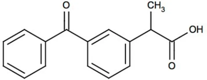
Ketoprofen [2-(3-benzoylphenyl)propionic acid] (KP) is known by its non-steroidal anti-inflammatory, antipyretic and analgesic features. Although KP has a short half-life of 2 hours, it reaches the highest plasma concentration in 1-2 hours. Due to the fast elimination property of KP from the body, frequent doses are needed to have therapeutic maintenance and to get the therapeutic efficacy for extended time. KP is generally implemented orally [6]. In every six or eight hours, KP must be taken for pain, that is three or four times in a day. Revealing of high doses may lead to damage in the stomach as ulceration or bleeding. In order to prevent this drawback, sustained release or enteric coating dosage forms have been improved [7]. The mechanism of action of KP is mainly associated to the inhibition of the body’s ability to synthesize prostaglandins [8]. The short half-life, low bioavailability and local or
systemic disturbance in the GI tract to cause withdrawal of treatment make KP a very good candidate for formulation of increased and of controlled release dosage forms [9,10]. Controlled drug delivery is more advantageous than the conventional dosage forms in terms of improvement of therapeutic efficacy, reducing toxicity and delivering at controlled rate, many researchers focused on controlled drug delivery systems more and more in the last decades [5].
Various attempts have been made to increase the efficacy of KP. Polymeric prodrugs containing profens were synthesized for the controlled release of KP and other profens in order to increase solubility, protect from deactivation, and improve pharmacokinetics which are also known as the advantages of polymeric prodrugs [11]. KP-PEG conjugates were prepared and their potentials as a sustained release system were also investigated [6]. Various molecular weights of PEG were studied to form conjugates with KP and the hydrolysis kinetics of the drug were examined. A gelatin-based pharmaceutical hydrogel was prepared by the use of konjac glucomannan cross-linker, which crosslinked and gelled within minutes [1]. KP release from biodegradable injectable thermo-sensitive hydrogels of PEG-PLGA-PEG triblock copolymers [12], polycaprolactone, polyurethanes and their blends prepared from electrospinning [13], polyions and gelatin encapsules were studied.
The focus of this work is to investigate the use of KP containing photopolymerized PEG-DA hydrogels. The release of KP from polymeric network with different polymer concentrations was studied in vitro in buffered solutions at different pH values and their obtained results were discussed.
Figure 1. Chemical structure of ketoprofen.
OH
MATERIALS AND METHODS
Materials
Poly(ethylene glycol)-diacrylate (PEG-DA, Mn: 700), cross-linking agent ethylene glycol dimethacrylate (EGDMA) and 2,2-dimethoxy-2-phenyl-aceto-phenone (DMPA) were obtained from Aldrich Chemical Company. Ketoprofen was a gift from Eczacıbaşı (Eczacıbaşı İlaç San. Tic. A.Ş., Turkey). All other solvents and reagents were of analytical grade.
Preparation of Hydrogels
The hydrogels of PEG-DA containing non-steroidal anti-inflammatory, antipyretic and analgesic drug, KP, and ethylene glycol dimethacrylate as crosslinking agent were prepared by the UV-curing polymerization method. Precursor solution was prepared by mixing 30 and 50 wt % PEG-DA and 0.5 wt % photoinitiator, DMPA (macromere based). The crosslinking agent, EGDMA, was added at a concentration of 1 wt %, based on macromere weight. KP was dissolved in distilled water and added to precursor solution up to the final volume. The final drug concentration in each hydrogel was kept as 2 mg in all experiments. PEG-DA hydrogel without drug was also synthesized as a control group. The final solution was purged with nitrogen for a few minutes. The solution was polymerized by exposure to 365 nm UV light with 10 mW/cm2
intensity for 5 min. [2]. Cylindrical glass petri dishes of diameter size of 17 mm were used to synthesize hydrogels. Then, they were removed gently after synthesis and stored at 4oC until use. The same
method was used to prepare hydrogels without drug by adding distilled water.
In Vitro Dissolution Studies
In vitro release studies were performed with 75 rpm speed at room temperature (25oC). The release
studies were tested for 180 minutes in 40 mL buffer of pH 1.2 ± 0.1, pH 4.5 ± 0.1, pH 7.5 ± 0.1 (citrate/
HCl and phosphate) and the absorbance was estimated at different time intervals spectro-photometrically at 259 and/or 260 nm, respectively [14-16]. The cumulative fractional release at time t was then calculated (n=3). The dissolution of pure drug of KP (P-KP) was investigated with the same procedure [17]. The drug (2 mg) was placed into the buffer solution and the absorbance of the sample taken at the same time intervals as done for hydrogels. The concentration of KP released from the networks was expressed as a percentage of the total KP available and plotted as a function of time. Each value was given as the average of the three experimental results.
Release Kinetics
Four kinetic models were chosen to describe the release profiles of the prepared systems. Thus, the following kinetic models represented in Eqs. (1-4) were investigated using a computer program developed in our laboratory for empirical analysis [18].
First-order model;
ln (100-W) = ln 100- kf t (1)
Higuchi square root of time model;
W = kHt1/2 (2)
Hixon and Crowell cube-root model;
(100-W)1/3 = 1001/3 - k
HCt (3)
Zero-order model;
m = 100 – k0t (4)
Where W is percent drug release rates at time t, and kf, kH , kHC and k0 are release rate constants, respectively [19-23].
RESULTS AND DISCUSSION
Preparation of drug loaded hydrogels and in vitro drug release studies
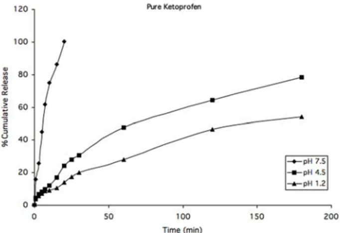
The use of acrylate-based hydrogels in controlled drug release devices was investigated. In this scope, two different compositions of acrylate-based hydrogels were synthesized and used in this study. The cumulative release of pure KP in three different pH medium, pH 1.2, 4.5, and 7.5, were shown in Figure 2. Pure drug KP dissolution was measured spectrophotometrically. The KP release was completed approximately after 20 minutes in pH 7.4 medium. On the other hand, drug release of 78% was observed in pH 4.5 and 54% was measured for pH 1.2. The drug dissolution was faster in neutral medium than acidic medium. KP is a white or offwhite, odorless, fine or granular powder, melting at about 95°C and practically insoluble in water at 20°C and at low pH [24]. The results indicated below are in agreement with literature.
The cumulative release of KP from 30% PEG-DA hydrogels in different pH medium was shown in Figure 3. All the cumulative results were given for 180 min solute release time. The drug release from hydrogel networks follows the same release trend as in the previous dissolution study. It was observed that the drug release was completed about 87% in pH 7.5. The second release was monitored in pH 4.5 and the last KP release was measured in high
acidic media which is pH 1.2. The cumulative release occurred as 72% in pH 4.5 and 54% in pH 1.2 after the release time indicated before.
The cumulative release of KP from 50% PEG-DA hydrogels in different pH medium was shown in Figure 4. In the experiments, drug release was investigated in the same pH values as before. The graph reveals the values referring to three different medium at different time intervals up to 180 min. The highest cumulative drug release was again observed at pH 7.5 with a cumulative release value of 58%.
Then, KP release in pH 4.5 and 1.2 was calculated as 48% and 31%, respectively. In neutral medium pH 7.5, the release of KP was again higher than other acidic medium when 50% PEG-DA was used in the hydrogel synthesis.
Figure 2. Dissolution of pure KP in three different pH mediums (n=3).
Figure 3. The release of KP from 30% PEG-DA hydrogels in three different pH buffers (n=3).
Figure 4. The release of KP from 50% PEG-DA hydrogels in different pH buffers (n=3).
~OPEG-OA 120 P\l'e Ketoproten 100 120
5
80 100:
..
..
~ 60 M 80..
1 ., .; o a: 40 .,*
~ 601
20 u 40*
o 20 o 50 100 150 200 Tlme (mln) o o so 100 1 so 200 Tlme (mln) 120 1650 PEG-OA 100 80J
t 60 ;; ( # •0 20 so 100 1sO 200 Tm:(mn)The cumulative drug release in phosphate buffer of pH 7.5 was bigger for both pure KP, 30 and 50% PEG-DA hydrogels which can be seen from the Figures 2-4. In general, drug release increases as pH gets high. This was an expected result. The main reason of this situation is greatly due to the drug release behaviour in different medium and different PEG concentrations in hydrogels. In the hydrogels; PEG diacrylates in high percentage (50%) leads to high pore formation. Thus, the network was more grift and the drug release becomes slowly and as a result the release profile was lowered. When Figures 3 and 4 were examined, because of the PEG acrylates concentrations, in the same pH, KP release was changed. In all graphs, the cumulative KP were approximately increased 20% with increasing pH. In pH 4.5; 30% PEG-DA KP release was 72% when 50% PEG-DA KP release was 48%. As seen in the Figures 2-4 dissolution profile of P-KP was not almost similar. The results showed that high concentration of PEG-DA cause the highly cross-linked network sturucture. Because in the same pH values 30% PEG-DA hydrogels release more than 50% PEG-DA. In the Figure 2, Pure-KP was released immediately in the pH 7.5 greatly due to its high dissolution as a chemical compound.
The results indicate that the cumulative KP release was lowered with decreasing pH. Arida and Al-Tabakha investigated KP release from KP particles micro-encapsulated with 41-111 nm thick gelatin/ polyanion multilayer shells in three different pH (pH: 1.4 (stomach), pH: 4.1 (intestine), pH 7.4 (blood)). It was reported that the results of the KP release indicated 107 times less release compared to uncoated KP. The fraction of KP release decreased when gelatin coating layer increased two and three fold [24]. The cumulative KP release increased with increasing pH. Jeong et al. synthesized PEG-PLGA-PEG triblock copolymer for loading model drug KP. Hydrophilic model drug, KP’s release were investigated at 37°C. The release was finished after
2 weeks. The initial polymer concentrations were changed as 20%, 25%, and 33% while drug concentration was fixed at 1.0 wt%. It was reported that the slower drug release was obtained when the initial polymer solution concentration was higher due to higher polymer-polymer contacts [12].
Non-steroidal anti-inflammatory drugs, ketoprofen, ibuprofen and naproxen were studied by Babazadeh. In three pH values, the release of these drugs were examined. According to the study, the cumulative drug releases were increased when the pH was raised [11]. Kenawy et al. were prepared electrospun fibers in different ratios of polyurethane/ polycaprolactane. These nanofibers were loaded with model drug KP and its release profiles were studied [13]. Gentamicin release from 30 and 50% PEG-DA containing hydrogels were examined in buffered solutions at pH 7.4 and 2.2. It was observed that the drug release depends on polymer concentration and less concentration leads to higher cumulative drug release. The pH of the release medium is reported as another delivery parameter and the drug release from the hydrogel with low polymer concentration concluded with higher cumulative release [25].
Release Kinetic Models
The first order (Eq.(1)) describes the release from systems where release rate is concentration dependent. Higuchi (1963) described the release of drugs from insoluble matrix as a square root of time dependent process based on Fickian diffusion (Eq. (2)). The Hixon–Crowell cube root law (Eq. (3)) describes the release from systems where there is a change in surface area and diameter of the particles or tablets. The zero order rate Eq. (4) describes the systems where the drug release rate is independent of its concentration.
The dissolution results were evaluated using a computer programme [19]. The obtained
determination coefficient (r2) values of release
kinetics for KP from PEG-DA hydrogel formulations were listed in Table 1. The abbreviations of 30 and 50% PEG-DA hydrogels were given as F1 and F2, respectively. The release profiles of this type of drug diffusion also obeyed Higuchi square root of time kinetic [26]. Dissolution data from F1 fit to Higuchian model while F2 in pH 1.2 obeys first order equation which suggests drug load dependent release. The dissolution data from all the products in pH 4.5 were plotted in accordance to the proposed models. The results fit to the first order model which is the loaded drug dependent release behaviour for both types of hydrogels. According to the kinetic evaluations, the best fitting equation with the highest determination coefficient is the Higuchi square root of time kinetic in pH 7.5 for both F1 and F2 formulations. The release at this pH is also based on Fickian diffusion according to the Higuchi model.
CONCLUSION
Ketoprofen loaded PEG-DA hydrogels were synthesized with free radical photopolymerization method. The release of KP from different
acrylate-based hydrogels depends on the amount of used macromere and immersion medium. High concentration of PEG-DA cause the higher cross-linked network structure which diminishes the cumulative release of the model drug KP. The release mechanism was usually found to be first order and Higuchi kinetics from the calculations of the release data in the different pH medium. Our results suggest that PEG-DA hydrogels could be an alternative method to the controlled release of KP in order to diminish the side effects of the drug and protect its stability.
REFERENCES
1. Yu, H., Xiao, C., Synthesis and properties of novel hydrojels from oxidized konjac glucomannan crosslinked gelatin for in vitro drug delivery, Carbohydrate Polym., 72, 479-489, 2008.
2. Scott, R.A., Peppas, N.A., Highly crosslinked, PEG-containing copolymers for sustained solute delivery, Biomater., 20:1371-1380, 1999.
3. Peppas, N.A., Bures, P., Leobandung, W., Ichikawa, H., Hydrogels in pharmaceutical formulations, Eur. J. Pharm. Biopharm., 50: 27–46, 2000.
4. Mellott, M.B., Searcy, K., Pishko, M.V., Release of protein from highly cross-linked hydrogels of poly(ethylene glycol) diacrylate fabricated by UV polymerization, Biomater., 22:929–941, 2001.
5. Gursel, I., Balcik, C., Arica, Y., Akkus, O., Akkas, N., and Hasirci, V., Synthesis and mechanical properties of interpenetrating networks of polyhydroxybutyrate- co-hydroxyvalerate and polyhydroxyethyl methacrylate, Biomat., 19:1137-1143, 1998.
6. Choi, Hoo-K., Chun, Myung-K., Lee, S.H., Jang, M.H., Kim, H.D., Jung, C.S., Oh, S.Y., In vitro and in vivo study of poly(ethylene glycol) conjugated ketoprofen to extend the duration of action, Int. J. Pharm., 341; 50-57, 2007. 7. Vueba, M.L., Batista de Carvalho, L.A.E., Veiga, F.,
Sousa, J.J., Pina, M.E., Influence of cellulose ether polymers on ketoprofen release term from hydrophilic matrix tablets, Eur. J. Pharm. Biopharm., 58: 51-59, 2004. Table 1. The determination coefficient (r2) of release
kinetics for hydrogels formulations.
Code* First orderHiguchi Zero order
Hixon-Crowell Zero order pH 1.2 F1 0.9888 0.9968 0.9807 0.9590 F2 0.9970 0.9594 0.9961 0.9930 pH 4.5 F1 0.9976 0.9909 0.9967 0.9575 F2 0.9945 0.9907 0.9899 0.9766 pH 7.5 F1 0.9847 0.9964 0.9684 0.9225 F2 0.9213 0.9761 0.9026 0.8612
8. Reynolds, J.E.F. (Ed.) Martindale The Extra Pharmacopeia, Vol.29, The Pharmaceutical Press, London, pp.25, 1989.
9. Liversidge, G.G. in: Florey K. (Ed.), Analytical Profiles of Drug Substance, Academic Press, Inc, New York, pp. 443-471, 1981.
10. Habib, M.J., Mesue, R., Development of Controlled Release Formulations of Ketoprofen for Oral Use, Drug Develop. Ind. Pharm., 21: 1463-1472, 1995.
11. Babazadeh, M., Design, synthesis and in vitro evaluation of vinyl ether type polymeric prodrugs of ibuprofen, ketoprofen and naproxen, Int. J. Pharm., 356: 167-173, 2008.
12. Jeong, B., Bae, Y.H., Kim, S.W., Drug Release from biodegradable injectable thermosensitive hydrogel of PEG-PLGA-PEG triblock copolymers, J. Control. Release, 63; 155-163, 2000.
13. El-Kenawy, R., Abdel-Hay, F.I., El-Newehy, M.H., Wnek., G.E., Processing of polymer nanofibers through electrospinning as drug delivery systems, Mater. Chem. Phys., 113; 296-302, 2009.
14. The United States Pharmacopeia, The National Formulary, USP XXIV, NF XIX, Supplement Two, Convention Inc., Rockville, 2000.
15. British Pharmacopeia (2005), Vol. I-II, The Stationery Office, London.
16. Clarke’s, Isolation and Identification of Drugs, 2ndEdition,
Ed. A.C. Moffat, London: The Pharmaceutical Press, pp.697-698, 1986.
17. The United States Pharmacopeia, The National Formulary, USP XXIV, NF XIX, Supplement Two, Convention Inc., Rockville, 2000.
18. Ege, M.A., Karasulu, H.Y., Karasulu, E., Ertan, G., 4th Central European Symposium on Pharmaceutical Technology, 23–25 September, Vienna, 2001, Sci. Pharm. 69 (3), <http://pharm.ege.edu.tr/applications/>.
19. Martin, A., Physical Pharmacy, Physical Chemical Principles In The Pharmaceutical Sciences, Fourth Edition, Lea and Febiger, Philadelphia, Pennsylvania, USA 1993, pp. 324-361.
20. Borodkin, S., Tucker, F.E., Drug release from hydroxpropyl cellulose-polyvinyl acetate films, J. Pharm. Sci., 63: 1359-1363, 1974.
21. Higuchi, T., Mechanism of sustained-action medication. Theoretical analysis of rate of release of solid drugs dispersed in solid matrices, J. Pharm. Sci. 52: 1145-1149, 1963.
22. Schwartz, B.J., Simonelli, A.P., Higuchi, W.I., Drug release from wax matrices I; Analysis of data with first-order kinetics and with the diffusion-controlled model, J. Pharm. Sci. 57: 274-277, 1968.
23. Wagner, J.G., Interpretation of percent dissolved time plots derived from in vitro testing of conventional tablets and capsules, J. Pharm. Sci., 58: 1253-1257, 1969. 24. Arida, A.I., Al-Tabakha, M.M., Encapsulation of ketoprofen
for controlled drug release, Eur. J. Pharm. Biopharm., 66; 48-54, 2007.
25. Ayhan F., Özkan S., Gentamicin Release from Photopolymerized PEG Diacrylate and PHEMA Hydrogel Discs and Their in vitro Antimicrobial Activities, Drug Delivery, 14: 433-439, 2007.
26. Schwartz, B.J., Simonelli, A.P., Higuchi, W.I., Drug release from wax matrices I: Analysis of data with first-order kinetics and with the diffusion-controlled model, J. Pharm. Sci. 57: 274-277, 1968.