Introduction
Angiogenesis, the formation of new blood vessels, is critical to physiological processes such as wound healing, tissue growth, intrauterine development, and reproductive functions. Wound healing presents the formation of granulation tissue, which may be characterized with fibrovascular tissue, including fi broblasts, collagen, and blood vessels. Angiogenesis is
regulated by the expression of various vascular growth factors and modulators, which are defi ned as engines driving wound repair. Among them, vascular endothelial growth factor (VEGF) is a potent, multifunctional cytokine that exerts some vital and independent actions on vascular endothelium. Being more potent than histamine, it serves as an inducer of vascular permeability. Increased vascular permeability occurs during the early phases of wound repair. Events such as increased vascular permeability and concentration of infl ammatory cells on the site of injury, produced by VEGF, actively provide tissue repair. Increasing the migration and proliferation of pre-existing endothelial cells also assists in this process.[1-3]
The VEGF gene encodes a protein that is often found as a disulfi de-linked homodimer. The VEGF protein is a glycosylated mitogen that specifically acts on
Topical Single-dose Vascular Endothelial Growth
Factor has No Effect on Soft Tissue Healing
Nilufer Bolukbasi, Huseyin Avni Balcioglu
1, Birkan T. Ozkan
2, Merva Soluk Tekkesin
3,
Duran Ustek
4Department of Oral Implantology, Faculty of Dentistry, 1Department of Anatomy, Faculty of Dentistry, 3Institute of Oncology, Istanbul University, Istanbul, 2Department of Oral Surgery, Faculty of Dentistry, 100 Yil University, Van,
4Medipol University, Istanbul, Turkey
Abstract
Background: Vascular endothelial growth factor (VEGF) production in dermal wounds has been evaluated for evidence that it plays
a probable role in wound healing. Events such as increased vascular permeability and concentration of infl ammatory cells on the site of injury, produced by VEGF, were linked to tissue repair. Aim: The present study aimed to evaluate the effects of single-dose topical administration of VEGF on wound healing. Materials and Methods: A total of 30 male Wistar albino rats weighing 250-280 g were used in this study. In addition, 2-cm-long skin incisions were created over bilaterally exposed skin of the tibia region in each rat. VEGF plasmid 2 μg was administered locally into the right side wound bed of each animal. No other procedure besides skin closure was administered on the left side. To determine histologic assessments, skin samples were obtained from six anesthetized rats at each interval (4, 8, 12, 16 and 30 days) through excisional biopsy. The tissues were fi xed in 10% neutral-buffered formalin for 1 week and then embedded in paraffi n wax. Transverse sections of the embedded tissue 5-7 μm thick were stained with hematoxylin and eosin (H and E). Results: There was no signifi cant difference regarding necrosis, epithelialization, infl ammation, fi broblast activity, ulcerative formation, or hemorrhage between experimental and control groups. No statistically signifi cant difference was found between the groups regarding granulation tissue formation and epidermal thickness. Conclusion: The administration method and dosage of VEGF is a major factor in terms of its effectiveness. The results of the present study did not evaluate the effectiveness of single-dose 2 μg topical administration of VEGF; however, various doses of VEGF plasmid should be tested in future studies in order to provide benefi cial effects from topical administration of VEGF.
Keywords:
Angiogenesis, Vascular endothelial growth factor, Vascular permeability, Wound healingAddress for correspondence: Dr. Huseyin Avni Balcioglu, Department of Anatomy, Faculty of Dentistry, Istanbul University, Istanbul, Turkey. E-mail: [email protected]
Access this article online
Quick Response Code:
Website:
www.najms.org
DOI:
10.4103/1947-2714.143281
endothelial cells and has various effects, including mediating increased vascular permeability; inducing angiogenesis, vasculogenesis, and endothelial cell growth; promoting cell migration; and inhibiting apoptosis. The identification of increased vascular permeability accompanied with increased VEGF production in dermal wounds provided evidence for a probable role of VEGF in wound healing. VEGF seems to be a direct angiogenic factor that stimulates endothelial cell migration. Furthermore, various animal studies have been published investigating the upregulation of VEGF production in wound healing.[4]
The present study was carried out to evaluate the effects of single-dose topical administration of VEGF plasmid on the healing of induced rat skin wounds.
Materials and Methods
The study was conducted at the Department of Genetics, Institute of Experimental Medicine, Istanbul University. The study protocol was approved by the Istanbul University Animal Care Ethical Committee. All procedures were conducted in accordance with the Istanbul University ethical guidelines for the treatment and welfare of experimental animals. In total, 30 male Wistar albino rats weighing 250-280 g were used in this study. The animals were housed at 21°C and were given tap water and standard rat food.
Plasmid construction
The VEGF-A expression plasmid was constructed by ligating full-length human VEGF-A (Gene ID: 7422) cDNA into pcDNA4 (Invitrogen, Carlsbad, USA). Briefl y, total RNA was isolated from cultured cells using a total RNA purifi cation kit according to the manufacturer’s recommendations (QiagenCo. Hilden, Germany). After reverse transcription, the VEGF-A cDNA PCR was amplifi ed with the forward primer 5’ AATTCGCCG ACATGACGGACAGACAG ACAGACACCGCC 3’ containing EcoRI sites and the reverse primer 5’ TCTAGATCAC CGCCTCGGCTTGTCA CATCTGC 3’ with an modified XbaI site at their 5’ and 3’ ends. The purified PCR fragments and pcDNA4 were double-digested with EcoRI and XbaI restriction enzymes. The digested fragments recovered from 1% low-melting agarose gel by cutting, and DNA was eluted from the sliced agarose gel using an extraction kit (Qiagen Co. Hilden, Germany). In addition, 50 ng of EcoRI and XbaI digested, dephosphorylated vector DNA and 1 μl of PCR product were incubated overnight with DNA ligase buffer containing T4 DNA ligase at 16°C. Further, 3 μl of ligation reaction and 100 μl of DH5 alpha competent cells were heat-shock transformed at 42°C and spread on agar plates containing 100 μg/ml ampicillin
(Invitrogen, Carlsbad, USA). Plates were incubated overnight at 37°C. PCR-tested and selected positive clones were confi rmed with sequencing (Refgen Co., Turkey). Plasmid was purifi ed using an Endo-free Maxi plasmid kit (Qiagen Co. Hilden, Germany). Then, 2 μg of the plasmid constructs, bearing a human VEGF-A DNA, were administered to the rat. Empty pcDNA4 plasmid (2 μg) was used as a control.[5,6]
Surgical procedure
The animals were anesthetized via intramuscular injection of ketamine hydrochloride (50–100 mg per kg of body weight). Bilateral tibial regions of the rats were shaved with electric clippers and cleansed with 10% povidone iodine and 70% alcohol swabs before manipulation. Next, 2-cm-long skin incisions were created over bilaterally exposed skin of the tibia region in each rat.
VEGF plasmid (2 μg) was administered locally into the right side wound bed of each animal (experimental group). No extra procedure besides skin closure was administered on the left side (control group). Incision lines were primarily sutured with 3.0 silk (Dogsan Medical Supplies Industry, Trabzon, Turkey) immediately after surgery in all rats.
To determine histologic assessments, skin samples were obtained from six anesthetized rats at each interval (4, 8, 12, 16 days and 1 month) through excisional biopsy, including incision lines, cleared of surrounding mesentery and fat and washed with saline. The anesthetized rats were then sacrifi ced using intraperitoneal 135 mg/kg sodium pentothal.
Histological analysis
The tissues were fixed in 10% neutral-buffered formalin for 1 week and then embedded in paraffi n wax. Transverse sections of the embedded tissue, 5-7 μm thick, were stained with hematoxylin and eosin (H and E). Histological assessment was carried out by an experienced pathologist.
The sections were examined under light microscopy regarding necrosis, epithelialization, inflammation, fi broblast activity, ulceration and hemorrhage. The mean area percentage of granulation tissue was detected in three randomly chosen fi elds for each section under microscope (X200 magnifi cation) and scored as the following: (+) for 0-10 %; (++) for 10-30%, and (+++) for 30-100 %. Epidermal thickness was calculated in millimeters.
Statistical study
The differences in epidermal thickness between the treatment and control groups at time intervals of the
Bolukbasi, et al.: VGEF on soft tissue healing 4, 8, 12, 16 and 30 days, based on all z-scores, were
compared using the Mann-Whitney U-test. At each time interval, the granulation scores of the treatment group were compared with the granulation scores of the control group using Pearson’s chi-square test across treatment groups or Fisher’s exact test, if 20% of the expected counts were < 5. The treatment comparison was performed at an α level of 0.05. All data were analyzed using SPSS for Windows version 21 (IBM SPSS Statistics, New York, USA).
Results
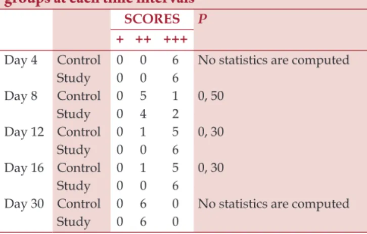
There was no mortality among the animals during the study period. The rats were macroscopically observed on each day of the study period. No complications, such as infection and exposure around the wound margin, which result in secondary healing, were observed. During the early period of wound healing, variables were evaluated and compared at intervals (4, 8, 12, 16 days and 1 month). There was no signifi cant difference with respect to necrosis, epithelialization, inflammation, fi broblast activity, ulcerative formation, or hemorrhage between experimental and control groups. No necrosis was found in any of the rats. The mean area percentage of granulation tissue was more than 30% in the 12th- and 16th-day groups (experimental and control), whereas the mean area percentage of granulation tissue was less than 30% in the remaining groups; however, there was no statistically signifi cant difference between the groups with respect to granulation tissue formation (P > 0.05) [Table 1]. Epidermal thickness scores were given in Table 2. No statistically signifi cant difference was found in epidermal thickness between the groups (P > 0.05) [Table 2].
4
thpostoperative day
In the experimental group, a few ulcerations and desiccate formations were observed on the surface of the epidermis. A number of vascular sections fi lled with red blood cells and surrounded by chronic infl ammatory cells infi ltration were observed. Fresh bloody focal points were present in one sample [Figure 1].
In the control group, ulcerative formation in epidermis was present in all rats. There was desiccate formation on the epidermal surface in two out of six rats. Vascular sections and intense infl ammatory cell infi ltration was observed in the edematous connective tissue under the desiccated areas [Figure 1].
8
thpostoperative day
Both groups showed similar wound-healing outcomes. Continuing ulcerative formation was observed in one rat in the experimental group and three rats in the control
Table 1: Granulation scores of the study and control groups at each time intervals
SCORES P
+ ++ +++
Day 4 Control 0 0 6 No statistics are computed
Study 0 0 6 Day 8 Control 0 5 1 0, 50 Study 0 4 2 Day 12 Control 0 1 5 0, 30 Study 0 0 6 Day 16 Control 0 1 5 0, 30 Study 0 0 6
Day 30 Control 0 6 0 No statistics are computed
Study 0 6 0
Table 2: Epidermal thickness of the study and control groups at each time intervals (mm)
Group Mean SD Z value P
Day 4 Control 0.0270 ,00179 −0.971 0.33 Study 0.0300 ,01789 Day 8 Control 0.0267 ,01633 −0.968 0.33 Study 0.0360 ,00179 Day 12 Control 0.0333 ,01211 −0.686 0.49 Study 0.0393 ,00103 Day 16 Control 0.0400 ,01095 0.000 1.0 Study 0.0400 ,01789 Day 30 Control 0.0400 ,01789 −0.490 0.62 Study 0.0450 ,01225
Figure 1: Histological sections of the study and control groups according to the time intervals
group. Abscess formation in dermis was observed in one rat in the experimental group; infl ammatory cell infiltration of the connective tissue was present in the remaining rats in the experimental group. Intense abscess formation and infl ammatory cell infi ltration of connective tissue was observed in all samples in the control group [Figure 1].
12
thpostoperative day
In the experimental group, the initial phase of epidermis regeneration was seen in all samples, and continuing abscess formation in connective tissue was seen in one individual. Dermal infi ltration of infl ammatory cells was reduced compared to 8th-day outcomes, and the initial phase of fi brotic activity was observed [Figure 1]. In the control group, there was no exposed area on the epidermal surface, except for one individual with continuing ulcerative formation on the surface. Fibrosis was observed in a few areas in the connective tissue [Figure 1].
16
thpostoperative day
Signifi cant reduction of infl ammatory infi ltration in connective tissue was observed in the experimental group. Epidermal healing was also observed in all rats in the control group. Fibrotic focal spots became marked in all samples in both groups [Figure 1].
30
thpostoperative day
Complete regeneration of epidermis and intense activity of dermis were both observed. Fibrosis penetration into deep tissues was observed in the experimental group [Figure 1].
In the control group, epidermal regeneration was almost completed in all samples, and fi brosis of superfi cial areas in connective tissue was clearly seen [Figure 1].
Discussion
VEGF has been proven to be an effective factor in wound healing immediately after injury. Acute infl ammation, re-epithelialization, formation of granulation tissue, and tissue remodeling are phases of wound healing that occur on an ongoing basis. VGEF has a role in wound healing by means of an angiogenic process. Angiogenic activity is maximally observed between days 3 and 7; VEGF is up-regulated to promote the early stages of angiogenesis.[1,7] In the present study, the fi ndings of early angiogenesis could be detected when 4th-day results and 8th-day results were examined, while the features of normal wound repair such as fi brovascular tissue, collagen and blood vessels were observed in all groups throughout the 30-day experiment. However, the ineffectiveness of the administration of VEGF on the following days seems unpromising.
Granulation tissue is eventually remodeled, as vessels are resorbed and fi broblasts disappear. As the wound is granulated, the process of angiogenesis stops, and blood vessels decline as endothelial cells undergo apoptosis.[1,7] In our study, granulation tissue was observed most
between days 12 and 16; area percentage of granulation tissue was detected to be less than 30% in the remaining groups. Contrary to our fi ndings, Corral et al., found that single-dose treatment at 30 μg per wound improved granulation tissue formation.[8] This confl ict may be due to the fact that receptor regulation may be more important than growth factor regulation during wound healing.[9] Wound surface is lined with macrophages that secrete VEGF. Collagen deposition is one of the responses of the endothelial cells to this. Collagen deposition is expected to be excessive in the fi rst 2 weeks and to regress by the end of the second week;[10] however, there was no difference between the groups.
In the present study, even though some infl ammation is needed for wound angiogenesis,[10] the utmost care was taken to avoid any possibility of trauma and infl ammation, which may cause cytokine release and thus affect wound healing. No deposition of unhealthy granulation tissue and delayed closure of the wound were observed. The epidermis regained its thickness in treated and non-treated groups during the phases of the experimental process. Nevertheless, no signifi cant differences were found between any of the groups with regard to epidermal thickness, meaning that a single dose of VEGF plasmid injection, known as a mitogen selective for endothelial cells, was not adequate to increase the capability of inhibiting apoptosis. Corral et al., also claimed that VEGF had no effect on new epithelium formation in either ischemic or nonischemic wounds, whereas Galiano reported that topical VEGF application accelerated cutaneous repair.[8,11] However, a short dosing interval of every-other-day administration for fi ve doses in total was reported to be used in Galiano’s study. VEGF induces interstitial collagenase expression in human endothelial cells.[12] When it is considered that the collagen deposition revealed no difference between the groups, it may be stated that single-dose topical administration of VEGF could neither induce endothelial cells grown on the surface of the collagen matrix nor stimulate the proliferative response.
The authors of this paper are aware that the effect of VEGF is highly dependent on its administration method and dosage.[8,11] While the results of the present study did not evaluate the effectiveness of single-dose 2 μg topical administration of VEGF plasmid, various doses of VEGF should be tested in future studies in order to provide benefi cial effects from topical administration of VEGF.
References
1. Bao P, Kodra A, Tomic-Canic M, Golinko MS, Ehrlich HP, Brem H. The role of vascular endothelial growth factor in wound healing. J Surg Res 2009;153:347-58.
Bolukbasi, et al.: VGEF on soft tissue healing
2. Kanno Y, Hirade K, Ishisaki A, Nakajima K, Suga H, Into T,
et al. Lack of alpha 2-antiplasmin improves cutaneous wound
healing via over-released vascular endothelial growth factor-induced angiogenesis in wound lesions. J Thromb Haemost 2006;4:1602-10.
3. Ozkan U, Osun A, Samancioglu A, Ercan S, Firat U, Kemaloglu S. The effect of bevacizumab and 5-Fluorouracil combination on epidural fi brosis in a rat laminectomy model. Eur Rev Med Pharmacol Sci 2014;18:95-100.
4. Nissen NN, Polverini PJ, Koch AE, Volin MV, Gamelli RL, DiPietro LA. Vascular endothelial growth factor mediates angiogenic activity during the proliferative phase of wound healing. Am J Pathol 1998;152:1445-52.
5. Karatepe HO, Kilincaslan H, Berber M, Ozen A, Saricoban HE, Ustek D, et al. The effect of vascular endothelial growth factor overexpression in experimental necrotizing enterocolitis. Pediatr Surg Int 2014;30:327-32.
6. Adas G, Percem A, Adas M, Kemik O, Arikan S, Ustek D,
et al. VEGF-A and FGF gene therapy accelerate healing of
ischemic colonic anastomoses (experimental study). Int J Surg 2011;9:467-71.
7. Hoeben A, Landuyt B, Highley MS, Wildiers H, Van Oosterom AT, De Bruijn EA. Vascular endothelial growth factor and angiogenesis. Pharmacol Rev 2004;56:549-80. 8. Corral CJ, Siddiqui A, Wu L, Farrell CL, Lyons D, Mustoe TA.
Vascular endothelial growth factor is more important than
basic fibroblastic growth factor during ischemic wound healing. Arch Surg 1999;134:200-5.
9. Brucker MJ, Gruskin E, Farrell CL, Siddiqui A, Mustoe TA. Differential expression of platelet-derived growth factor receptor-beta in an aging model of wound repair. Wound Repair Regen 1996;4:219-23.
10. Hunt TK, Gimbel M, Sen CK. Revascularization of wounds. In: Figg W, Folkman J. Eds. Angiogenesis: An Integrative
Approach from Science to Medicine. 1st ed. New York:
Springer; 2008:541-60.
11. Galiano RD, Tepper OM, Pelo CR, Bhatt KA, Callaghan M, Bastidas N, et al. Topical vascular endothelial growth factor accelerates diabetic wound healing through increased angiogenesis and by mobilizing and recruiting bone marrow-derived cells. Am J Pathol 2004;164:1935-47. 12. Unemori EN, Ferrara N, Bauer EA, Amento EP. Vascular
endothelial growth factor induces interstitial collagenase expression in human endothelial cells. J Cell Physiol 1992;153:557-62.
How to cite this article: Bolukbasi N, Balcioglu HA, Ozkan BT, Tekkesin MS, Ustek D. Topical single-dose vascular endothelial growth factor has no effect on soft tissue healing. North Am J Med Sci 2014;6:505-9.