The powdery mildews of Kıbrıs Village Valley
(Ankara, Turkey)
Tuğba EKİCİ , Makbule ERDOĞDU1 2
1 1
, Zeki AYTAÇ , Zekiye SULUDERE 1
2
Gazi University, ,
Department of Biology, Teknikokullar, Ankara-TURKEY
Ahi Evran University, ,
Department of Biology, Kırsehir-TURKEY Abstract
Key words: :
Özet:
Anahtar Kelimeler:
A search for powdery mildews present in Kıbrıs V
N T
Kıbrıs Köyü Vadisi' nde (Ankara, Türkiye) bulunan külleme mantarlarının araştırılması 2009-2010 yılları yapılmıştır. Külleme mantarlarına ait toplam 10 tespit edilmiştir:
konukçu bitki üzerinde küllemeye sebep olan ajan tespit edilmiştir.
yeni konukçu olarak kaydedilmiştir. Teşhis edilmiş mantarların ışık ve taramalı elektron mikroskobuna dayalı morfolojik verileri sunulmuştur.
Yeni konukçu, Taksonomi, Türkiye
illage Valley (Ankara,Turkey) was carried out during the period 2009-2010. A total of ten fungal taxa of powdery mildews was observed:
(Griffon & Maubl.) U. Braun & S. Takam. U. Braun DC. R.Y. Zheng & G.Q. Chen DC. var DC. var. (S. Blumer) U.
Braun (Lév.) G. Arnaud (Wallr.) Lév. and
(Duby) U. Braun They were determined as the causal agents of powdery mildew on 13 host plant species. Schreber. for (Duby) U. Braun reported as new host plant. Microscopic data obtained by light and scanning electron microscopy of identified fungi are presented.
ew host, axonomy, Turkey
nda taxa
(Griffon & Maubl.) U. Braun & S. Takam. U. Braun DC.
R.Y. Zheng & G.Q. Chen DC. var DC. var. (S. Blumer) U. Braun
DC (Lév.) G. Arnaud (Wallr.) Lév. (Duby) U.
Braun 3 Rubus sanctus türü
Phyllactinia mali için
Erysiphe alphitoides , E. buhrii , E. heraclei , E. lycopsidis , E. pisi . pisi, E. pisi cruchetiana
, E. polygoni DC., Leveillula taurica , Phyllactinia guttata P. mali .
Rubus sanctus Phyllactinia mali is Erysiphales,
Erysiphe alphitoides , E. buhrii , E. heraclei , E. lycopsidis , E. pisi . pisi, E. pisi cruchetiana , E. polygoni ., Leveillula taurica , Phyllactinia guttata ve P. mali
. 1
Kıbrıs Köyü Vadisi' nin (Ankara, Türkiye)
Külleme Mantarları
Erysiphales,
Faculty of Science
Faculty of Science and Literature
11.06.2013 21.10.2013
Corresponding author:[email protected]
Introduction
The powdery mildews ( ) are one of the most conspicuous and most studied groups of plant pathogens currently comprising 873 recognized species infecting more than 1500 plant genera (Amano 1986; Braun and Cook 2012). The taxonomy and identification of
different powdery mildew taxa were based largely on their host range and the morphological characteristics of their ascomata formerly known as cleistothecia, but recently re-named as chasmothecia (Braun et al. 2002).
This causes problem when a powdery mildew increases its host range or geographical area, because the teleomorph may not be formed for some years, or may even not be produced at all (Cook et al. 1997). So, the classical morphological criteria and host range data have been supplemented with additional taxonomic features such as scanning electron microscope (SEM) studies of conidial surfaces. In addition, the discovery of additional features based on SEM has provided useful support for identification purposes when crucial characters are not clear only using LM (Cook et al. 1997).
-Turanian phytogeographic region and according to the grid square system adopted by Davis
(1965-detail and illustrated.
Plant specimens infected with powdery mildew were collected from
in Ankara province of Turkey. The host specimens were prepared according to established herbarium techniques. Host plants were identified using the Flora of Turkey and East Aegean Islands (Davis 1965-1985). The fungal specimens were isolated from the host plants by obtaining thin sections or scraping. Microscopic examination and microphotographs were done by means of a Leica DM E light microscope. A Leica EZ4D stereo microscope was used for close-up photo of the chasmothecia on the leaf surface. The powdery mildews were identified using relevant literature (Karaca 1961; Dennis 1981; Ellis and Ellis 1987;
Heluta 1989; Fakirova 1991; Braun 1995; Braun and Cook 2012). All specimens examined were deposited in the mycological collection of the Department of Biology, Faculty of Science, Gazi University, in Ankara province of Turkey.
For scanning electron microscopy (SEM), 8 10-mm-square pieces of infected leaves bearing conidia and/or chasmothecia were mounted on the SEM stubs with double-sided adhesive tape. They were coated with gold using a Polaron SC 502 Sputter Coater and were examined with a JEOL JSM 6060 scanning electron microscope operated at 5-10 kV in the Electron Microscopy Unit, Faculty of Science, Gazi University (Turkey).
The was chosen as a
research area, because its climatic conditions and plant distributions are suitable for the growth of numerous microfungi. But the plants are usually completely covered by a dense dust mass caused by the activities of a stone quarries in the research area. This dust mass is a mechanical barrier for the penetration and distribution of leaf-inhabiting fungi. This was detected as a factor for decreasing fungal diversity and rate of contamination.
Ten powdery mildews were identified in the research area. Morphological data obtained by light and scanning electron microscopy of these fungi are provided. The author abbreviations of fungi are according to Kirk and Ansell (1992). The systematics of taxa follow Kirk et al. (2008) and Index Fungorum (www.speciesfungorum. org, accessed 2013). Family and species names are listed in alphabetical order in the text.
(Griffon & Maubl.) U. Braun & S. Takam., 4: 5. 2000.
Mycelium: amphigenous, mainly epiphyllous, in white patches or effuse, persistent on the upper leaf surface.
This research was carried out in valley of Kıbrıs village belonging to Mamak district which is about 20 km southeast of Ankara province. Kıbrıs Village Valley is situated in the Irano
1985), it is located in the squares B4. The climate of the province is Mediterranean. Kıbrıs Village Valley is 1st degree field of natural sites and its three areas are 1st archaeological sites. The powdery mildews on plants in Kıbrıs Village Valley were investigated and classified in this paper. 10 taxa of powdery mildews in Kıbrıs Village Valley are described in
Kıbrıs Village Valley
Kıbrıs Village Valley
Materials and methods
Results and discussion
-Erysiphe alphitoides
Conidiophores erect, straight, rarely curved or flexuous. Conidia: ellipsoid-ovoid to doliiform, with squared wrinkling, 20-26
10-Chasmothecia: amphigenous, mainly epiphyllous, scattered to gregarious,
78-each with multiple asci. Appendages: more or less equatorial, straight to somewhat curved, 0.5-1 times as long as the chasmothecial diam., wall almost smooth to verruculose, colourless or only pigmented at the very base, apically (3-)4-6 times closely and regularly branched, branched part 35- Asci: broadly ellipsoid-ovoid, saccate, short-stalked, 55-62.5 37.5-45(-47.5) µm, containing (6-)8 ascospores. Ascospores: broadly ellipsoid-ovoid, colourless, guttulate, 15-22.5 (7.5-)10-12.5 µm (Fig. 1).
B4 Ankara: Kıbrıs Village, 1100-1150 m,
roadside, on Willd.,
24.09.2009, TE 1097.
Several species of R. Hedw. ex DC. are known to infect oaks. These include (Peck) U. Braun & S.
Takam. (syn. Peck),
(Griffon & Maubl.) U. Braun & S. Takam. (syn. Griffon & Maubl.),
(G.F. Atk.) U. Braun & S. Takam.
(syn. G.F. Atk.), S.
Takam. & U. Braun, (Cooke & Peck) U. Braun & S. Takam. (syn. Cooke & Peck), S. Takam. & U. Braun,
(Nevod.) U. Braun & Cunningt. (syn.
Nevod.), and S.
Takam. & U. Braun (Braun 1987; Braun and Takamatsu 2000; Braun et al. 2003; Braun and Cook 2012). Until now, three species
including , and
have been reported to cause powdery mildews on spp. in Turkey. s. lat. is common, widespread in Turkey on L.,
L., L., Olivier,
Olivier subsp. (Reuter) O.
Schwarz Decne. subsp.
(Kotschy) Hedge & Yalt.,
Ehrb., Willd.,
L. subsp. (Ten.) Schwarz (Göbelez 1963;
Tamer et al. 1990a; Braun 1995; Bahçecioğlu et al. 2006; Erdoğdu and Hüseyin 2008).
on sp. and on
(Mattuschka) Liebl. var. were reported from Turkey as well (Bahçecioğlu et al. 2006; Kabaktepe and Bahçecioğlu 2006).
U. Braun, 32(2): 80. 1978.
Mycelium: amphigenous, white, dense, irregular patches or effuse Conidiophores: straight, cylindrical, erect. Conidia: single-celled, cylindrical, ellipsoid, 23-30 9-15 µm. Conidia viewed with SEM characterized by randomly orientated reticulated wrinkling. Chasmothecia: , numerous, scattered, dark brown to black, 110-150 µm diam., each with multiple asci. Appendages: numerous, 0.5-1.5 times as long as the chasmothecial diam., mycelium-like, septate, thin-walled, brown when mature, simple or irregular branched. Asci: per ascoma sessile or shortl stalked, 62.5-)65-75 (30-)32.5-35 µm, containing 3-5 ascospores. Ascospores: ellipsoid, ovoid, hyaline, 15-22.5 10-15 µm (Fig. 2).
B4 Ankara: Kıbrıs Village, around Durhasanın Kayası, 1300 m, steppe, on
subsp. (Boiss.) Mc Neill &
H.C. Prent. ( Boiss.),
01.08.2010, TE 1189.
is common on
throughout the world, especially Asia and Europe. It is known from Turkey on
L., Boiss.
L., Sibth. & Sm., and (Desf.) Bieb. (Braun 1995; Bahçecioğlu and Yıldız 2005; Bahçecioğlu et al. 2006 )
DC., , Edn 3
(Paris) 6: 107. 1815.
Mycelium: amphigenous, irregular white patches, sometimes effuse to covering the whole leaf surface. Conidiophores: straight, cylindrical. Conidia: cylindrical, oval to fusiform, 26-31 12-15 µm in size. : Quercus pubescens Erysiphe Erysiphe abbreviata Microsphaera abbreviata E. alphitoides M. alphitoides E. calocladophora M. calocladophora E. epigena E. extensa M. extensa E. hypogena E. hypophylla M. hypophylla E. quercicola Erysiphe E. abbreviata, E. alphitoides E. hypophylla Quercus Erysiphe alphitoides Quercus alba Q.
cerris Q. ilex Q. infectoria Q.
infectoria boussieri
, Q. ithaburensis
macrolepis Q.
pedinculata Q. pubescens Q. robur
brutia
Erysiphe
abbreviata Quercus E. hypophylla
Q. petraea petraea . amphigenous 3-10 , ( Silene pratensis eriocalycina Silene eriocalycina E. buhrii Caryophyllaceae Dianthus
caryophyllus Gypsophila libanotica , G.
paniculata Silene discolor
S. spergulifolia Fl. franç. 14 μm. 100 μm diam., 72 μm long. Erysiphe buhrii Erysiphe heraclei Česká Mykol.
Conidia viewed with SEM characterized by randomly orientated reticulated wrinkling. Chasmothecia: spherical, gregarious, 73-130 µm diam., each with multiple asci. Appendages: 0.5-1.5 times as long as the chasmothecial diam., myceloid with branched tips, septate, thin-walled, brown when mature. Asci: 4-6 per ascoma, sessile or short stalked, round to ovoid, 57.5-65 40-45 µm, containing 3-5 ascospores. Ascospores: ellipsoid to ovoid, hyaline, 25-27.5 12.5-15 µm (Fig. 3).
B4 Ankara: Kıbrıs Village, around Cehrelik,
1300-1360 m, on Bernh.,
01.08.2010, TE 1188.
was reported on several
host plants belonging to on
was recorded from Austria, Bulgaria, Czechia, Slovakia, France, Germany, Hungary, Iran, Israel, Poland, Romania, Russia, Turkey, Ukraine, Yugoslavia (Amano 1986; Braun 1995).
R.Y. Zheng & G.Q. Chen, 34: 234. 1981.
Mycelium: amphigenous, white, effuse, persistent or evanescent. Conidiophores:
straight, erect, cylindrical. Conidia: single-celled, ellipsoid, doliiform or cylindrical, 20-32 11-16 µm. Conidia viewed with SEM characterized by randomly orientated and reticulated wrinkling. Chasmothecia: gregarious, numerous, scattered, dark brown to black, 110-140 µm diam. Appendages: 0.5-1.5 times as long as the chasmothecial diam., mycelium-like, hyaline or brown in the lower half, septate, simple or irregular branched. Asci: 4-6 per ascoma, short stalked or subsessile, 72.5-75 42.5-50 µm, containing 3-5 ascospores. Ascospores: ellipsoid, ovoid, hyaline, 15-25(-27.5) 11-15 µm (Fig. 4).
B4 Ankara: Kıbrıs Village, around Kavak
Stream, 1000 m, on Roemer
& Schultes subsp. 01.08.2010, TE 1180.
infects
Desf. in Germany, (L.) Bieb. in Austria, Denmark, France, Germany, Italy, Poland, Romania, Spain, Switzerland, United Kingdom, Miller in Germany, Romania,
Ledeb. in Ukraine, M.
Bieb. in Romania, Boiss & Spruner in
Ukraine, (L.) Johnston. Falcaria vulgaris Erysiphe heraclei Apiaceae. E. heraclei Falcaria vulgaris Sydowia Anchusa leptophylla leptophylla,
Erysiphe lycopsidis Anchusa
altissima A. arvensis A. azurea A. gmelinii A. ochroleuca A. thessala Buglossoides arvensis Erysiphe lycopsidis
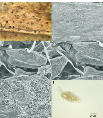
Fig. 1. a - general appearance of powdery mildew; b conidia (SEM). c chasmothecia (SEM); d appendage (SEM); e ascus; f ascospores
Erysiphe alphitoides:
-
-- -
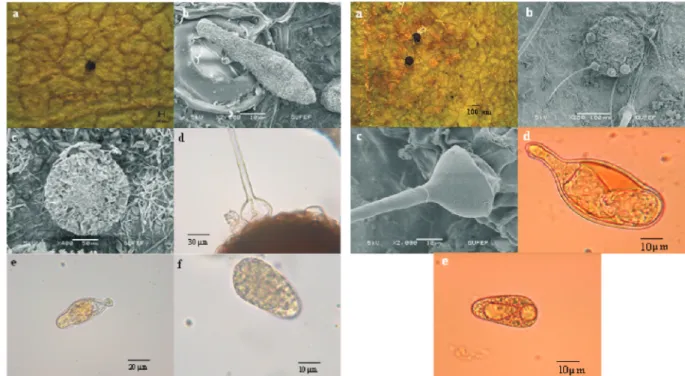
-Fig. 2. a - general appearance of powdery mildew; b conidia (SEM); c chasmothecia (SEM); d chasmothecia and appendages; e ascus and ascospores
Erysiphe buhrii:
-
-in Bulgaria, Ukra-ine,
(L.) Tausch ex L.H. Bailey in Ukraine (Braun 1995), (L.) Bieb. subsp. (L.) Nordh. in China (Amano 1986), Romania, Ukraine (Braun 1995), Lam. in Switzerland (Bolay 2005), Thunb. in Poland (Mulenko et al. 2008), Switzerland (Bolay 2005),
L. in Belarus (Girilovich et al. 2005), Bulgaria, Germany, Norvay (Braun 1995), Poland (Ruszkiewicz-Michalska & Michalski 2005), Russia (Gasich & Berestetskij 1997), Switzerland (Bolay 2005), in Iran (Khodaparast et al.
2000), (Lacaita) Dobrocz. in
Ukraine (Dudka et al. 2004),
L. in Poland (Mulenko et al. 2008), Ukraine (Braun 1995), L. in Russia (Rusanov and Bulgakov 2008),
L. in Russia (Rusanov and Bulgakov 2008), Ukraine (Dudka et al. 2004), L. in China (Braun 1987), Ukraine (Dudka et al. 2004) and sp. in Ukraine (Dudka et al. 2004). It
was observed on L.,
Roemer & Schultes, and sp. in Turkey (Braun 1995; Bahçecioğlu and Yıldız 2005; Bahçecioğlu et al. 2006). Pentaglottis sempervirens A. arvensis orientalis A. caespitosa A. capensis A. officinalis A. ovata Echium biebersteinii Lithospermum arvense L. officinalis Lycopsis arvensis L. orientalis Onosma Anchusa officinalis A. leptophylla Alkanna Erysiphe pisi pisi DC., . 2: 274. 1805 var.
Mycelium: on stems and leaves, amphigenous, white, effuse, sometimes covering the whole leaf surface, persistent or evanescent. Conidiophores: straight, erect, cylindrical. Conidia: single-celled, ellipsoid to cylindrical, 23-29 12-15 µm. Surface ornamentation of conidia viewed with SEM consisting of low reticulate ridges. Chasmothecia: gregarious, numerous, scattered, dark brown to black, 75-150 µm in diam. Appendages: in the lower half, 1.5-3 times as long as the chasmothecial diam., mycelium-like, hyaline or brown, septate, simple or irregular branched. Asci: 4-8 per ascoma, short stalked or subsessile, 60-75 20-30 µm in size, containing 3-6 ascospores.
Fl. franç
Fig. 4. a, b - general appearance of powdery mildew; c conidia (SEM); d chasmothecia and appendages (SEM); e ascus and ascospores
Erysiphe lycopsidis:
-
-Fig. 3. a, b - general appearance of powdery mildew; c conidia (SEM); d chasmothecia and appendages (SEM); e asci and ascospores
Erysiphe heraclei:
-
-Ascospores: ellipsoid, ovoid, hyaline, 22.5-30 10-17.5 µm (Fig. 5).
B4 Ankara: Kıbrıs Village, around
Dipsizgöl, 1050 m, on L.,
24.09.2009, TE 1090.
var was reported on several host plants belonging to It is
known from Turkey on L.,
Lam., DC.,
Pall., Stev.,
sp., L., L.,
L., (L.) All.,
subsp. (L.) Arcangeli, x Martyn, Ten ex Guss.,
L., L.,
L., Huds.,
L., L., (L.) Willd.,
. L., Bornm., L.,
Reuter ex Boiss., L., and Roth (Braun 1995; Bahçecioğlu and Yıldız 2005; Bahçecioğlu et al. 2006; Kabaktepe and Bahçecioğlu 2006). var on was recorded from Austria, Czechia, Slovakia, Finland, France, Germany,
Hungary, Italy, Netherlands, Poland, Romania, Spain, Sweden, Switzerland, Turkey, United Kingdom, Yugoslavia (Braun 1995; Kabaktepe and Bahçecioğlu 2006).
DC. var. (S.
Blumer) U. Braun, (3-4): 692. 1981.
Mycelium: amphigenous, white, effuse, sometimes covering the whole leaf surface, persistent or evanescent. Conidiophores: straight, erect, cylindrical. Conidia: single-celled, ellipsoid to cylindrical, 35-44 15-17.5 µm. Conidia: viewed with SEM characterized by randomly orientated reticulated wrinkling and end of conidium with wart-like structure. Chasmothecia: gregarious, numerous, scattered, dark brown to black, 120-150 µm diam. Appendages: in the lower half, 1-3 times as long as the chasmothecial diam., mycelium-like, hyaline or brown, septate, simple or irregular branched. Asci: 4-8 per ascoma, short stalked or subsessile, 60-75 30-35 µm in size, containing 3-6 ascospores. Ascospores: ellipsoid, ovoid, hyaline, 20-25 12.5-15 µm (Fig. 6).
Medicago lupina
Erysiphe pisi . pisi
Fabaceae. Arachis hypogaea
Astragalus odoratus A. oleifolius A.
ponticus Lathyrus roseus Lathyrus
Medicago falcata M. lupina M.
polymorpha M. rigidula M. sativa
falcata M. varia
Mellilotus neapolitana Phaseolus
vulgaris Pisum sativum Sophora
alopecuriodes Trifolium ochroleucum
T. pratense Vicia cracca V. ervilia
V faba V. feyniana V. lutea V.
noeana V. sativa V.
tenuifolia
Erysiphe pisi . pisi
Medicago lupina
Nova Hedwigia
Erysiphe pisi cruchetiana
34
Fig. 5. var. a, b - general appearance of powdery mildew; c conidia (SEM); d chasmothecia and appendages (SEM); e ascus and ascospores
Erysiphe pisi pisi:
-
-Fig. 6. var. a, b - general
appearance of powdery mildew; c conidia (SEM); d end of conidium showed wart-like structure (SEM); e chasmothecia and appendages (SEM); f ascus and ascospores
Erysiphe pisi cruchetiana:
-
-B4 Ankara: Kıbrıs Village, 1000-1100 m, steppe, on L., 01.07.2010, TE 1185.
Braun (1995) indicated that
var. is distinguished from var. by frequently irregular branching chasmothecial appendage. However, he did not describe other characteristics of this variety. According to our investigations the conidia of
var.
var. conidia and wart-like structures were observed at the end of the conidia when viewed by SEM.
var. is known
from Turkey on L.,
L., Huds., L.
(Braun 1995; Bahçecioğlu and Yıldız 2005; Bahçecioğlu et al. 2006). var
on was recorded
from Hungary, Italy, Switzerland, Ukraine, Yugoslavia (Braun 1995).
DC., Fl. franç. 2: 273. 1805.
Mycelium: amphigenous, white, dense, irregular patches or effuse, sometimes covering the entire surface of leaves. Conidiophores: straight, cylindrical, erect. Conidia: single-celled, cylindrical, doliiform, 24-38 10-14 µm. Conidia viewed with SEM characterized by randomly o r i e n t e d w r i n k l i n g . C h a s m o t h e c i a : hypophyllous, numerous, scattered, dark brown to black, 80-140 µm diam., each with multiple asci. Appendages: numerous, 0.5-1.5 times as long as the chasmothecial diam., mycelium-like, septate, thin-walled, brown when mature, simple or irregular branched. Asci: per ascoma short stalked, sometimes sessile, 45-75 30-45(-50) µm, containing 3-5 ascospores. Ascospores ellipsoid, ovoid, hyaline, (20-) 25-32.5 10-12.5 µm in size (Fig. 7).
B4 Ankara: Kıbrıs Village, 1000-1200 m, roadside, 39°52'439''N, 32°59'830''E, on
L , 20.09.2009, TE 1084. is common on hosts of various genera of numerous plant families throughout the world It is known from Turkey on
L., Willd.,
var. (Post) Chamberlain,
sp., sp.,
Moench, Bolus
ex Hook. (Willd.) Poiret,
L., (Rydb.)
Munz., L., L.,
Waldst & Kit.,
L. L. L.,
L., L., Lois.
L.,
L., L., Campd.
L., Murray,
subsp. (Schur) Celak
L., L., L.,
subsp. (Koch) Rech.,
sp. (Göbelez 1963; Uçar and Öner 1977; Tamer and Öner 1978; Sezgin et al. 1981; Tamer et al. 1987; Tamer et al. 1989; Tamer et al. 1990b; Braun 1995; Bahçecioğlu and Yıldız 2005; Bahçecioğlu et al. 2006).
(Lév.) G. Arnaud, 7: 92. 1921.
Mycelium: amphigenous, dense, white, confluent, sometimes effuse or evanescent. Conidiophores: simple or occasionally branched, cylindrical, septate. Conidia: hyaline, primary conidia lanceolate with narrowed apex and relatively broad base, secondary conidia elongate to cylindrical, germ tube arising near end of conidium. Surface ornamentation of conidia viewed with SEM consisting of low reticulate ridges between which were scattered low wart-like punctuations. Wart-like punctuations concentrated at the ends. Chasmothecia: gregarious to subscattered, often immersed in the dense mycelium, dark brown to black, 80-150 µm diam., each with multiple asci. Appendages: arising from the lower half of the ascoma, 0.5-1.5 times as long as the chasmothecial diam., myceloid, simple or irregularly branched, septate, hyaline or light brown. Asci: clavate-ovoid, short-stalked, 50-90 25-37.5 µm in size, containing 2( 4) ascospores. Ascospores: ellipsoid-ovoid, subhyaline to pale yellow, 20-37.5 17.5-25 µm in size (Fig. 8).
Ononis pusilla
Erysiphe pisi
cruchetiana E. pisi
pisi
E.
pisi cruchetiana are bigger than those of E.
pisi pisi
Erysiphe pisi cruchetiana
Ononis arvensis O. spinosa
Trifolium ochroleucum T. pratense
Erysiphe pisi .
cruchetiana Ononis pusilla
3-8 ,
:
Polygonum aviculare . Erysiphe polygoni
.
Astragalus christianus A. elongatus A.
leporinus hirsitus
Dahliae Dianthus Fagopyrum
esculentum Gerbera jasmesonii
, Hesperis bicupidata
Mentha spicata Oenothera latifolia
Ononis pusilla Papaver rhoeas
Polygonum arenarium P. aviculare
, P. bistorta , P. hydropiper P. lapathifolium
P. maritimum P. pulchellum ,
Raphanus raphanistrum Rumex acetosella
R. acetosa R. angustifolius , R.
crispus R. conglomeratus R.
obtusifolius subalpinus , R.
patientia R. pulcher R. scutatus R.
tuberosus horizontalis
Trifolium
Annls Épiphyt.
Erysiphe polygoni Leveillula taurica
-B4 Ankara: Kıbrıs Village, around Kartal Kayası, 1000-1100 m, on
L., 24.09.2009, TE 1086; B4 Ankara: Kıbrıs Village, around Cellinin Kayası, 1100 m, on
(L.) F.W. Schmidt, 24.08.2009, TE 1101; B4 Ankara: Kıbrıs Village, around Dipsizgöl, 1100
m, on Ivan., 01.08.2010, TE
1178; B4 Ankara: Kıbrıs Village, around Dipsizgöl,
1010 m, on subsp.
(Nab.) Cullen, 01.08.2010, TE 1165. The powdery mildew fungus,
is a unique foliar pathogen in its ability to infect a large and diverse number of plant species (Correl et al. 1987). Hirata (1968) reported
on some 710 host species from 59 plant families. Additional reports indicate that the host range of includes a minimum of 750 plant species including 27 economically important crop host (Palti 1974). This fungus is common on hosts of various genera of numerous plant families in Turkey. We collected this fungus
on leaves of subsp.
and in our research area.
(Wallr.) Lév., , Bot., Sér. 3 15: 144. 1851.
M y c e l i u m : a m p h i g e n o u s , m o s t l y hypophyllous, white to greyish. Conidiophores: straight and cylindrical. Conidia: single-celled, clavate, fusiform-clavate, 50-75 15-25 µm. Conidia viewed with SEM characterized by squared serpentine wrinkles between which were s c a t t e r e d l o w w a r t - l i k e p u n c t u a t i o n s . Chasmothecia: hypophyllous, scattered, dark brown to black, 150-250 µm diam., each with multiple asci, ca. 8-25, mostly 15-20. Appendages: equatorial, acicular, with bulbous swelling, 1-2.5 times as long as the chasmothecial diam. Asci: broadly clavate, saccate, 60-70 25-30 µm, containing 2(-3) ascospores. Andrachne telephioides Scariola viminea Digitalis lamarckii Glaucium corniculatum refractum Leveillula taurica, L. taurica Leveillula taurica
Digitalis lamarckii, Glacium
corniculatum refractum, Scariola viminea,
Andrachne telephioides
Annls Sci. Nat.
Phyllactinia guttata
Fig. 7. a, b - general appearance of powdery mildew; c conidia (SEM); d chasmothecia and appendages (SEM); e ascospores
Erysiphe polygoni:
-
-Fig. 8. a, b - general appearance of powdery mildew on ; c conidia (SEM)
on ; d end of conidium showed
dense wart-like punctuations (SEM) on
e chasmothecia and appendages (SEM)
on f ascus on Leveillula taurica: Digitalis lamarckii -Glacium corniculatum -Glacium corniculatum;
-Digitalis lamarckii; - Andrachne telephioides
Ascospores: ellipsoid-ovoid, hyaline, 37.5-42.5 15-20 µm (Fig. 9).
B4 Ankara: Kıbrıs Village, between Dipsizgöl and Kale, 1200 m, on
Gand., 24.09.2009, TE 1123. The most common species of these
powdery mildews is ,
occurring on hosts of various genera of numerous plant families (Ellis and Ellis 1987; Farr et al. 1989; Braun 1995). In Turkey the fungus has been recorded on (
L., L.), ( L.), ( Roth), ( L.), ( L.), ( Miller, L., Lipsky), ( L., L.), ( Boiss.), ( Miller, sp.), ( sp., Pojark., L., Pallas subsp. L.), ( L., Miller) (Karel 1958; Göbelez 1963; Braun 1995; Bahçecioğlu and Yıldız 2005; Bahçecioğlu et al. 2006).
(Duby) U. Braun, 88: 657. 1978.
Mycelium: hypophyllous, effuse or in irregular patches, evanescent. Conidiophores: long and slender. Conidia: hyaline, clavate, 57.5 75 17.5 22.5 µm. Chasmothecia: scattered to gregarious, dark brown to black, 75-180 µm diam., each with multiple asci. Appendages: 4 12, equatorial, 160-180 µm long, bulbous base 25-30 µm diam. Asci: ca. 8 20 per ascoma, stalked, 60-75 (27.5-)30-35 µm, containing 2 ascospores. Ascospores: ellipsoid-ovoid, hyaline, 22.5-35(-37.5) 15-17.5 µm (Fig. 10).
Crataegus rhipidophylla
Phyllactinia guttata
Aceraceae Acer
campestre A. negundo Anacardiaceae
Pistacia terebinthus Betulaceae Betula
pendula Buxaceae Buxus sempervirens
Corylaceae Corylus avellana Fagaceae
Castanea sativa Fagus sylvatica F.
orientalis Moraceae Morus alba M.
nigra Oleaceae Fraxinus syriaca
Rhamnaceae Paliurus spina-cristi
Rhamnus Rosaceae Cerasus
Crataegus szovitsii Pyrus communis
Pyrus elaegnifolia elaegnifolia,
Rubus fruticosus Ulmaceae Ulmus
campestris U. minor Feddes Repert. Phyllactinia mali
-Fig. 9. a general appearance of powdery mildew; b conidia (SEM); c chasmothecia (SEM); d appendage; e ascus; f ascospores
Phyllactinia guttata:
--
-- -
-Fig. 10. a general appearance of powdery mildew; b chasmothecia (SEM); c appendage (SEM); d ascus; e ascospores
Phyllactinia mali:
--
-B4 Ankara: Kıbrıs Village, around Kavak Stream, 1080 m, on Schreber., 24.09.2009, TE 1127.
This species is widespread in Europe on
, , ., , and
spp All examined collections on L. were characterized by large ascomata (150-250 µm diam.) and previously referred to
(Braun 1995), but our examined collection on is characterized by smaller
chasmothecia, 75-180 µm diam. agreeing with
. is a new host
for
Uwe Braun (Martin-Luther-Universität, Halle/Saale, Germany) for confirmation of identification of
(Duby) U. Braun.
Rubus sanctus
Amelanchier Crataegus Mespilus Malus
Pyrus . Rubus
Phyllactinia guttata s. lat.
Rubus
Phyllactinia mali Rubus sanctus Phyllactinia mali.
Phyllactinia mali
Acknowledge
We would like to thank
References
Amano K., Host range and geographical distribution of the powdery mildew fungi, Japan Scientific Societies Press, Japan(1986).
Bahçecioğlu Z., Yıldız B., A study on the microfungi of Sivas province, Turkish Journal of Botany, 29:23-44(2005). Bahçecioğlu Z., Kabaktepe Ş., Yıldız B., Microfungi isolated from plants in Kahramanmaraş province, Turkey, Turkish
Journal of Botany, 30:419-434(2006).
Bolay A., Powdery mildews of Switzerland (Erysiphaceae), Cryptogamica Helvetica, 20:1-176(2005). Braun U., A monograph of the Erysiphales (powdery mildews), Verlagsbuchhandlung, Berlin(1987). Braun U., The powdery mildews (Erysiphales) of Europe, Gustav Fischer Verlag, New York(1995).
Braun U., Cook R.T.A., Taxonomic Manual of the Erysiphales (Powdery Mildews), CBS, Utrecht, The Netherlands(2012).
Braun U., Takamatsu S., Phylogeny of Erysiphe, Microsphaera, Uncinula (Erysipheae) and Cystotheca, Podosphaera, Sphaerotheca (Cystotheceae) inferred from rDNA ITS sequences: Some taxonomic consequences, Schlechtendalia, 4:1-33(2000).
Braun U., Cook R.T.A., Inman A.J., Shin D., The taxonomy of the powdery mildew fungi, In: Bélanger R.R., Bushnell W.R., Dik A.J., Carver T.L.W., (eds). The Powdery Mildews: A Comprehensive Treatise, pp. 13-55, American Phytopathogical Society, St. Paul(2002).
Braun U., Cunnington J.H., Brielmaier-Liebetanz U., Ale-Agha N., Heluta V., Miscellaneous notes on some powdery mildew fungi, Schlechtendalia, 10:91-95(2003).
Cook R.T.A., Inman A.J., Billings C., Identification and classification of powdery mildew anamorphs using light and scanning electron microscopy and host range data, Mycological Research, 101:975-1002(1997).
Correll J.C., Gordon T.R., Elliott V.J., Host range, specificity, and biometrical measurements of Leveillula taurica in California, Plant Disease, 71:248-251(1987).
Davis P.H., Flora of Turkey and East Aegean Islands. Vols. 1-9, Edinburgh University Press, Edinburgh(1965-1985). Dennis R.W.G., British Ascomycetes, J. Cramer, Stuttgart(1981).
Dudka I.O., Heluta V.P., Tykhonenko Y.Y., Andrianova T.V., Hayova V.P., Prydiuk M.P., Dzhagan V.V., Isikov V.P., Fungi of the Crimean Peninsula, M.G. Kholodny Institute of Botany, Ukraine(2004).
Ellis M.B., Ellis J.P., Microfungi on Land Plants, Croom Helm, London, Sydney(1987).
Erdoğdu M., Hüseyin E., Microfungi of Kurtboğazı Dam (Ankara) and its environment. - Ot Sistematik Botanik Dergisi 14(1)131-150(2008).
Fakirova V.I., Order Erysiphales, In: Vanev S., (ed.). Fungi Bulgaricae. Vol. 1. Bulgarian Academy of Sciences Publishing House, Sofia(1991).
Farr D.F., Bills G.F., Chamuris G.P., Rossman A.Y., Fungi on plants and plant products in the United States, APS Press, St. Paul(1989).
Gasich E.L., Berestetskij A.O., Mycobiota of weeds in vicinities of Saratov and Engels, Mikologia i Fitopatologia, 31:18-22(1997).
Girilovich I.S., Gulis V.I., Khramtsov A.K., Poliksenova V.D., Micromycetes of state national park of republik Belarus II. Powdery mildew fungi, Mikologia i Fitopatologia, 39:24-30(2005).
Göbelez M., La Mycoflore de Turquie. I, Mycopathologia et Mycologia Applicata, 19:296-314(1963). Heluta V.P., Flora of fungi of Ukraine. Powdery mildews, Naukova Dumka, Kiev(1989).
Hirata K., Notes on host range and geographical distribution of the powdery mildew fungi, Transactions of the Mycological Society of Japan, 9:73-88(1968).
Kabaktepe Ş., Bahçecioğlu Z., Microfungi identified from the flora of Ordu province in Turkey, Turkish Journal of Botany, 30:251-265(2006).
Karaca I., A research on Erysiphaceae of Turkey, Atatürk Üniversitesi Yıllığı, Erzurum(1961). Karel G.A., Preliminary list of plant diseases in Turkey, Ayyıldız Matbaası, Ankara(1958).
Khodaparast S.A., Hedjaroude G.A., Ershad D., Zad J., Termeh F., A study on the identification of Erysiphaceae in Gilan Province, Iran (I), Rostaniha, 1:53-63(2000).
Kirk P.M., Ansell A.E., Authors of fungal names, CAB International, Wallingford(1992).
Kirk P.M., Cannon P.F., Minter D.W., Stalpers J.A., Ainsworth and Bisby's Dictionary of the Fungi, 10 edn, CAB International, Wallingford(2008).
Mulenko W., Majewski T., Ruszkiewicz-Michalska M., A preliminary checklist of micromycetes in Poland. Vol. 9, W. Szafer Institute of Botany, Polish Academy of Sciences, Poland(2008).
Palti J., Striking divergences in the distribution of Leveillula taurica (Lév.) Arn. on some major crop hosts, Phytopathology Mediterranean, 13:17-22(1974).
Rusanov V.A., Bulgakov, T.S., Powdery mildew fungi of Rostov region, Mikologia i Fitopatologia, 42:314-322(2008). Ruszkiewıcz-Michalska M., Michalski M., Phytopathogenic micromycetes in Central Poland. I. Peronosporales and
Erysiphales, Acta Mycologica, 40:223-250(2005).
Sezgin E., Karcılıoğlu A., Esentepe M., Onan E., Determinations of fungal diseases on the commercially grown ornamental plants in Aegean Region, Journal of Turkish Phytopathology, 10(1)53-61(1981).
Tamer A.Ü., Öner M., The parasitic fungi of Aydın province, Mycopathologia, 64(2)87-90(1978).
Tamer A.Ü., Gücin F., Altan Y., Some parasitic fungi determined in plants living in Pötürge district of Malatya, VIII. Biological Congress Botanical Information, Vol. 2, 3-5 September 1986, Ege University Press, Pp. 202-217, İzmir(1987).
Tamer A.Ü., Altan Y., Gücin F., Some parasitic fungi determinated on the flora of Gülveren Village (Erzurum-Şenkaya), Anadolu Üniversitesi Fen-Edebiyat Fakültesi Dergisi, 1(2)45-55(1989).
Tamer A.Ü., Altan Y., Gücin F., Parasitic fungi determined in plants living in Hazar mountain, X. Biological Congress Botanical Information, Vol. 2, 18-20 July 1990, Atatürk Üniversitesi, Fen Edebiyat Fakültesi Ofset Tesisleri, Pp. 173-181, Erzurum(1990a).
Tamer A.Ü., Altan Y., Gücin F., Some parasitic fungi from the flora of Eastern Anatolia, Turkish Journal of Botany, 14:83-86(1990b).
Uçar F., Öner M., A taxonomical investigation on parasitic fungi living on various plants from the vicinity of Izmir, Ege Üniversitesi Fen Fakültesi Dergisi, B1(3)221-240(1977).