OLGU SUNUMU/CASE REPORT
Magnetic resonance imaging findings of extraskeletal mesenchymal
chondrosarcoma of wrist: a case report
El bileğinde ekstraskeletal mezenkimal kondrosarkomun manyetik rezonans
görüntüleme bulguları: olgu sunumu
Aysin Pourbagher
1, Hülya Aslan
1, Nebil Bal
11Baskent University Faculty of Medicine, Department of Radiology, Adana, Turkey
Cukurova Medical Journal 2016;41(2):374-378.
Abstract Öz
Extraskeletal mesenchymal chondrosarcomas (EMC) are high grade malignant tumors that rarely involves wrist. Published reports about them are mostly based on conventional radiography, tomography and conventional magnetic resonance imaging (MRI) findings. We did not recognize any report of dynamic-contrast enhanced MRI so far. Herein, we present dynamic contrast enhanced and Diffusion Weighted MRI findings of a case with extraskeletal mesenchymal chondrosarcoma of the wrist. In conclusion, EMC might show peripheral enhancement at early arterial phase and restricted diffusion on MRI. The knowledge of the imaging spectrum of the EMC might help us suggesting the diagnosis.
Ekstraskeletal mezenkimal kondrosarkomlar (EMK) el bileğini nadiren tutan yüksek gradeli malign tümörlerdir. Literatürde bildirilen yayınlarda çoğunlukla direk grafi, bilgisayarlı tomografi ve konvansiyonel manyetik rezonans görüntüleme (MRG) bulguları esas alınmıştır. Şimdiye kadar literatürde dinamik kontrastlı MRG bulgularını bildiren yayın bulunmamaktadır. Bu olgu sunumunda EMK’ un dinamik kontrastlı ve difüzyon ağırlıklı MRG bulgularını sunduk. Sonuç olarak EMK MRG’de erken arteriyel fazda periferal kontrastlanma ve difüzyon kısıtlaması gösterebilir. EMK’nın görüntüleme bulgularının bilinmesi tanının akla gelmesinde önemlidir.
Key words: Extraskeletal mesenchymal chondrosarcoma;
dynamic contrast enhanced, MRI. Anahtar kelimeler: kondorsarkom; dinamik kontrastlı MRGEkstraskeletal mezenkimal
INTRODUCTION
Extraskeletal chondrosarcomas are rare tumors
counting 2% of all soft-tissue sarcomas 1. Myxoid
and mesenchymal chondrosarcomas are the histologic subtypes. Extraskeletal mesenchymal chondrosarcomas (EMC) are rare than myxoid
subtype and commonly involve head and neck 1. If
it involves the extremities, it affects particularly
thigh 1. Wrist is a rare location for EMC. EMC has
two peaks. First peak is in 3rd decade, commonly involving head and neck. Second peak is in the 5th
decade and it occurs most frequently in the thigh 2.
Published reports about EMC are mostly based on conventional radiography, tomography and conventional magnetic resonance imaging (MRI)
findings. The largest studies evaluating imaging features of EMC were reported by Hashimato et al
in 10 patients and Shapero et al in 7 patients 3,4. To
the best of our knowledge we did not recognize any report of MRI with dynamic contrast-enhanced MRI so far.
CASE
A-53-year old man presenting with painful swelling of the right wrist was admitted to our hospital 3 years ago. He had been operated for Kienbock’s disease six months before the administration. Physical examination revealed a soft-tissue swelling on the volar surface of the right wrist. Then we performed X-Ray, conventional, dynamic enhanced Yazışma Adresi/Address for Correspondence: Dr. Hülya Aslan, Baskent University Faculty of Medicine, Department of Radiology, Adana, Turkey. E-mail: [email protected]
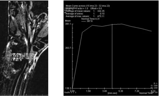
and diffusion weighted MRI. The mass did not contain calcification on X-Ray (Fig. 1). T1 and T2 weighted MRI revealed a well defined, lobulated mass within the carpal tunnel, arising from the distal forearm, extending to the carpometacarpal joint, measuring 2.5x3.4x5.7 cm (Figs.2 and 3). On T1 and T2 weighted images the tumor did not cause any change in the adjacent osseous bony cortex and the medulla. On T1 weighted images the lesion was isointense to muscle (Fig. 2). On T2-weighted images the lesion had heterogenous intermediate signal intensity (Fig. 3). The heterogenous signal intensity on T2 weighted images and deep location of the tumor suggested malignancy. There weren’t any necrosis in the tumor. The T1-weighted fat-saturated volumetric interpolated gradient echo sequence was obtained for dynamic contrast enhanced MRI. Contrast enhanced images were subtracted from the precontrast scan on the console. Subtracted images depicted peripheral enhancement at early arterial phase (Fig. 4). In the late phase the central portion of the mass did not enhanced (Fig.5). Time-signal intensity (TSI) curve showed a steep rise to an early peak followed by slightly washout and it suggested malignancy (Figs. 6A and 6B). Single shot spin echo planar imaging
sequence at b values of 0, 500, and 1000 s/mm (2)
were obtained for diffusion-weighted MRI and the apparent diffusion coefficient (ADC) map of the
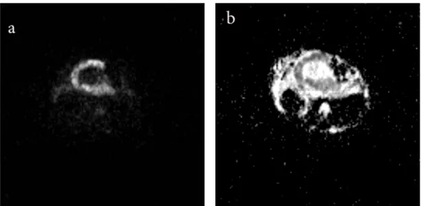
lesion. The peripheral portions of the mass were hyperintense at all b values and the mass showed low ADC values on ADC maps corresponding with restricted diffussion of the peripheral portions of the mass (Figs.7A and 7B). Mean ADC value was
0.98 ± 0.06 x10⁻³ mm2 /sec. On diffusion-weighted
MRI, the areas showing restricted diffusion corresponded with the enhancing areas on dynamic-contrast enhanced images.
The differential diagnosis of the tumor includes; synovial sarcoma, malignant fibrous histiocytoma which has more recently classified as pleomorphic
undifferentiated sarcoma. Extraskeletal
chondrosarcoma and osteosarcoma were less likely in the differential diagnosis because no matrix calcification was found in the mass. Patient underwent gross total resection of the tumor. The specimen demonstrated 2.3x3x6 cm lobulated solid mass including hemorrhage. Microscopically, lesion showed atypical pleomorphic mesenchymal cells with increased mitotic activity, chondroid matrix and necrosis (Figs. 8 and 9). The mass did not contain calcification on X-Ray and histology. Non-calcified chondroid matrix was found on histology. The final diagnosis was a grade 3 EMC. No bony involvement was identified at histological assessment. He received radiotherapy after surgery. After three years of surgery local recurrence was
occurred.
Figure 1. On X-Ray the mass did not contain calcification. There were postoperative changes on lunate bone and distal radius due to Kienbock’s disease
Figure 2 Axial T1-weighted MRI depicts a lobulated mass, isointense to muscle, adjacent to the flexor tendons, within the wrist.
Figure 3. On a coronal T2-weighed MRI, the mass had heterogenous intermediate signal intesity. The low T2 signal intensity area within the central portion of the mass may due to chondroid matrix with high collagen components
Figure 4. Subtracted images at early arterial phase showed peripheral enhancement pattern of the mass.
Figure 5. Subtracted images at delayed phase showed no central enhancement pattern
Figures 6A and 6B. Time to signal intensity curve showed a steep rise to an early peak followed by slightly washout.
Figures 7A and 7B. Diffusion weighted images at 1000 b value and ADC maps shows restricted diffusion of the peripheral portions of the mass
Figure 8. (H&E, X200) grade 3 chondrosarcoma characterized by atypical pleomorphic mesenchymal cells with prominent nucleolus
Figure 9. (H&E, X400) High power view reveals increased mitotic activity, pleomorphism and necrosis in the dedifferantiated areas of chondrosarcoma
DISCUSSION
EMC are high grade malignant tumors that most frequently affect head and neck and also thigh
1.Wrist is a very rare site for extraskeletal
mesenchymal chondrosarcomas. X-Ray findings of these lesions are nonspecific soft tissue masses which may include chondroid matrix mineralization
1. Imaging findings of EMC were rarely reported
and there weren’t any specific findings. EMC shows
weighted MR images 1.Some of the cases showed
lobulation in the previous reports similar with this case 3,5,6,7.
Peripheral enhancement pattern on
contrast-enhanced CT images were also described 4. The
gadolinium enhancement pattern of EMC was very rarely reported. Inhomogeneous enhancement pattern and contrast enhancement except low signal intensity areas on T1 weighted MRI were previously
the dynamic enhancement pattern of EMC have not been reported previously. According to our case early arterial rapid enhancement and wash-out pattern of the mass may suggest a malignant process. The differential diagnosis of the tumor includes; synovial sarcoma and malignant fibrous histiocytoma which has more recently classified as pleomorphic undifferentiated sarcoma. Synovial sarcomas showed typical intermixed areas of low, intermediate and high signal intensity defining as
‘triple sign’ on T2 weighted images 8. Our case
shows predominantly intermediate signal intensity
on T2 weighted images. Pleomorphic
undifferentiated sarcoma is more likely in the differential diagnosis with low or intermediate T1 and high T2 signal intensities. Razek et al proposed
using a threshold mean ADC value of 1.34×10⁻³mm
2 /sec to distinguish benign soft tissue neoplasms of
extremities from malignant neoplasms 9 . We found
a mean ADC value of 0.98 ± 0.06 x10⁻³ mm 2 /sec
which is highly suspicious for malignancy.
In conclusion, EMC is a rare soft tissue tumor that can be unusually origin in wrist and might show peripheral enhancement at early arterial phase and restricted diffusion on MRI. The knowledge of the imaging spectrum of the EMC might help us suggesting the diagnosis.
REFERENCES
1. Murphey MD, Walker EA, Wilson AJ, Kransdorf MJ, Temple HT et al. From the archives of the AFIP: imaging of primary chondrosarcoma: radiologic-pathologic correlation. Radiographics. 2003;23:1245-78.
2. Kransdorf MJ, Meis JM. From the archives of the AFIP: Extraskeletal osseous and cartilaginous tumors of the extremities. Radiographics. 1993;13:853-84. 3. Hashimoto N, Ueda T, Joyama S, Araki N, Beppu Y,
Tatezaki S et al: Extraskeletal mesenchymal chondrosarcoma: an imaging review of ten new patients. Skeletal Radiol. 2005;34:785-92.
4. Shapeero LG, Vanel D, Couanet D, Contesso G, Ackerman LV. Extraskeletal mesenchymal chondrosarcoma. Radiology. 1993;186:819-26. 5. Guccion JG, Font RL, Enzinger FM, Zimmerman
LE. Extraskeletal mesenchymal chondrosarcoma. Arch Pathol. 1973;95:336- 40.
6. Salvador AH, Beabout JW, Dahlin DC. Mesenchymal chondrosarcoma: observations on 30 new cases. Cancer. 1971;28:605-15.
7. Pringle J, Stoker DJ. Case report 127. Skeletal Radiol .1980;5:263-6.
8. Jones BC, Sundaram M, Kransdorf MJ. Synovial sarcoma: MR imaging findings in 34 patients. AJR Am J Roentgenol. 1993;161:827–30.
9. Razek A, Nada N, Ghaniem M, Elkhamary S. Assessment of soft tissue tumours of the extremities with diffusion echoplanar MR imaging. Radiol Med (Torino). 2012;117:96–101.