*Corresponding author: [email protected]
Sibel Demir Kanmazalp orcid.org/0000-0002-5896-0966 Muharrem Dinçer orcid.org/0000-0003-3960-9991
Sharma et al. 1998). In addition, it has been proved that 3-substituted cyclobutane carboxylic acid derivatives exhibit anti-inflammatory and antidepressant activities Jaen et al. 1990 and also liquid crystal properties (Tsuji et al. 1994). In this study, we present the results of a detailed investigation of the synthesis and structural characterization of a N-[4-(3- methyl-3-phenyl-cyclobutyl)-thiazol-2-yl]-N´-thiophen-2ylmethylene-Chloro-acetic acid hydrazide (NNT2CAH) compound by using single crystal X-ray diffraction, IR and NMR spectroscopy and quantum chemical methods. The geometrical parameters, fundamental frequencies and GIAO 1H and 13C NMR chemical shift values of the title compound in the ground state have been calculated by using the B3LYP method. A comparison of the experimental and theoretical spectra can be very useful in making correct assignments and understanding the basic chemical
shift-1. Introduction
The chemistry of aminothiazoles and their derivatives has attracted the attention of chemists, because they exhibit an important biological activity in medicinal chemistry Pozharskii et al. 1997, Wipf and Wang 2007 such as antibiotic, anti-inflammatory, antihelmintic, or fungicidal properties (Metzger 1979, Koike et al. 1999, Walczynski et al. 1999). 2-Aminothiazoles are mainly known as biologically active compounds with a broad range of activities and as intermediates in the synthesis of antibiotics, well-known sulfa drugs, and some dyes (Patt et al. 1992,
Spectral and Theoretical Analysis of
N-[4-(3-methyl-3-phenyl-cyclobutyl)-thiazol-2-yl]-N´-thiophen-2ylmethylene-Chloro-acetic Acid Hydrazide by DFT Method
N-[4-(3-metil-3-fenil-siklobutil)-tiazol-2-yl]-N´-tiyofen-2ylmetilene-Khloro-Asetik Asit Hidrazid’in
DFT Metodu Kullanılarak Spektral ve Teorik Analizi
Sibel Demir Kanmazalp
1*, Muharrem Dinçer
2, Alaaddin Çukurovalı
3, İbrahim Yılmaz
4 1 Gaziantep University, Technical Science Vocational School, Gaziantep, Turkey2 Ondokuz Mayıs University, Faculty of Arts and Sciences, Department of Physics, Samsun, Turkey 3Firat University, Faculty of Science, Department of Chemistry, Elaziğ, Turkey
4Karamanoğlu Mehmet Bey University, Faculty of Science, Department of Chemistry, Karaman, Turkey
Öz
Bu çalışma N-[4-(3-metil-3-fenil-siklobutil)-tiazol-2-yl]-N´-tiyofen-2ylmetilene-Khloro-asetik asit hidrazid bileşiğinin deneysel ve teorik çalışmasını gösterir. Optimize edilmiş moleküler yapı, 1H ve 13C kimyasal kayma değerleri ve titreşim frekansları DFT metodu ile birlikte 6-31G(d), 6-31G(d,p) ve 6-311G(d,p) baz setleri kullanılarak incelenmiştir. İlaveten moleküldeki yük delokalizasyonu incelemek için HOMO-LUMO analizi ve moleküler elektrostatik potansiyeli araştırılmıştır.
Anahtar Kelimeler: Sınır moleküler orbitaller, Moleküler elektrostatik potansiyel, IR ve NMR spektroskopisi, X-ışını yapı tanımı
Abstract
This study presents a combined experimental and theoretical research on an N-[4-(3-methyl-3-phenyl-cyclobutyl)-thiazol-2-yl]-N´-thiophen-2ylmethylene-Chloro-acetic acid hydrazide compound. The optimized molecular structure, 1H and 13C chemical shift values and vibrational assignments of the titled compound were examined by depending on the density functional method and by using 6-31G(d), 6-31G(d,p) and 6-311G(d,p) basis sets. Moreover, HOMO–LUMO analysis and molecular electrostatic potential were carried out in order explore charge delocalization on this molecule.
Keywords: Frontier molecular orbitals, Molecular electrostatic potential, IR and NMR spectroscopy, X-ray structure determination
molecular structure relationship. Furthermore, the molecular electrostatic potential, and frontier molecular orbitals, were studied at the B3LYP/6-311G(d, p) level.
2. Materials and Method
2.1 Crystallographic AnalysisSingle crystal X-ray data were collected on an STOE diffractometer with an IPDS(II) image plate detector (Mo Kα radiation) at 296 K. The crystal was solved by direct methods by using SHELXS-97 and refinement was carried out with full-matrix least-squares methods based on F2 with the SHELXL-97 software package (Sheldrick 1997). All hydrogen atoms were refined in isotropic approximation in a riding model with the Uiso(H) parameters which equal to 1.2 Ueq(Ci), for methyl groups which equal to 1.5 Ueq(Cii), where U(Ci) and U(Cii) are, respectively the equivalent thermal
parameters of the carbon atoms to which corresponding H atoms are bonded. The crystal data and experimental parameters are given in Table 1.
2.2. Instrumentation
FT-IR spectra were measured by ATI Unicam-Mattson 1000 FT-IR spectrometer in the frequency range of 4000–400 cm−1 by using KBr disc. The FT-IR spectra were recorded at room temperature at the spectral resolution of 1 cm−1. 1H and 13C NMR spectra were recorded with Bruker Avance III 400 MHz spectrometer. Chemical shifts were reported on a scale which correlate with TMS.
2.3. Synthesis
The synthesis of the title compound was carried out in the following reaction Scheme. A solution of 0.3535 gram (1 mmol) of N-thiophen-2-ylmethylene-N´-[4-(3-methyl-3-phenyl-cyclobutyl)-thiazol-2-yl]-hydrazine was dissolved in 20 mL of dioxane containing 1 mmol triethylamine. To this solution, 90 μL (1 mmol) of chloroacetyl chloride solution in 20 mL 1,4-dioxane was added dropwise in a two hour period at room temperature by stirring. The mixture was stirred for another two hours, then was neutralized with 5% aqueous ammonia (if necessary, but generally it is necessary). Thus, the precipitated compound was filtered, washed with copious water and crystallized from ethanol. The reaction sequence of synthesis of the title compound is shown in Scheme.
Scheme. Reaction sequence of synthesis of NNT2CAH. 2.4. Computational details
Initial geometry generated from standard geometrical parameters was minimized. The optimized structural parameters were used in the vibrational frequency calculations at the DFT level to characterize all the stationary points as minima. The calculated NMR, molecular electrostatic potential and frontier orbitals were calculated using the same method and then vibrational frequencies of normal modes scaled 0.9613 for 6-31G(d) (Foresman et al. 1996, Merrick et al. 2007) 0.9611 for 6-31G(d,p) (Schlegel 1982, Hehre et al. 1972) and 0.9670 6-311G(d, p) (Andersson et al. 2005) with B3LYP. Additionally, we calculated the 1H and 13C NMR chemical shifts for this structure using
Table 1. Crystallographic data for NNT2CAH
Formula CCDC Formula Weight Temperature[K] Wavelength [Å] Crystal system Space group a [Å] b [Å] c [Å] α [o] β [o] γ [o] V[Å3] Z Dcalc [g/cm3] F(000) h, k, l Range Reflections collected Independent reflections Rint
Reflections observed [I≥2σ(I)] R [I>2σ(I)]
Rw [I>2σ(I)]
Goodness-of-fit on Indicator Structure determination Refinement
(∆σ)max, (∆σmin) [e/ Å3]
C21H20ClN3OS2 775739 430 296 0.71073 Orthorhombic P 212121 5.9330 (3) 17.2192(12) 20.2868(12) 90 90 90 2072.53(2) 4 1.38 895.9 -7≤h≤7 -21≤k≤21 -17≤l≤25 11938 4357 0.053 2649 0.046 0.0691 0.891 Shelxs-97 Full matrix 0.16, −0.16
GIAO model at the same level. These calculations were performed at DFT (B3LYP) method using the Gaussian 03W program package (Frisch et al. 2003).
3. Results
3.1 Description of The Crystal Structure
The molecular structure of the title compound is shown in Figure 1(A). Thermal ellipsoids are drawn at 40% probability level. The crystal data and experimental parameters are given in Table 1. Besides, hydrogen bonding geometry is given in Table 2.
In the asymmetry unit, the thiazole ring and acetic acid hydrazide group are coplanar. The thiazole ring makes the dihedral angle with the acetic acid hydrazide group of 11.96(0.22). There is a significant intramolecular C15– H15…N1 hydrogen bonds forming the corresponding pseudo five-membered ring. The crystal packing diagram of NNT2CAH is shown in Figure 1(B). The molecular conformation was stabilized by intra and inter molecular C—H…N/O/Cl and C—H…π (C4…B ring 3.772(3)
Å) hydrogen bonds (Figure 1(B) and Table 2). The short C20–C21 distance [1.508(4) Å] may be due to a collective interaction of C21 with the nearby hydrazide group and O1 atom, an intramolecular close contact interaction with the nearby nitrogen atom in thiazole (C15–H15…N1; see Table 2), and an C2–H2…O1 intermolecular interaction (H2…O1= 2.86 Å; symmetry code: -1/2+x, 1/2-y, 1-z; that may influence the bond strength of the C20–C21. Other hydrogen bonds are listed in Table 2. The hydrazide group lies in a trans configuration along the N(3)–C(15) bond (torsion angles C15–N3–N2–C21= 169.3(3)˚ and C15– N3–N2–C1= -14.4(4)˚, respectively).
3.2 Optimized Molecular Geometry
Listed in Table 3 are, the optimized geometrical parameters of NNT2CAH, calculated at B3LYP/6-31G(d)/6-31G(d,p)/6-311G(d,p) basis sets in the gas phase which are in accordance with the atom numbering scheme are given in Figure 1(A). These indicate a slight underestimate or overestimate of some bond lengths, as a result, 6-31G(d,p) basis set predicts bond lengths in an excellent agreement
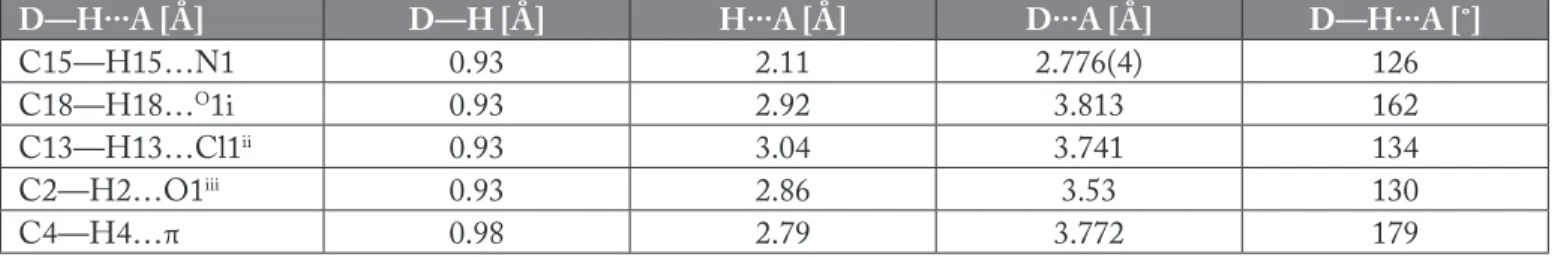
Figure 1. (A) Molecular structure with atomic numbering for NNT2CAH (B) Packing diagram of NNT2CAH. Table 2. Hydrogen bonding geometry [Å, ˚] for the NNT2CAH.
D—H···A [Å] D—H [Å] H···A [Å] D···A [Å] D—H···A [˚]
C15—H15…N1 0.93 2.11 2.776(4) 126
C18—H18…O1i 0.93 2.92 3.813 162
C13—H13…Cl1ii 0.93 3.04 3.741 134
C2—H2…O1iii 0.93 2.86 3.53 130
C4—H4…π 0.98 2.79 3.772 179
i:5/2-x,-y,-1/2+z, ii: 3/2-x, -y, -1/2+z iii: -1/2+x,1/2-y,1-z
and the observed fundamental modes. The resulting vibrational wave numbers for the optimized geometry and the proposed assignments are given in Table 4. Based on the results of the normal coordinate calculations the vibrational spectral data obtained from the solid phase FT-IR spectra are assigned. All vibrational calculations were performed in the harmonic approximation.
In the heteroaromatic compounds, the C–H stretching vibrations normally occur at 3100–3000 cm-1 (Stuart 2008). These vibrations were not found to be the influence to the nature and the position of the substituent and they typically exhibit weak bands when compared with the aliphatic stretching vibrations. In infrared spectra, most of the aromatic compounds have nearly four peaks in the region 3080–3010 cm-1 due to the ring C–H stretching bands. IR frequencies of C–H band are a function of sp hybridization (Pavia et al. 2001). Four IR bands were observed at 3115, 3104, 3080 and 2925 cm-1 were assigned to the C–H stretching vibration for NNT2CAH. The second band belongs to the symmetric and the third to the asymmetric stretching band. In the higher frequency region, almost all of the vibrations belong to CH3 and CH2 stretching vibrations. with X-ray data. However, for the bond angles of 6-31G(d)
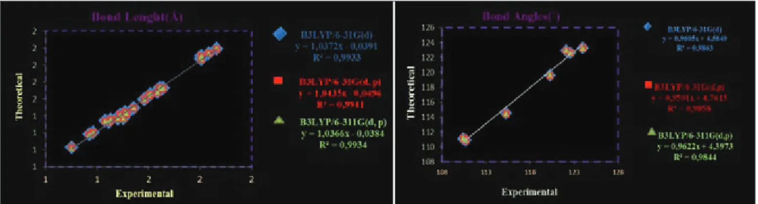
basis set are better when compared with experimental results. We present correlation graphics in Figure 2 based on these calculations. Considering the comparison above, although the theoretical and experimental values slightly differentiate from each other the optimized structural parameters can reproduce the experimental one well and they are the basis for following discussion.
A logical method for globally comparing of the structures obtained with the theoretical calculations is to superimpose the molecular skeleton with that obtained from X-ray diffraction, giving an RMSE of 0.366 Å for 6–31G(d), 0.362 Å for 6–31G(d,p) and 0.383 Å for 6–311G(d,p) calculated by using B3LYP method (Figure 3). Consequently, the 6–31G(d,p) basis set correlates well with the geometrical parameters when compared with other basis sets. Figure 3 represents atom-by-atom superimposition of the structures were calculated (black) in the X-ray structure (red) of the title compound.
3.3. Vibrational Analysis
Assignment of organic systems can be proposed on the basis of frequency agreement between the computed harmonics
Figure 2. Correlation graphics of calculated and experimental geometric parameters of NNT2CAH.
Figure 3. Atom-by-atom superimposition of the structures calculated (black) as (B3LYP/6–31G(d), 6–31G(d,p) and as 6–311G(d,p)) on the X-ray structure (red) of NNT2CAH hydrogen atoms have been omitted for clarity.
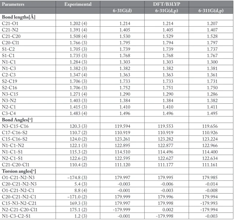
Table 3. Selected optimized and experimental geometries parameters of NNT2CAH.
Parameters Experimental DFT/B3LYP
6-31G(d) 6-31G(d,p) 6-311G(d,p) Bond lengths[Å] C21-O1 1.202 (4) 1.214 1.214 1.207 C21-N2 1.391 (4) 1.405 1.405 1.407 C21-C20 1.508 (4) 1.530 1.529 1.528 C20-Cl1 1.766 (3) 1.795 1.794 1.797 S1-C2 1.705 (3) 1.739 1.739 1.737 S1-C1 1.735 (3) 1.768 1.768 1.767 N1-C1 1.284 (3) 1.303 1.303 1.300 N1-C3 1.382 (3) 1.382 1.382 1.381 C2-C3 1.347 (4) 1.363 1.363 1.361 S2-C19 1.706 (3) 1.733 1.733 1.731 S2-C16 1.706 (3) 1.752 1.751 1.750 N3-C15 1.271 (4) 1.290 1.290 1.286 N3-N2 1.403 (3) 1.384 1.384 1.382 N2-C1 1.415 (3) 1.410 1.410 1.411 C3-C4 1.483 (4) 1.496 1.496 1.495 Bond Angles[o] N3-C15-C16 120.3 (3) 119.594 119.553 119.656 C17-C16-S2 110.7 (2) 110.919 110.919 110.926 C15-C16-S2 124.0 (2) 123.261 123.282 123.224 N1-C1-N2 122.1 (3) 122.895 122.877 122.966 N1-C1-S1 115.3 (2) 114.510 114.496 114.400 N2-C1-S1 122.6 (2) 122.595 122.627 122.634 C21-C20-Cl1 110.4 (2) 111.120 111.177 111.161 Torsion angles[o] O1-C21-N2-N3 −174.8 (3) 179.997 179.995 179.985 C20-C21-N2-N3 5.4 (3) -0.003 -0.006 -0.014 O1-C21-N2-C1 8.8 (4) -0.001 -0.003 -0.008 C20-C21-N2-C1 −171.0 (2) 179.999 179.996 179.994 C15-N3-N2-C21 169.3 (3) 179.997 179.998 -179.993 N2-C21-C20-Cl1 175.1 (2) -179.999 0.002 -179.994 N1-C3-C2-S1 1.2 (3) -0.001 -179.998 -0.003
If a CH3 group presents in a compound, it can give rise to one asymmetric and one symmetric stretching vibration. In this molecule, these vibrations were observed at 2959 and 2563 cm-1, respectively. The range of frequencies obtained by B3LYP method in this region are 2987/2921 cm-1 for 6-31G(d) and 2986/2916 cm-1 for 6-31G(d,p) 2982/2919 cm-1 for 6-311G(d,p) basis sets, respectively. CH
2 stretching vibration modes are observed at 3036, 3021, 2975 and 2862 cm-1. The first of these modes is the asymmetric vibration of the cyclobutane ring and chloro-asetic acid group and
the others are a symmetric vibrations of same groups, respectively. Belonging to CH2 other calculated vibrations can also be seen in Table 4. Generally, the C=C stretching vibrations in aromatic compounds compose six bands in the region of 1650–1430 cm-1. These bands show the intention to shift to the lower wavenumber with heavy substituents and an increase in a number of substituents on the compound gives rise to these vibrations, and were observed in the wide region of FT-IR spectrum (Roeges 1994). The characteristic band of C=C stretching vibration of the
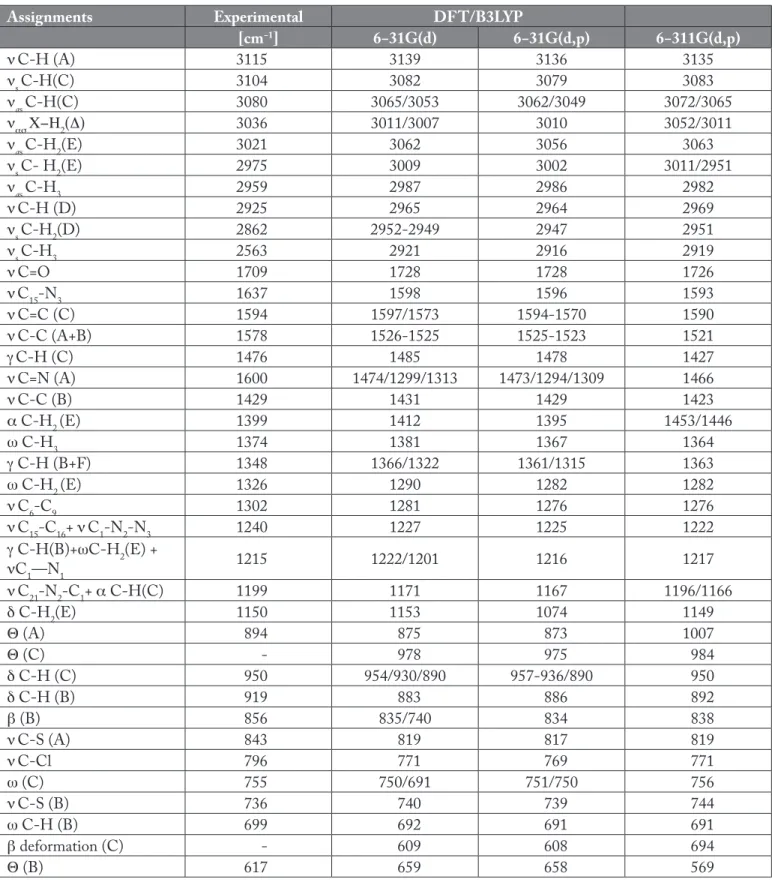
Table 4. Comparison of the observed and calculated vibrational spectra of NNT2CAH.
Assignments Experimental DFT/B3LYP
[cm−1] 6−31G(d) 6−31G(d,p) 6−311G(d,p) νC-H (A) 3115 3139 3136 3135 νs C-H(C) 3104 3082 3079 3083 νas C-H(C) 3080 3065/3053 3062/3049 3072/3065 νασ C-H2(D) 3036 3011/3007 3010 3052/3011 νas C-H2(E) 3021 3062 3056 3063 νs C- H2(E) 2975 3009 3002 3011/2951 νas C-H3 2959 2987 2986 2982 νC-H (D) 2925 2965 2964 2969 νs C-H2(D) 2862 2952-2949 2947 2951 νs C-H3 2563 2921 2916 2919 νC=O 1709 1728 1728 1726 νC15-N3 1637 1598 1596 1593 νC=C (C) 1594 1597/1573 1594-1570 1590 νC-C (A+B) 1578 1526-1525 1525-1523 1521 γC-H (C) 1476 1485 1478 1427 νC=N (A) 1600 1474/1299/1313 1473/1294/1309 1466 νC-C (B) 1429 1431 1429 1423 α C-H2 (E) 1399 1412 1395 1453/1446 ω C-H3 1374 1381 1367 1364 γ C-H (B+F) 1348 1366/1322 1361/1315 1363 ω C-H2 (E) 1326 1290 1282 1282 νC6-C9 1302 1281 1276 1276 νC15-C16+ νC1-N2-N3 1240 1227 1225 1222 γ C-H(B)+ωC-H2(E) + νC1—N1 1215 1222/1201 1216 1217 νC21-N2-C1+ α C-H(C) 1199 1171 1167 1196/1166 δ C-H2(E) 1150 1153 1074 1149 Θ (A) 894 875 873 1007 Θ (C) - 978 975 984 δ C-H (C) 950 954/930/890 957-936/890 950 δ C-H (B) 919 883 886 892 β (B) 856 835/740 834 838 νC-S (A) 843 819 817 819 νC-Cl 796 771 769 771 ω (C) 755 750/691 751/750 756 νC-S (B) 736 740 739 744 ω C-H (B) 699 692 691 691 β deformation (C) - 609 608 694 Θ (B) 617 659 658 569
Vibrational modes: ν, stretching; s, symmetric; as, asymmetric; α, scissoring; γ, rocking; ω, wagging; δ, twisting; θ, ring breathing; β, in-plane bending. Abbreviations: A, thiazole ring; B, furan ring; C, phenyl ring; D, cyclobutane ring; E, chloro acetic acid group; F, hydrazide group.
311G(d,p), 817 and 769 cm-1 with B3LYP/6-31G(d,p), respectively.
3.4. Nuclear Magnetic Resonance
Since NMR chemical shifts bear witness to the electron density which is close to nuclei, the use of core pseudopotentials is in principle precluded. For this reason, these chemical shifts have been calculated with the GIAO (Gauge Invariant Atomic Orbitals) method (Wolinski et al. 1990) and these results relative to the TMS value are given in Table 5. The NNT2CAH compound has 21 different carbon atoms, which is consistent with the structure based on molecular symmetry. Calculated 1H and 13C isotropic chemical shielding values for TMS at the B3LYP level were obtained 32.10 ppm and 189.40 ppm for 6-31G(d) and 32.37 ppm and 193.97 ppm for 6-31G(d, p), and 31.99 ppm and 185.06 ppm for 6-311G(d, p) basis sets, respectively. The experimental values for 1H and 13C isotropic chemical shifts for TMS were 30.84 ppm and 188.1 ppm, respectively. Since experimental 1H chemical shift values were not available for an individual hydrogen, we have presented the average values for aromatic CH2 and CH3 hydrogen atoms. In our present study the methyl protons at C8 appears as a singlet with three protons integral at 1.60 ppm. The singlet observed at 6.85 ppm was assigned to H2(C2) atoms that were calculated at 2.92 ppm (6-31G(d)) and 4 ppm (6-31G(d,p)) and 6.93 ppm (6- 311G(d,p)) for the B3LYP level. The CH2 signals of the cyclobutane were observed at 2.52–2.68 ppm. Another CH2 signal, C20(H20) in the chloro-acetic acid was observed at 4.82 ppm. Due to deshielding by the electronegative property of N1, N2, and S1 atoms, the chemical shift value of C1 which has a larger value than the others, was observed at 167.29 ppm. Besides, due to the shielding effect of the non-electronegative property of the hydrogen atom, the chemical shift value of C8 atom is lower than the others carbon peak (Karakurt et al. 2011). As shown in Table 5, theoretical 1H and 13C chemical shift results of the title compound were generally closer to the experimental 1H and 13C shift data.
3.5. Molecular Electrostatic Potential
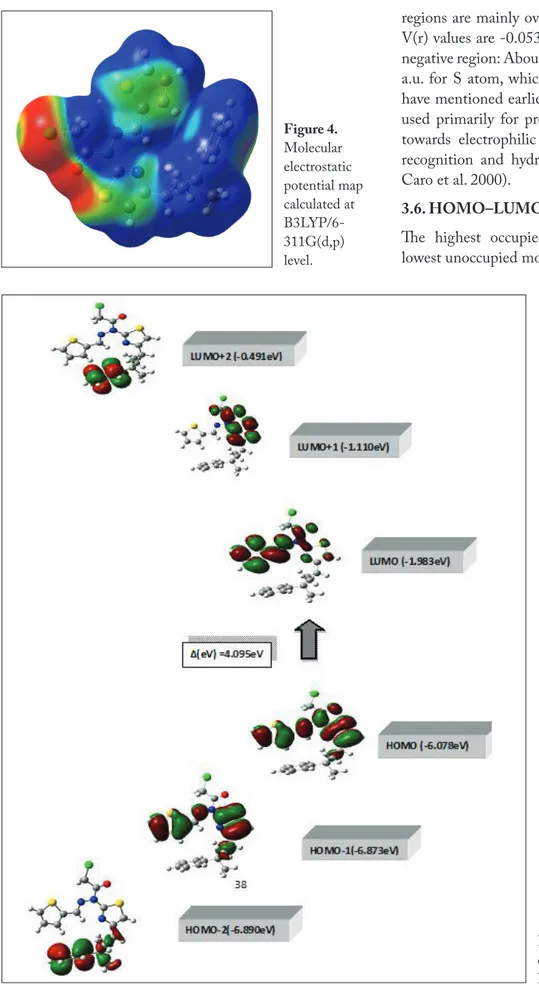
The molecular electrostatic potential (MEP) is widely used as a reactivity map displaying most probable regions for the electrophilic attack of charged point-like reagents on organic molecules (Politzer 1981). The MEP of NNT2CAH is obtained based on the B3LYP/6-11G(d,p) optimized result and shown in Figure 4. The calculated 3D MEP contour map (red is negative, blue is positive) shows that the negative phenyl ring of NNT2CAH appears at 1594 cm-1. Stretching
vibration of C=O group is expected to appear at 1715–1680 cm-1 (Roeges 1994). The very strong C=O experimental bands which were observed at 1709 cm-1 for NNT2CAH correspond to the stretching vibration of C=O group of chloro-acetic acid, which lie in a higher frequency region in the present case indicating the weak delocalization of a lone pair of electrons. The increase in conjugation, generally, leads to the intensification of infrared bands. The conjugation and the influence of intermolecular hydrogen bonding result in the lowering of the stretching wavenumbers of C=O vibration (Sıdır et al., 2010). Frequencies of C=O stretching for a DFT method vibrations are in good agreement with the experimental results.
Any deviation of the calculated wavenumber for this mode can be attributed to π-electron delocalization due to the conjugation or formation of hydrogen bonds (Panicker et al.2007). The identification of C–N vibrations is a very challenging task since the mixing of several bands could be possible in this region. However, with the help of theoretical calculations and percentage relative weight of vibrations the C–N stretching vibrations are identified and assigned in this study. The C–N stretching vibrations are assigned in the region 1382–1266 cm-1 for aromatic amines (Silverstein 2003). The C–N stretching modes appear at 1637, 1600, 1240 and 1199 cm-1 for NNT2CAH in the solid-state FT-IR spectra. The FT-IR band observed at 1637 cm-1 is the most intensive band in this region. For B3LYP level, calculated wavenumbers of these bands show excellent agreement with corresponding experimental ones for NNT2CAH. These results are detailed in Table 4. Here, it is important to conclude the vibrations of C-halogen bonds in the aromatic ring, because, the mixture of vibrational modes are possible due to a lack of symmetry and the presence of heavy atoms such as F, Cl, Br, I and S in the molecule (Yadav and Sing 1985). The C–S and S–H bonds are highly polarizable and hence produce stronger spectral activity. The C–S stretching vibration is expected in the region 710–685cm−1 (Coates 2000).The C–S stretching vibrations were observed at 843 and 736 cm-1 for NNT2CAH. The infrared band appearing at 796 cm−1 was designated to C–Cl stretching mode in this molecule. This C–Cl vibrational mode was reported at 795 cm-1 for N’-benzylidene-N-[4-(3-methyl-3-phenyl-cyclobutyl)-thiazol-2-yl]-Chloro-acetic acid hydrazide (Demir et al. 2012). On the other hand, C–S stretching in thiazole (ring A) and C–Cl stretching bands were calculated 819 and 771 cm-1 with 31G(d) and
B3LYP/6-Table 5. Theoretical and experimental 13C and 1H isotropic chemical shifts (with respect to TMS all values in ppm) for NNT2CAH.
Atom Experimental DFT/B3LYP
6-31G(d) 6-31G(d, p) 6−311G(d, p) C1 167.29 148.91 153.56 166.81 C2 111.31 98.59 102 119.73 C3 155.30 145.29 150.04 159.9 C4 30.71 16.4 19.66 36.01 C5 41.04 25.15 27.75 46.19 C6 38.75 29.82 34.06 45.58 C7 41.04 22.7 25.27 46.2 C8 30.06 7.32 9.44 34.51 C9 152.26 142.95 147.98 164.4 C10 124.70 108.88 112.22 131.86 C11 127.69 111.78 115.36 134.93 C12 125.34 106.89 110.43 131.51 C13 127.69 110.8 114.38 134.94 C14 124.70 109.06 112.49 131.86 C15 146.02 134.19 137.44 150.22 C16 138.49 129.54 134.61 151.11 C17 128.27 121.74 125.27 140.66 C18 132.17 113.72 117.39 133.25 C19 129.47 118.8 122.32 141.64 C20 43.66 37.08 40.22 56.75 C21 156.27 156.8 161.3 174.25 H2 6.85 2.92 4 6.93 H4 3.79 0.26 0.94 3.7 H5* 2.68 0.82 0.16 2.71 H7* 2.52 0.85 0.255 2.71 H8* 1.60 3.21 2.61 1.54 H10 6.87 4.47 5.41 7.54 H11 6.98 4.64 5.57 7.74 H12 6.96 4.65 5.58 7.68 H13 7.05 4.87 5.81 7.74 H14 7.03 4.58 5.53 7.55 H15 9.40 6.96 8.14 10.95 H17 7.17 2.52 3.51 5.89 H18 7.20 3.86 4.84 7 H19 7.24 4.21 5.24 7.64 H20* 4.82 1.34 2.09 4.98 *: Average.
regions are mainly over the Cl and O atoms. The negative V(r) values are -0.0530 a.u. for O atom, which is the most negative region: About -0.0272 a.u. for Cl atom and -0.0217 a.u. for S atom, which is the least negative region. As we have mentioned earlier, the electrostatic potential has been used primarily for predicting sites and relative reactivities towards electrophilic attack, and in studies of biological recognition and hydrogen bonding interactions (Munoz-Caro et al. 2000).
3.6. HOMO–LUMO Analysis
The highest occupied molecular orbital (HOMO) and lowest unoccupied molecular orbital (LUMO) are the main
Figure 4. Molecular electrostatic potential map calculated at B3LYP/6-311G(d,p) level.
Figure 5. The atomic orbital composition
of the frontier molecular orbital for NNT2CAH.
Demir, S., Dincer, M., Cukurovali, A., Yilmaz, I. 2012.
Experimental and Theoretical Investigation of the Molecular and Electronic Structure of N´-Benzylidene-N-[4-(3-methyl-3-phenyl-cyclobutyl)-thiazol-2-yl]-chloro-acetic acid hydrazide. Int. J. Quantum Chem. 112, 2: 1016-1028.
Frisch, MJ., Trucks, GW., Schlegel, HB., Scuseria, G. E., Robb, MA., Cheeseman, J. R., Scalmani, G., Barone, V., Mennucci, B., Petersson, GA., Nakatsuji, H., Caricato, M., Li, X., Hratchian, HP., Izmaylov, AF., Bloino, J., Zheng, G., Sonnenberg, JL., Hada, M., Ehara, M., Toyota, K., Fukuda, R., Hasegawa, J., Ishida, M., Nakajima, T., Honda, Y., Kitao, O., Nakai, H., Vreven, T., Montgomery, JAJr., Peralta, JE., Ogliaro, F., Bearpark, M., Heyd, JJ., Brothers, E., Kudin, KN., Staroverov, VN., Kobayashi, R., Normand, J., Raghavachari, K., Rendell, A., Burant, JC., Iyengar, S. S., Tomasi, J., Cossi, M., Rega, N., Millam, JM., Klene, M., Knox, JE., Cross, JB., Bakken, V., Adamo, C., Jaramillo, J., Gomperts, R., Stratmann, RE., Yazyev, O., Austin, AJ., Cammi, R., Pomelli, C., Ochterski, JW., Martin, RL., Morokuma, K., Zakrzewski, VG., Voth, GA., Salvador, P., Dannenberg, JJ., Dapprich, S., Daniels, AD., Farkas, Ö., Foresman, JB., Ortiz, JV., Cioslowski, J., Fox, DJ. 2004.
Gaussian 03, Revision E.01, Gaussian, Inc., Wallingford CT.
Foresman, JB., Frisch, A. 1996. Exploring Chemistry with
Electronic Structure Methods, second ed., Gaussian Inc., Pittsburgh, PA.
Fukui, K. 1975. Theory of Orientation and Stereo Selection,
Springer-Verlag, New York.
Gunasekaran, S., Balaji, RA., Kumerasan, S., Anand, G., Srinivasan, S. 2008. Experimental and theoretical
investigations of spectroscopic properties of N-acetyl-5-methoxytryptamine. Can. J. Anal. Sci. Spectrosc. 53: 149-160.
Hehre, WJ., Ditchfield, R., Pople, JA. 1972. Self—consistent
molecular orbital methods. XII. Further extensions of gaussian—type basis sets for use in molecular orbital studies of organic molecules. J. Chem. Phys. 56(5): 2257-2261.
Jaen, JC., Wise, L.D., Caprathe, B.W., Tecle, H., Bergmeier, H., Humblet, CC., Heffner, TG., Meltzner, LT., Pugsley, TA. 1990.
4-(1,2,5,6-Tetrahydro-1-alkyl-3-pyridinyl)-2-thiazolamines: a novel class of compounds with central dopamine agonist properties. J. Med. Chem., 33: 311-7.
Karakurt, T., Dincer, M., Cukurovali A., Yilmaz, I. 2011. Ab
initio and semi-empirical computational studies on 5-hydroxy-4-methyl-5, 6-di-pyridin-2-yl-4, 5-dihydro-2H-[1, 2, 4] triazine-3-thione. J. Mol. Struct., 991:186-201.
Khalil, AM., Berghot, MA., Gouda, MA. 2009. Synthesis and
antibacterial activity of some new heterocycles incorporating phthalazine. Eur. J. Med. Chem., 44 (11): 4434-4440.
orbital and take part in chemical stability (Gunasekaran et al. 2008). The HOMO represents the ability to donate an electron, while LUMO as an electron acceptor represents the ability to obtain an electron. This electronic transition absorption corresponds to the transition from the ground to the first excited state and is mainly described by an electron excitation from the highest occupied molecular orbital (HOMO) to the lowest unoccupied molecular orbital (LUMO). Owing to the interaction between HOMO and LUMO orbital of a structure, the transition state of π–π* type is observed with regard to the molecular orbital theory (Fukui 1975, Fukui 1982). Therefore, while the energy of the HOMO is directly related to the ionization potential, LUMO energy is directly related to the electron affinity. The energy difference between HOMO and LUMO orbital is called as energy gap that is an important stability for structures (Lewis et al. 1994). The value of the energy separation between the HOMO and LUMO is 4.095 eV for B3LYP/6-311G(d,p) level and this gap demonstrates that the title compound is stable (Demir et al. 2012). In addition, 3D plots of frontier molecular orbitals and energy values are shown in Figure 5.
4. Conclusion
In this work, we calculated the geometrical parameters and vibrational frequencies and some molecular properties of the monomer form of the N-[4-(3-methyl-3-phenyl- cyclobutyl)-thiazol-2-yl]-N’-thiophen-2ylmethylene-Chloro-acetic acid hydrazide molecule by using B3LYP method for 6-31G(d), 6-31G(d,p) and 6-311G(d,p) basis sets. To support the solid state structure, the geometric parameters, vibrational frequencies and 1H and 13C NMR chemical shifts were theoretically determined through comparision with experimental data. A good correlation between the experimental and computed values was observed. The conformational stability was determined to find the most stable form of the title compound.
5. References
Andersson, MP., Uvdal, P. 2005. New Scale Factors for Harmonic
Vibrational Frequencies Using the B3LYP Density Functional Method with the Triple-ζ Basis Set 6-311+G(d,p). J. Phys. Chem. A, 109, 2937-2941.
Coates, J. 2000. Interpretation of Infrared Spectra, A Practical
Approach. In Encyclopedia of Analytical Chemistry, Meyers, R.A., Ed., John Wiley & Sons, Ltd: Chichester, UK, pp. 10815-10837.
Sharma, PK., Sawnhney, SN., Gupta, A., Singh, GB., Bani, S. 1998. Synthesis and antiinflammatory activity of some
3-(2-thiazolyl)-1,2-benzisothiazoles. Indian J. Chem. 37B: 376-381.
Sheldrick, GM. 1997. SHELXS-97 and SHELXL-97, Program
for Crystal Structure Determination; University of Göttingen: Germany.
Sıdır, I., Sıdır, YG., Tasal, E., Ogretir, C. 2010. density functional
theory inestigations on the conformational stability, molecular structure and vibrational spectra of 3-(2-(4-methylpiperazin-1-yl)-2-oxeoethyl) benzo [d] thiazol-2 (3H)-one. J. Mol. Struct., 980: 230-244.
Silverstein, M. 2003. Webster FX: Spectrometric Identification
of Organic Compounds. Sixth ed., John Wiley, Asia.
Stuart, B. 2008. Infrared Spectroscopy: Fundamentals and
Applications. John Wiley and Sons, Ltd.
Tsuji, K., Ishikawa, H. 1994. Synthesis and anti-pseudomonal
activity of new 2-isocephems with a dihydroxypyridone moiety at C-7. Bioorg. Med. Chem. Lett., 4: 1601-1606.
Walczynski, K., Guryn, R., Zuiderveld, OP., Timmermann, H. 1999. Non-imidazole histamine H3 ligands. Part I. Synthesis
of 2-(1-piperazinyl)- and 2-(hexahydro-1H-1,4-diazepin-1-yl)benzothiazole derivatives as H3-antagonists with H1 blocking activities. Farmaco, 54: 684-694.
Wipf, P., Wang, Z. 2007. Total synthesis of N14
-desacetoxytubulysin H. Org. Lett., 98: 1605–1607.
Wolinski, K., Hilton, JF., Pulay, P. 1990. Efficient implementation
of the gauge-independent atomic orbital method for NMR chemical shift calculations. J. Am. Chem. Soc., 112: 8251-8260.
Yadav, RA., Sing, IS. 1985. Intermolecular hydrogen-bonding in
o-ethyl and m-ethyl phenols. Indian J. Pure Appl. Phys., 23: 626-627.
Koike, K., Jia Z., Nikaido, T., Liu, Y., Zhao, Y., Guo, D. 1999.
Echinothiophene, a Novel Benzothiophene Glycoside from the Roots of Echinops grijissii. Org. Lett., 1: 197-198.
Metzger, JV. 1979. Thiazole and Its Derivatives, 1st ed.; John
Wiley and Sons: New York, NY, USA, p 612.
Munoz-Caro, C., Ni˜no, A., Senent, ML., Leal, JM., Ibeas, S. 2000. Modeling of protonation processes in acetohydroxamic
acid. J. Org. Chem., 65: 405-410.
Panicker, CY., Varghese, HT., Philip, D., Nogueira, HIS., Kastkova, K. 2007. Raman, IR and SERS spectra of
methyl(2-methyl-4,6-dinitrophenylsulfanyl)ethanoate. Spectrochim. Acta A, 67: 1313-1320.
Patt, WC., Hamilton, HW., Taylor, MD., Ryan, MJ., Taylor, Jr. DG., Connolly, CJC., Doherty, AM., Klutchko, SR., Sircar, I., Steinbaugh, BA., Batley, BL., Painchaud, CA., Rapundalo, ST., Michniewicz, BM., Olson, SCJ. 1992.
Structure-activity relationships of a series of 2-amino-4-thiazole-containing renin inhibitors. J. Med. Chem., 35: 2562-2572.
Pavia, DI., Lampman, GM., Kriz, GS. 2001. Physics in: J.
Vondeling (Ed.), Introduction to Spectroscopy: A Guide for Student of Organic Chemistry, third ed., Thomson Learning, p 579.
Politzer, P. 1981. Truhlar (Eds), D.G. Chemical Applications of
Atomic and Molecular Electrostatic Potentials. Plenum Press, New York.
Pozharskii, AF., Soldatenkov, AT., Katritzky, AR. 1997.
Heterocycles in life and society, 1st. ed., 301 p., John Wiley & Sons.
Schlegel, HB. 1982. Optimization of equilibrium geometries and