Andrologia. 2020;52:e13412. wileyonlinelibrary.com/journal/and
|
1 of 11 https://doi.org/10.1111/and.13412© 2019 Blackwell Verlag GmbH Received: 10 June 2019
|
Revised: 26 July 2019|
Accepted: 6 August 2019DOI: 10.1111/and.13412 O R I G I N A L A R T I C L E
Sperm miR‐15a and miR‐29b are associated with bull fertility
Erika S. B. Menezes
1,2| Peres Ramos Badial
3| Hazem El Debaky
1,4| Asma Ul Husna
1,5|
Muhammet Rasit Ugur
1| Abdullah Kaya
6,7| Einko Topper
6| Camilo Bulla
3|
Kamilah E. Grant
8| Olga Bolden‐Tiller
9| Arlindo A. Moura
2| Erdoğan Memili
1 1Department of Animal and DairySciences, Mississippi State University, Mississippi State, MS, USA
2Department of Animal Sciences, Federal University of Ceara, Fortaleza, Brazil 3Department of Pathobiology and Population Medicine, College of Veterinary Medicine, Mississippi State University, Mississippi State, MS, USA
4National Research Center, Cairo, Egypt 5Department of Zoology, Pir Mehr Ali Shah Arid Agriculture University, Rawalpindi, Pakistan
6URUS Group LP, Madison, WI, USA 7Department of Reproduction and Artificial Insemination, Selcuk University, Konya, Turkey 8Center for Biotechnology and Department of Agriculture School of Agriculture & Applied Sciences, Alcorn State University, Lorman, MS, USA 9Department of Agricultural and Environmental Sciences, Tuskegee University, Tuskegee, AL, USA Correspondence
Erdoğan Memili, Department of Animal and Dairy Sciences, Mississippi State University, Mississippi State, MS, USA. Email: [email protected] Funding information Mississippi Agricultural Experiment Station; Alta Genetics, Inc; Coordenação de Aperfeiçoamento de Pessoal de Nível Superior (CAPES) of Brazil; Higher Education Commission of Pakistan under International Research Support Program; Conselho Nacional de Desenvolvimento Científico e Tecnológico (CNPq) of Brazil; Arab Republic of Egypt; The Republic of Turkey, Ministry of National Education, Directorate General for Higher and Foreign Education
Abstract
MicroRNAs modulate male fertility by regulating gene expression. In this study, dy‐ namics of sperm miR‐15a, miR‐29b and miR‐34a from high fertility (HF) and low fer‐ tility (LF) bulls using RT‐qPCR were evaluated. Bioinformatic tools were employed to ascertain genes of interest of the sperm miRNAs. The expression levels of p53, BCL2, BAX and DNMT1 in bull spermatozoa were determined by immunoblotting. MicroRNA levels of miR‐15a and miR‐29 were higher in LF sires when compared with those present in HF bulls. Expression levels of miR‐34a did not differ between the two groups. We found an inverse correlation between miR‐15a and bull fertility. MiR29‐b was also negatively associated with fertility scores. BCL2 and DNMT1 were higher in HF bulls while BAX was higher in the LF group. Our data showed a positive correlation between BCL2 and bull fertility. In addition, DNMT1 was positively asso‐ ciated with bull fertility. Furthermore, levels of BAX were negatively linked with bull fertility scores. Identification of miRNAs found in the spermatozoa of sires with dif‐ ferent in vivo fertility helps understand the alterations in the fertilising capacity from cattle and other mammals. These potential biomarkers can be used in reproductive biotechnology as fertility markers to assess semen quality and predict male fertility.K E Y W O R D S
1 | INTRODUCTION
Bull fertility is the most critical factor in dictating economic poten‐ tial in both dairy and meat production systems. Fertilisation failure and early embryo loss are the two major elements contributing to reduced reproductive efficiency in the breeding system (Han & Penagaricano, 2016). One possible explanation can be attributed to the identification of sub‐fertile bulls since the conventional semen evaluation is not always sufficient for assessment of accurate sperm function and male fertility (Fair & Lonergan, 2018). Moreover, studies show that 20%–40% of bulls might be afflicted with subpar fertility (Kastelic & Thundathil, 2008). Despite abundant investigations, the mechanism of the reduced male fertility remains poorly understood since the route of fertilisation is a multifactorial and complex process. MicroRNAs (miRNA) are among the most significant biological dis‐ coveries of the past decades as they regulate expression of many genes and consequently their functions (Gomes, Nolasco, & Soares, 2013; Tulay & Sengupta, 2016). Mature miRNAs (ranging 17–25 nucleotides in length) regulate gene expression through the post‐transcriptional and post‐translational modifications of mRNA (Catalanotto, Cogoni, & Zardo, 2016). Spermatozoon not only transfers paternal DNA but also an entire RNA population, including miRNAs, to the oocyte (Krawetz, 2005). Several miRNAs have been identified in mature bull spermato‐ zoa, and a few studies have sought to characterise the differences in sperm microRNA between males of differing bull fertility (Fagerlind, Stalhammar, Olsson, & Klinga‐Levan, 2015; Govindaraju et al., 2012). Recent evidence suggests that miRNA signatures are different in sires of contrasting fertility status (Govindaraju et al., 2012). Through targeting multiple genes, miRNAs play vital roles in cell differentiation, proliferation and apoptosis (Chen, Li, Guo, Zhang, & Zeng, 2017). Recently, a study used small RNA sequencing analysis to map 40 miRNAs that were related to apoptosis in bull spermato‐ zoa (Capra et al., 2017). In humans, certain miRNAs were decreased in infertile oligoasthenoteratozoospermic men with varicocele, and specific miRNA levels had a positive correlation with sperm param‐ eters but negative correlation with oxidative stress (Mostafa et al., 2016). Early life stress causes changes in the levels of sperm miRNAs in mice and men (Dickson et al., 2018). Although several transcripts have been found in spermatozoa, the complexity of the sperm miR‐ NAs and its target genes associated with apoptosis and male fertility are not well studied. To investigate the significance of miRNA and its target genes linked to apoptosis in male fertility, in this study, we revealed expression dynamics of three mature miRNAs (miR‐15a, miR‐29b and miR‐34a) in bull spermatozoa from Holstein bulls with varying field fertility phenotypes. In addition, we performed bioin‐ formatics analysis to identify target genes of the selected miRNAs.
2 | MATERIALS AND METHODS
2.1 | Study design
We determined the expression of miR‐15a, miR‐29b and miR‐ 34a miRNAs in spermatozoa from high fertility (HF; n = 5) and
low fertility (LF; n = 5) bulls. Following the isolation of total RNA from spermatozoa, this material was subjected to the enrichment of miRNAs and analysed by real‐time qRT‐PCR. We also employed computational biology tools to ascertain the target genes of those miRNAs. Moreover, gene ontology annotations of the miRNA tar‐ gets were performed using STRAP®, and the expression of p53,
BCL2, BAX and DNMT1 in bull spermatozoa were determined using immunoblotting.
2.2 | Determination of bull fertility
Cryopreserved semen samples were provided by Alta Genetics. Sires (n = 10) were selected according to their field fertility scores (Table 1) calculated based on parameters such as breeding events. Factors such as management of the herd as well as environmental that are likely to influence the fertility of the bulls were adjusted using a model previously described (Zwald, Weigel, Chang, Welper, & Clay, 2004a, 2004b). Sire conception rates were confirmed by ul‐ trasound or veterinary palpation according to established methods (Fagerlind et al., 2015; Killian, Chapman, & Rogowski, 1993; Kumar, Kroetsch, Blondin, & Anzar, 2015; Moura, Chapman, & Killian, 2007).
The Probit.F90 software was used to calculate the average con‐ ception of breeding records and conception rates (Chang, Gianola, Heringstad, & Klemetsdal, 2004) where the per cent deviation in the conception rates was employed to determine the bull fertil‐ ity score. While the animals with per cent difference of the con‐ ception rate above average were categorised as high fertility (HF) sires, those bulls with per cent difference of their conception rate below average were grouped as low fertility (LF) animals (Table 1). Bull age is highly related to miRNA expression, semen quality, and male fertility. Sperm samples analyzed for the expression dynamics of the miRNAs in this study were from mature bulls with similar age. Thus, the age effects on the three miRNAs of the spermatozoa are expected to be minimal. TA B L E 1 Fertility scores of Holstein bulls used in this study
Bulls Fertility status Conception rate (% difference from average fertility) SD
1 HF 6.8 2.4 2 5.2 1.8 3 5 2.9 4 4.2 2.2 5 4.2 2.1 6 LF −3.7 −2.5 7 −4.2 −2.6 8 −6.4 −2.9 9 −12.41 −3.4 10 −12.82 −4.5 Note: Alta Genetics provided all bulls and their fertility scores. Probit. F90 software was used to estimate fertility. According to their fertility scores, bulls (1–5) were classified as high fertility (HF) and bulls (6–10) were grouped as low fertility (LF).
2.3 | Isolation of spermatozoa, total RNA extraction,
quantification and quality analysis
Ten cryopreserved semen straws (0.5 ml, 10 × 106 sperm per dose)
of each animal were carefully thawed in a water bath previously set at 37°C for 30 s. Isolation of spermatozoa was carried out according to Govindaraju et al. (2012). Total RNA isolation from bull spermato‐ zoa was performed out as previously described (Rosas‐Cardenas Fde et al., 2011), with modifications. Briefly, sperm pellet (108 cells) was resuspended in 1 ml of LiCl extraction buffer (100 mM Tris‐HCl [pH 8.0], 100 mM LiCl, 10 mm EDTA, and 1% SDS) freshly supplemented with 1% 2β‐mercaptoethanol. The cell suspension was grinded using a nitrogen liquid‐cooled pestle and mortar. Soon after, samples were transferred to sterile microcentrifuge tubes and 1 ml of phenol (pH 4.3) was added and mixed. The samples were incubated on a dry bath at 65°C for 10 min followed by centrifugation at 12,000 g at 4°C for 10 min. The aqueous phase was then carefully transferred to a new sterile tube without disturbing the interphase. An equal vol‐ ume of phenol: chloroform: isoamyl alcohol (25:24:1) was added. The mixture was vortexed and then centrifuged (12,000 g; 4°C; 10 min). The aqueous phase containing RNA was carefully transferred to a new RNA‐free tube, and an equal volume of chloroform was added followed by centrifugation at 12,000 g at 4°C for 15 min. Following centrifugation, sperm total RNA was precipitated with 0.1 volume of 3 M sodium acetate (pH 5.2) and two volumes of absolute ethanol. The sample was incubated at −80°C overnight and then centrifuged at 12,000 g at 4°C for 10 min. The supernatant was discarded, and the pellet was washed twice with 1 ml of 70% ethanol. The washed RNA pellet was dried out at 37°C for 10 min. The pellet contain‐ ing nucleic acids was resuspended in RNase‐free water at 65°C. Digestion of genomic DNA was carried using 10 units of DNase I (CAT# 2270A, Clontech Laboratories Inc.) at 37°C for 30 min. Sperm total RNA extractions were resolved on 1.5% agarose gels and vis‐ ualised by staining with SYBR Safe DNA Gel Stain (CAT# S33102, Thermo Fisher Scientific) prior and after DNase I treatment. Quality of the total RNA was also assessed by a 260/280 ratio of 2.0 for RNA samples calculated by ND‐1000 spectrophotometer (NanoDrop Technologies). Sperm total RNA samples were processed immedi‐ ately for cDNA synthesis and then stored at −80°C.
2.4 | MicroRNA real‐time quantitative reverse
transcriptase PCR amplification
Reverse transcription of the total sperm RNA into cDNA was car‐ ried out using the Qiagen miScript II RT Kit (CAT# 218161) according to the manufacturer's instructions. The cDNA was prepared using MicroScript HiSpec buffer (5×) for quantification of sperm mature miRNA. The RT reaction mixture was incubated at 37°C for 60 min, followed by an additional step at 95°C for 5 min to inactivate miS‐ cript Reverse Transcriptase Mix, and then kept on ice. The cDNA was further stored at −80°C.Quantitative PCR amplification was carried out using the miS‐ cript SYBR Green PCR Kit (CAT# 218073, Qiagen) according to the
manufacturer's instructions. In order to prepare the reaction mix for bovine miRNA qPCR, we used 2X QuantiTect SYBR Green PCR Master Mix, 10X miScript Universal Reverse Primer, RNase‐free water and template cDNA. Soon after, 25 µl of the reaction mix was added to each well of a 96‐well plate. Real‐time qPCR analysis was conducted using a 7500 Fast Real‐Time PCR system (Applied Biosystems®, Life Technologies), and a total of 45 cycles of three‐
step cycling were conducted. The thermal cycling conditions for qPCR experiments were as follows: in the activation step, the plate was heated at 95°C for 15 min. The next step included denaturation at 94°C for 15 s, followed by annealing at 55°C for 30 s. and the extension and acquisition at 70°C for 30 s. Fluorescence data were generated during the extension step. Baseline and threshold were set for all PCR runs. The bull sperm RT‐PCR products were also separated on 1.5% agarose gels and stained with SYBR Safe DNA Gel Stain for visualisation. Three miRNA‐specific primers selected for our RT‐qPCR analyses including miR‐15a (CAT# MS00044814, Qiagen), miR‐29b (CAT# MS00053382, Qiagen) and miR‐34a (CAT# MS00044807, Qiagen). The RNU6B (CAT# 218193, Qiagen) was used as an internal control for normalisation of miRNA expression (Abu‐Halima et al., 2013; Govindaraju et al., 2012). All procedures were done in triplicates and bull testis was used as a tissue of reference.
2.5 | Determination of expression levels of p53,
BAX, BCL2 and DNMT1 in bull spermatozoa by
immunoblotting
2.5.1 | Extraction and quantification of
sperm proteins
Sperm protein extraction was performed as described earlier (de Menezes et al., 2016). Briefly, cryopreserved spermatozoon was washed using PBS supplemented with protease inhibitor three times (700 g; 4°C; 10 min). Soon after, spermatozoa were disrupted me‐ chanically using an ultrasonic cell disruptor homogeniser (20 times; 15 s per cycle). Freeze‐thaw cycles were carried out to enhance cell disruption. The freeze and thaw procedures were performed three times, and it consists of freezing spermatozoa using liquid nitrogen vapour (30 min) and thawing at room temperature (RT) for 30 min. Bull spermatozoa were then washed three times with cold PBS with protease inhibitor (1,500 g; 4°C; 15 min). Following the last centrif‐ ugation, the supernatant was discarded and the pellet was resus‐ pended in 2 ml cold PBS containing 1% Triton X‐100 and samples were then incubated at 4°C for 2 hr with mild agitation, followed by sonication using a bath ultrasonicator (Fisher Scientific™ CPXH Series Ultrasonic Baths). The bath ultrasonicator was set at 120 W and 40 kHz frequency. The samples were sonicated in ice‐cold water for 30 min. Total sperm protein was centrifuged at 5,000 g at 4°C for 60 min, and the supernatant was precipitated at −30°C for 2 hr with ice‐cold acetone supplemented with 10% trichloroacetic acid and 0.07% 2‐mercaptoethanol. Following incubation, samples were centrifuged at 5,000 g at 4°C for 60 min. Then, the upper layer was
discarded, and the protein sediment was carefully washed with ice‐ cold acetone containing 0.07% 2‐mercaptoethanol (5,000 g; 4°C; 60 min). Sperm protein samples were kept at 4°C overnight to elimi‐ nate any residual acetone. The extracted proteins were resuspended in sample buffer (7 M urea, 2 M thiourea, 40 mM dithiothreitol [DTT], 4% 3‐[(3‐Cholamidopropyl)dimethylammonio]‐1‐propanesul‐ fonate hydrate). Sperm proteins were then aliquoted and stored at −30°C. Concentrations of the proteins were measured in triplicates by using the Bradford method (Bradford, 1976).
2.5.2 | Immunoblotting analysis of sperm proteins
Total sperm protein (20 µg) was separated on 4%–20% SDS‐PAGE using a Mini‐Protean TGXTM device (Bio‐Rad Laboratories). The proteins were then transferred on to an Immobilon®‐P membrane
polyvinylidene difluoride using HEP‐1 semi‐dry electroblotting (Thermo Fisher Scientific) set at 46 mA for 2.5 hr. Membranes were washed with PBS for 5 min with gentle agitation. Binding sites were blocked with 5% bovine serum albumin (BSA) diluted in the appropriated blocking buffer (PBS‐0.1% Tween 20 [PBS‐T] plus 1% BSA) at RT with mild agitation for 60 min. The blocked membranes were incubated with primary antibodies within PBS‐T overnight at 4°C. The primary antibodies used in our study were p53 (DO‐1; Santa Cruz; CAT# sc‐126; 1:1,000 dilution; monoclonal IgG2a), BAX (N‐20; Santa Cruz; CAT# sc‐493; 1:500 dilution; poly‐ clonal IgG), Bcl‐2 (C‐2; Santa Cruz; CAT# sc‐7382; 1:500 dilution; monoclonal IgG1) and DNMT1 (H‐12; Santa Cruz; CAT# sc‐271729; 1:1,000 dilution; monoclonal IgG1). β2C Tubulin (1A9; Santa Cruz; CAT# sc‐134230; 1:1,000 dilution, monoclonal IgG1) was used as a loading control. Following incubation with the primary antibody, the membranes were washed using PBS‐T for 5 min with gentle and constant shaking (three times). Thereafter, immunoblots were incu‐ bated with secondary antibodies conjugated to horseradish peroxi‐ dase. Donkey anti‐mouse IgG‐HRP (CAT# sc‐2090) was used when membranes were previously incubated with p53, BCL2, DNMT1 and β2C as primary antibodies. Donkey anti‐rabbit IgG‐HRP (CAT# sc‐2313) was employed when membranes were incubated with antibodies raised against BAX protein. Finally, blots were washed once again (three times, 5 min, PBS‐T). Protein bands were re‐ vealed using chemiluminescence reagents (Clarity™ Western ECL Substrate, Bio‐Rad Laboratories). Image Laboratory software (Bio‐ Rad Laboratories) was used for signal detection. ImageJ (https :// imagej.nih.gov/ij/) was employed for densitometric analysis. Blot images were filtered and normalized and background was reduced, and intensities of band signals were quantified according to the manufacturer's instruction.
2.6 | Bioinformatics
TargetScan 7.2 (http://www.targe tscan.org/vert_72/) was cho‐ sen to detect genes of interest of miR‐15a, miR‐29b and miR‐34a (Friedman, Farh, Burge, & Bartel, 2009; Lewis, Burge, & Bartel, 2005). Following the identification of miRNA targets, STRAP® (ver‐
sion 1.1.0.0; Bhatia, Perlman, Costello, & McComb, 2009) software was run to define gene ontology terms of cellular localisation, mo‐ lecular function and biological process based on UniProtKB and EBI GOA databases.
2.7 | Statistical analysis
RT‐qPCR data were analysed based on the comparative CT (ΔΔCt).
A one‐factor ANOVA was carried out to evaluate the difference in
F I G U R E 1 Expression dynamics
of miR‐15a, miR‐29b and miR‐34a in spermatozoa from 10 bulls with different fertility scores. Mean Ct values from HF bulls were used as reference points and Ct values for groups, HF (n = 5) and LF (n = 5) were used to calculate the expression levels from the reference points for high fertility group using reference point Ct values according to the 2−ΔΔCt method.
Data were presented as mean value ± SEM for each miRNA and expression ratios were significantly different at p < .05
expression of selected miRNAs identified in the two groups (HF bulls vs. LF sires). We also employed Student's t test to analyse the results from immunoblotting. In addition, Pearson's correlation analysis was used to establish the strength of the relationship involving sperm miRNA, protein targets and bull fertility scores. Differences associ‐ ated with a p < .05 were considered significant. We used the statisti‐ cal software GraphPad Prism, version 5 (GraphPad) to evaluate the data.
3 | RESULTS
3.1 | Quantitative reverse transcription PCR (RT‐
qPCR)
The candidate miRNAs (miR‐15a, miR‐29b and miR‐34a) were evalu‐ ated by RT‐qPCR in spermatozoa from bulls with varying field fertility phenotypes. Expression of miR‐15a (p = .01, Figure 1a) and miR‐29b (p = .02, Figure 1b) was higher in spermatozoa from LF animals than in HF sires. There is no significant difference in the expression of miR‐34a (p = .12, Figure 1c) between HF and LF bulls. Moreover, the expression of miR‐15a (r = −.60; p = .004) and miR‐29b (r = −.46; p = .03) in bull spermatozoa was negatively associated with fertility scores of LF bulls.3.2 | Detection of proteins whose transcripts are
targeted by the miRNA via immunoblotting
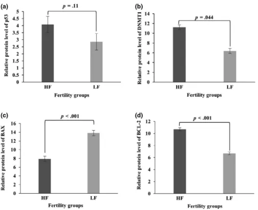
The expression of p53, BCL2, BAX and DNMT1 in bull spermato‐ zoa was determined using immunoblotting (Figure 2). The average expression of p53 protein in HF and LF bulls was 4.08 ± 0.58 and
2.85 ± 0.51, respectively, with no statistical difference (p = .11; Figure 2a) between the two groups. Expression of DNMT1 protein was greater in spermatozoa of HF sires (11.22 ± 0.49) when com‐ pared with LF bulls (6.37 ± 1.8; p = .044; Figure 2b) as well as the expression of BAX protein (7.88 ± 0.6 in HF bulls vs. 13.81 ± 3.1 in LF bulls; p < .001; Figure 2c). Moreover, levels of BCL2 protein were increased in HF bulls (10.7 ± 0.25; p < .001) as compared to those found in the LF individuals (6.7 ± 0.29; p < .001; Figure 2d). Based on Pearson's method, sperm BCL2 (r = .90, p < .001) and DNMT1 (r = .56 p = .01) were positively associated with bull fertility scores while BAX had a negative correlation with field fertility phenotypes of bulls (r = −.79 p = .01).
3.3 | Gene ontology
We performed a gene ontology enrichment analysis of miR‐15a, miR‐29b and miR‐34a using STRAP® software (Figure 3a–c). By this
means, biological processes of miR‐15a targets were mainly regula‐ tion (29.15%) and developmental process (11.55%). Molecular func‐ tion of miR‐15a targets was mostly binding (53.77%) and catalytic activity (23.8%), while cellular components of miR‐15a targets in‐ cluded intracellular organelles (23.73%), nucleus (21.71%) and cy‐ toplasm (13.14%), among others (Figure 3a). Biological processes linked to miR‐29b targets were mainly regulation (32.08%), cellular process (23.39%), developmental process (14.64%), among others (Figure 3b). Gene ontology analysis indicated the main molecular functions of miR‐29b targets as binding (58.31%), catalytic activity (25.22%) and structural molecular activity (14.2%), while cellular component‐related terms included nucleus (22.5%), extracellular (22.5%) and cytoplasm (17.1%; Figure 3b). F I G U R E 2 Expression of miRNA targets in spermatozoa from HF (n = 5) and LF (n = 5) bulls via immunoblotting. Bar graphs show the expression levels of (a) p53, (b) DNMT1, (c) BAX, and (d) BCL2 proteins. Data are expressed mean ± standard error of the mean. Statistical differences among band intensities in the blots were evaluated by t test (p < .05)
F I G U R E 3 Biological process, cellular component, and molecular function of miRNA targets. Protein data were analysed using the
4 | DISCUSSION
MicroRNA transcripts in bull spermatozoa are thought to be linked to the reproductive performance of sires (Abu‐Halima et al., 2014; Fagerlind et al., 2015; Govindaraju et al., 2012). In the present study, we hypothesised that variations in fertility status of bulls have sta‐ tistical associations with differences in levels of certain miRNAs in ejaculated spermatozoa associated with apoptosis. We demonstrate that the expression of miR‐15a and miR‐29 was higher in the sper‐ matozoa of LF bulls as compared to HF sires, and inverse correla‐ tions were identified between levels of both miR‐15a and miR29‐b and fertility scores. Considering miRNA targets, the average levels of both BCL2 and DNMT1 were higher in HF animals than in LF sires, and BAX expression was increased in spermatozoa from LF sires when compared to HF bulls. Moreover, we identified that the expression of BCL2 and DNMT1 had a positive correlation with bull fertility scores. Furthermore, we found strong negative correlations between BAX expression and bull fertility.
Growing evidence suggests that miRNAs play an epigenetic role in many physiological processes through regulating multiple target genes in post‐transcriptional process, including development, cell proliferation (Clotaire, Du, Wei, Yang, & Hua, 2018), differentiation (Gao et al., 2019) and apoptosis (Capra et al., 2017). Several miRNAs have been identified in spermatozoa that have implications as crit‐ ical regulators of apoptosis (Capra et al., 2017; Dai et al., 2019; Lv et al., 2019; Ma et al., 2018). Apoptosis regulates spermatogenesis (Wang, Li, Gong, Zhao, & Wu, 2019) and spontaneous programmed cell death in mature spermatozoa (Aitken & Baker, 2013; Engel, Springsguth, & Grunewald, 2018). Through analysis of miRNAs in sperm samples, researchers demonstrated their expression patterns in spermatozoa in different mammals including human (Heidary, Zaki‐Dizaji, Saliminejad, & Khorram Khorshid, 2019; Krawetz et al., 2011), boar (Godia et al., 2019), mouse (Hutcheon et al., 2017) and bull (Fagerlind et al., 2015). However, changes in miRNA levels and the expression of target genes of those miRNAs controlling apoptosis in bull spermatozoa remain poorly understood. A recent report has shown that miR‐15a levels had a negative correlation with HSPA1B in the spermatozoa of men with varicocele (Ji et al., 2014). Recently, it was demonstrated that the elevated expression of semenogelin‐1 and the reduced expression of miR‐525‐3p are associated with as‐ thenozoospermia and male infertility in human (Zhou et al., 2019). More recently, a study showed evidence that miRNA‐31‐5p regu‐ lates the proliferation, DNA synthesis and apoptosis of human sper‐ matogonial stem cells by the PAK1‐JAZF1‐cyclin A2 pathway (Fu et al., 2019).
The regulation of apoptosis or programmed cell death is complex and involves a balance between the pro‐ and antiapoptotic‐related proteins. Protein p53 tumour suppressor gene encodes a transcrip‐ tion factor that is critical apoptosis regulator in many cell types, including spermatozoa. Recently, a study showed that the upregu‐ lation of P53 is associated with infertility in bulls (Liu et al., 2018). In human, the increased level of p53 was negatively associated with motility (Silva et al., 2015) and in sperm samples from older men (Silva
et al., 2019). BAX and BCL2 are also regulators of apoptosis that helps maintain equilibrium within spermatozoa. The relative levels of these two apoptotic proteins determine whether a cell survives or undergoes apoptosis (Westphal, Dewson, Czabotar, & Kluck, 2011). It has been shown that the expression level of seminal BAX was sig‐ nificantly decreased, and BCL2 level was significantly increased in fertile men when compared to fertile men with varicocele (Mostafa, Rashed, Nabil, & Amin, 2014). DNA methyltransferases are a group of proteins responsible for transferring methyl groups to cytosine in CpG dinucleotides in the genome (Tajima, Suetake, Takeshita, Nakagawa, & Kimura, 2016). Methylated regions of imprinted genes are established during spermatogenesis and maintained in ma‐ ture spermatozoa by DNMT1 (Tang et al., 2018). Recently, a study demonstrated that abnormal DNA methylation‐mediated genomic imprinting is associated with oligozoospermia in men (Louie et al., 2016). In addition, DNA methylation levels in human spermatozoa from patients with unexplained infertility had different methylation patterns when compared to those of fertile men (Urdinguio et al., 2015).
Dynamic expression of highly conserved miR‐15 members is associated with cellular pro‐apoptotic activity (Cimmino et al., 2005). It was also shown in rodents that when miR‐15 family are inhibited, they were protected against cardiomyocyte apoptosis after myocardial infarction (Hullinger et al., 2012), and miR‐15a was also linked to the control of responses of cells to stress (Santosa, Castoldi, Paluschinski, Sommerfeld, & Haussinger, 2015). The mo‐ lecular mechanisms of how miR‐15a modulates sperm function in the bovine are yet unknown. The expression level of miR‐15a was downregulated in human spermatozoa with varicocele (Ji et al., 2014), suggesting that protection against hyperthermia or oxidative stress damage through decreased miR‐15a expression. Decreased levels of miR‐15a were also identified in oligoasthenozoospermic and asthenozoospermic men (Abu‐Halima et al., 2013). These find‐ ings led to the hypothesis that sperm miR‐15a expression can be linked to mechanisms controlling the apoptotic progression and cell response to stress (Mostafa et al., 2016). As miRNAs are thought to maintain the physiological balance within a cell, it is possible that the increased expression of miR‐15a may trigger apoptosis pathways in bull spermatozoa. In our study, spermatozoa from LF bulls had higher miR‐15a levels when compared to those of HF sires indicating that sperm miR‐15a is a potential biomarker for reduced male fertility. The negative correlation between miR‐15a and fertility scores was also evaluated in this study. Our finding is supported by a study in the human report where miR‐15a levels in the ejaculated sperma‐ tozoa were decreased in subjects with varicocele and infertility (Ji et al., 2014). Recently, a study showed evidence that miR‐15a trig‐ gers apoptosis by directly targeting BCL2 protein (Pekarsky, Balatti, & Croce, 2018). In our study, expression of BCL2 was significantly increased in HF sires compared to those of LF animals, suggesting that bull spermatozoa inhibit apoptosis by downregulating miR‐15a and upregulating BCL2. Family of miR‐29 miRNA includes miR‐29a, miR‐29b and miR‐29c. The miR‐29 family members function as a positive regulator of cell
proliferation, differentiation, cell cycle, senescence and apoptosis (Li, Wang, et al., 2013). It was reported that miR‐29 plays a role as a mo‐ lecular regulator of the meiosis in mice (Hilz, Fogarty, Modzelewski, Cohen, & Grimson, 2017), and it is underexpressed in the testis of sexually immature mouse (Yan et al., 2007). A recent study demon‐ strated that exposure to estradiol benzoate in newborns caused an increased expression of miR‐29b and apoptosis in adult germ cells (Meunier et al., 2012). Levels of miR‐29b are negatively correlated with superoxide dismutase and glutathione (Grant et al., 2015; Wu, Tang, Liu, Gan, & Zhou, 2016). Our results showed that the expres‐ sion of miR‐29b is higher in the spermatozoa of LF bulls as com‐ pared to the spermatozoa of HF sires. The importance of miR‐29b in male fertility is still unknown but it is plausible that miR‐29b has a vital role in apoptosis in spermatozoa (Meunier et al., 2012). It has been reported that miR‐29 expression is inversely correlated to DNA methyltransferase 1 (DNMT 1) by targeting transcription fac‐ tor 1 (Sp1), a transactivator of the DNMT1 gene (Yan et al., 2015). Recently, a study showed that an increased expression of miR‐29b was associated with a decrease in DNMT 1 in adult rats, resulting in apoptosis and an elevated risk of infertility (Meunier et al., 2012). DNA methylation is an epigenetic control of gene expression in male fertility (Takeda et al., 2017), and DNA methyltransferases mediate the DNA methylation process by transferring a methyl group to ge‐ nomic DNA sequence. In this study, the relative levels of DNMT1 were higher in spermatozoa from HF sires compared to those from LH bulls suggesting that spermatozoa of HF bulls maintain proper DNA methylation (Zhou et al., 2018).
Members of the miR‐34 family comprise miR‐34a, miR‐34b and miR‐34c. Previous studies showed evidence that miR‐34b and miR‐34c play significant roles in spermatogenesis, sperm function, fertilisation and early embryo development. Evolutionarily con‐ served family of the miR‐34 miRNAs have been identified in bull spermatozoa (Tscherner et al., 2014), and the downregulation of miR‐34b and miR‐34c in spermatozoa is linked to low fertility in humans (Comazzetto et al., 2014). Recently, a study demonstrated that miR‐34 may be crucial to bovine male and female gametes (Tscherner et al., 2014). Sperm miR‐34c levels were associated with intracytoplasmic sperm injection outcomes (Cui, Fang, Shi, Qiu, & Ye, 2015). It has been also showed that miR‐34c play roles in devel‐ opment and apoptosis of caprine and murine gametes (Li, Yu, et al., 2013; Liang et al., 2012). The previous report showed that miR‐34c is required for spermatogenesis in mice (Yuan et al., 2015). In humans, increased expression of miR‐34c was positively associated with high levels of p53 (Rahbar et al., 2017). The mechanisms underlying the importance of miR‐34a in spermatozoa physiology are still known. A recent study showed that miR‐34a is ubiquitous across spermato‐ genesis (Comazzetto et al., 2014). It has been shown that miR‐34a is associated with reduced cell motility and oxidative phosphorylation (Chang et al., 2011). In zebrafish, enhanced sperm motility and the influence on in vitro fertilisation rates were achieved when miR‐34a was inhibited (Guo et al., 2017). The authors showed evidence that miR‐34a downregulates glycogen synthase kinase‐3a (gsk3a), and miR‐34a/gsk3a interaction affects the motility of zebrafish
spermatozoa. We detected miR‐34a in bull spermatozoa with no significant difference between HF and LF groups. MiR‐34a miRNA may be an important regulator of male fertility. It has been sug‐ gested that members of the miR‐34 family, including miR‐34a, is a direct transcriptional target of the tumour suppressor p53 (He et al., 2007). It has been demonstrated that p53 regulates the expression of miR‐34a, by binding to a specific p53 binding site located within the gene that encodes miR‐34a (Raver‐Shapira et al., 2007). A study reported that loss of p53 in female but not in male mice significantly decreases fertility (Hu, Feng, Teresky, & Levine, 2007), and this may explain why we found no statistical differences in p53 levels in sper‐ matozoa from bulls of contrasting fertility status. BAX, a small 23‐kDa pro‐apoptotic member of the Bcl‐2 family, is known to be associated with spermatogenesis regulation (Metzler‐ Guillemain et al., 2015). In our study, BAX was increased in sperma‐ tozoa from LF bulls as compared with HF sires. Dogan et al. (2013) also showed that the expression of BAX protein was increased in low fertile animals when compared with high fertile sires. Levels of BAX in human seminal plasma were significantly higher in men with varicocele and infertile (Mostafa et al., 2016). An increase in BAX levels promotes the formation of mitochondrial pores in bovine spermatozoa (Martin et al., 2007) and contribute to permeabilisation of the mitochondrial membrane which releases mitochondrial pro‐ apoptotic factors (Crompton, 2000). It is possible that an increase in sperm membrane permeability could be associated with either early cell death or a premature acrosomal reaction (Medeiros, Forell, Oliveira, & Rodrigues, 2002) leading to low fertility. Our study also identified a strong negative correlation between expressions of BAX and bull fertility. This type of negative correlation has been reported previously in bulls (Dogan et al., 2013) and humans (Mostafa et al., 2014, 2016). It could be interpreted as BAX initiating the apoptotic pathway that subsequently leads to apoptosis in the mature sperma‐ tozoa, resulting in a decreased fertilisation capacity.
5 | CONCLUSION
We conclude that sperm miR‐15a, miR‐29b and miR‐34a are ex‐ pressed in bull spermatozoa with contrasting fertility scores. The combination of miR‐15a and miR‐29b represents a promising bio‐ marker of bull male fertility. Quantifications of sperm miR‐15a and miR‐29b miRNAs in a larger scale study are warranted to confirm their utility as reliable and accurate fertility indicators. Our results are important for deepening our understanding of the specific mechanisms regulating miR‐15a and miR‐29b and their targets in sperm physiology. In addition, it will enable promising new strategies to overcome reduced fertility rates in both cattle and other mam‐ mals including humans and endangered species.
ACKNOWLEDGEMENTS
This project was supported by Mississippi Agricultural Experiment Station, Alta Genetics Inc., and by Conselho Nacional de
Desenvolvimento Científico e Tecnológico (CNPq) of Brazil. Erika Menezes was funded by a competitive fellowship from Coordenação de Aperfeiçoamento de Pessoal de Nível Superior (CAPES) of Brazil. Hazem El Debaky, was funded by the Arab Republic of Egypt. Asma Ul Husna was supported by the Higher Education Commission of Pakistan under the International Research Support Program. Muhammet Rasit Ugur was founded by a competitive fellowship from Ministry of National Education of Turkey.
ORCID
Erdoğan Memili https://orcid.org/0000‐0002‐8335‐5645
REFERENCES
Abu‐Halima, M., Hammadeh, M., Backes, C., Fischer, U., Leidinger, P., Lubbad, A. M., … Meese, E. (2014). Panel of five microRNAs as potential biomarkers for the diagnosis and assessment of male in‐ fertility. Fertility and Sterility, 102(4), 989–997.e981. https ://doi. org/10.1016/j.fertn stert.2014.07.001
Abu‐Halima, M., Hammadeh, M., Schmitt, J., Leidinger, P., Keller, A., Meese, E., & Backes, C. (2013). Altered microRNA expression pro‐ files of human spermatozoa in patients with different spermatogenic impairments. Fertility and Sterility, 99(5), 1249–1255.e1216. https :// doi.org/10.1016/j.fertn stert.2012.11.054
Aitken, R. J., & Baker, M. A. (2013). Causes and consequences of apopto‐ sis in spermatozoa; Contributions to infertility and impacts on devel‐ opment. The International Journal of Developmental Biology, 57(2–4), 265–272. https ://doi.org/10.1387/ijdb.130146ja
Bhatia, V. N., Perlman, D. H., Costello, C. E., & McComb, M. E. (2009). Software tool for researching annotations of proteins: Open‐source protein annotation software with data visualization. Analytical
Chemistry, 81(23), 9819–9823. https ://doi.org/10.1021/ac901 335x
Bradford, M. M. (1976). A rapid and sensitive method for the quantita‐ tion of microgram quantities of protein utilizing the principle of pro‐ tein‐dye binding. Analytical Biochemistry, 72, 248–254. https ://doi. org/10.1016/0003‐2697(76)90527‐3 Capra, E., Turri, F., Lazzari, B., Cremonesi, P., Gliozzi, T. M., Fojadelli, I., … Pizzi, F. (2017). Small RNA sequencing of cryopreserved semen from single bull revealed altered miRNAs and piRNAs expression between High‐ and Low‐motile sperm populations. BMC Genomics, 18(1), 14. https ://doi.org/10.1186/s12864‐016‐3394‐7
Catalanotto, C., Cogoni, C., & Zardo, G. (2016). MicroRNA in control of gene expression: An overview of nuclear functions. International
Journal of Molecular Sciences, 17(10), 1712. https ://doi.org/10.3390/
ijms1 7101712
Chang, S. J., Weng, S. L., Hsieh, J. Y., Wang, T. Y., Chang, M. D., & Wang, H. W. (2011). MicroRNA‐34a modulates genes involved in cellular motility and oxidative phosphorylation in neural precursors derived from human umbilical cord mesenchymal stem cells. BMC Medical
Genomics, 4, 65. https ://doi.org/10.1186/1755‐8794‐4‐65
Chang, Y. M., Gianola, D., Heringstad, B., & Klemetsdal, G. (2004). Effects of trait definition on genetic parameter estimates and sire evalua‐ tion for clinical mastitis with threshold models. Animal Science, 79(3), 355–363. https ://doi.org/10.1017/S1357 72980 0090226
Chen, X., Li, X., Guo, J., Zhang, P., & Zeng, W. (2017). The roles of mi‐ croRNAs in regulation of mammalian spermatogenesis. Journal of
Animal Science and Biotechnology, 8, 35. https ://doi.org/10.1186/
s40104‐017‐0166‐4
Cimmino, A., Calin, G. A., Fabbri, M., Iorio, M. V., Ferracin, M., Shimizu, M., … Croce, C. M. (2005). miR‐15 and miR‐16 induce apoptosis by
targeting BCL2. Proceedings of the National Academy of Sciences of
the United States of America, 102(39), 13944–13949. https ://doi.
org/10.1073/pnas.05066 54102
Clotaire, D. Z. J., Du, X., Wei, Y., Yang, D., & Hua, J. (2018). miR‐19b‐3p integrates Jak‐Stat signaling pathway through Plzf to regulate self‐ renewal in dairy goat male germline stem cells. The International
Journal of Biochemistry & Cell Biology, 105, 104–114. https ://doi.
org/10.1016/j.biocel.2018.10.010
Comazzetto, S., Di Giacomo, M., Rasmussen, K. D., Much, C., Azzi, C., Perlas, E., … O'Carroll, D. (2014). Oligoasthenoteratozoospermia and infertility in mice deficient for miR‐34b/c and miR‐449 loci.
PLoS Genetics, 10(10), e1004597. https ://doi.org/10.1371/journ
al.pgen.1004597
Crompton, M. (2000). Bax, Bid and the permeabilization of the mitochon‐ drial outer membrane in apoptosis. Current Opinion in Cell Biology,
12(4), 414–419. https ://doi.org/10.1016/S0955‐0674(00)00110‐1
Cui, L., Fang, L., Shi, B., Qiu, S., & Ye, Y. (2015). Spermatozoa micro ri‐ bonucleic acid‐34c level is correlated with intracytoplasmic sperm injection outcomes. Fertility and Sterility, 104(2), 312–317.e311. https ://doi.org/10.1016/j.fertn stert.2015.05.003
Dai, D.‐H., Qazi, I., Ran, M.‐X., Liang, K., Zhang, Y., Zhang, M., … Zeng, C.‐J. (2019). Exploration of miRNA and mRNA profiles in fresh and frozen‐thawed boar sperm by transcriptome and small RNA sequenc‐ ing. International Journal of Molecular Sciences, 20(4), 802. https ://doi. org/10.3390/ijms2 0040802
de Menezes, E. B., van Tilburg, M., Plante, G., de Oliveira, R. V., Moura, A. A., & Manjunath, P. (2016). Milk proteins interact with goat Binder of SPerm (BSP) proteins and decrease their binding to sperm. Cell
and Tissue Research, 366(2), 427–442. https ://doi.org/10.1007/
s00441‐016‐2438‐2
Dickson, D. A., Paulus, J. K., Mensah, V., Lem, J., Saavedra‐Rodriguez, L., Gentry, A., … Feig, L. A. (2018). Reduced levels of miRNAs 449 and 34 in sperm of mice and men exposed to early life stress.
Translational Psychiatry, 8(1), 101. https ://doi.org/10.1038/
s41398‐018‐0146‐2
Dogan, S., Mason, M. C., Govindaraju, A., Belser, L., Kaya, A., Stokes, J., … Memili, E. (2013). Interrelationships between apoptosis and fertility in bull sperm. Journal of Reproduction and Development, 59(1), 18–26. https ://doi.org/10.1262/jrd.2012‐068
Engel, K. M., Springsguth, C. H., & Grunewald, S. (2018). What happens to the unsuccessful spermatozoa? Andrology, 6(2), 335–344. https :// doi.org/10.1111/andr.12467
Fagerlind, M., Stalhammar, H., Olsson, B., & Klinga‐Levan, K. (2015). Expression of miRNAs in bull spermatozoa correlates with fertility rates. Reproduction in Domestic Animals, 50(4), 587–594. https ://doi. org/10.1111/rda.12531
Fair, S., & Lonergan, P. (2018). Review: Understanding the causes of vari‐ ation in reproductive wastage among bulls. Animal, 12(s1), s53–s62. https ://doi.org/10.1017/S1751 73111 8000964
Friedman, R. C., Farh, K. K., Burge, C. B., & Bartel, D. P. (2009). Most mammalian mRNAs are conserved targets of microRNAs. Genome
Research, 19(1), 92–105. https ://doi.org/10.1101/gr.082701.108
Fu, H., Zhou, F., Yuan, Q., Zhang, W., Qiu, Q., Yu, X., & He, Z. (2019). miR‐ NA‐31‐5p mediates the proliferation and apoptosis of human sper‐ matogonial stem cells via targeting JAZF1 and cyclin A2. Molecular
Therapy ‐ Nucleic Acids, 14, 90–100. https ://doi.org/10.1016/j.
omtn.2018.11.004
Gao, H., Wen, H., Cao, C., Dong, D., Yang, C., Xie, S., … Dong, W. (2019). Overexpression of MicroRNA‐10a in germ cells causes male infertil‐ ity by targeting Rad51 in mouse and human. Frontiers in Physiology,
10, 765. https ://doi.org/10.3389/fphys.2019.00765
Gòdia, M., Estill, M., Castelló, A., Balasch, S., Rodríguez‐Gil, J. E., Krawetz, S. A., … Clop, A. (2019). A RNA‐Seq analysis to describe the boar sperm transcriptome and its seasonal changes. Frontiers in Genetics,
Gomes, A. Q., Nolasco, S., & Soares, H. (2013). Non‐coding RNAs: Multi‐tasking molecules in the cell. International Journal of Molecular
Sciences, 14(8), 16010–16039. https ://doi.org/10.3390/ijms1
40816010
Govindaraju, A., Uzun, A., Robertson, L. S., Atli, M. O., Kaya, A., Topper, E., … Memili, E. (2012). Dynamics of microRNAs in bull spermato‐ zoa. Reproductive Biology and Endocrinology, 10, 82. https ://doi. org/10.1186/1477‐7827‐10‐82
Grant, K. E., de Oliveira, R. V., Hennington, B. S., Govindaraju, A., Perkins, A., Stokes, J., … Memili, E. (2015). Sperm superoxide dismutase is as‐ sociated with bull fertility. Reproduction, Fertility and Development,
28, 1405. https ://doi.org/10.1071/RD14399
Guo, W., Xie, B., Xiong, S., Liang, X., Gui, J. F., & Mei, J. (2017). miR‐34a regulates sperm motility in zebrafish. International Journal of Molecular
Sciences, 18(12), 2676. https ://doi.org/10.3390/ijms1 8122676 Han, Y., & Penagaricano, F. (2016). Unravelling the genomic architecture of bull fertility in Holstein cattle. BMC Genetics, 17(1), 143. https :// doi.org/10.1186/s12863‐016‐0454‐6 He, L., He, X., Lim, L. P., de Stanchina, E., Xuan, Z., Liang, Y. U., … Hannon, G. J. (2007). A microRNA component of the p53 tumour suppressor network. Nature, 447(7148), 1130–1134. https ://doi.org/10.1038/ natur e05939
Heidary, Z., Zaki‐Dizaji, M., Saliminejad, K., & Khorram Khorshid, H. R. (2019). MicroRNA profiling in spermatozoa of men with unex‐ plained asthenozoospermia. Andrologia, 51(6), e13284. https ://doi. org/10.1111/and.13284
Hilz, S., Fogarty, E. A., Modzelewski, A. J., Cohen, P. E., & Grimson, A. (2017). Transcriptome profiling of the developing male germ line identifies the miR‐29 family as a global regulator during meio‐ sis. RNA Biology, 14(2), 219–235. https ://doi.org/10.1080/15476 286.2016.1270002 Hu, W., Feng, Z., Teresky, A. K., & Levine, A. J. (2007). p53 regulates ma‐ ternal reproduction through LIF. Nature, 450(7170), 721–724. https ://doi.org/10.1038/natur e05993 Hullinger, T. G., Montgomery, R. L., Seto, A. G., Dickinson, B. A., Semus, H. M., Lynch, J. M., … van Rooij, E. (2012). Inhibition of miR‐15 pro‐ tects against cardiac ischemic injury. Circulation Research, 110(1), 71–81. https ://doi.org/10.1161/CIRCR ESAHA.111.244442 Hutcheon, K., McLaughlin, E. A., Stanger, S. J., Bernstein, I. R., Dun, M. D., Eamens, A. L., & Nixon, B. (2017). Analysis of the small non‐pro‐ tein‐coding RNA profile of mouse spermatozoa reveals specific en‐ richment of piRNAs within mature spermatozoa. RNA Biology, 14(12), 1776–1790. https ://doi.org/10.1080/15476 286.2017.1356569 Ji, Z., Lu, R., Mou, L., Duan, Y.‐G., Zhang, Q., Wang, Y., … Cai, Z. (2014). Expressions of miR‐15a and its target gene HSPA1B in the spermato‐ zoa of patients with varicocele. Reproduction, 147(5), 693–701. https ://doi.org/10.1530/REP‐13‐0656
Kastelic, J. P., & Thundathil, J. C. (2008). Breeding soundness evalua‐ tion and semen analysis for predicting bull fertility. Reproduction
in Domestic Animals, 43(Suppl 2), 368–373. https ://doi.
org/10.1111/j.1439‐0531.2008.01186.x
Killian, G. J., Chapman, D. A., & Rogowski, L. A. (1993). Fertility‐associ‐ ated proteins in Holstein bull seminal plasma. Biology of Reproduction,
49(6), 1202–1207.
Krawetz, S. A. (2005). Paternal contribution: New insights and future challenges. Nature Reviews Genetics, 6(8), 633–642. https ://doi. org/10.1038/nrg1654
Krawetz, S. A., Kruger, A., Lalancette, C., Tagett, R., Anton, E., Draghici, S., & Diamond, M. P. (2011). A survey of small RNAs in human sperm.
Human Reproduction, 26(12), 3401–3412. https ://doi.org/10.1093/
humre p/der329
Kumar, A., Kroetsch, T., Blondin, P., & Anzar, M. (2015). Fertility‐as‐ sociated metabolites in bull seminal plasma and blood serum: 1H nuclear magnetic resonance analysis. Molecular Reproduction and
Development, 82(2), 123–131. https ://doi.org/10.1002/mrd.22450
Lewis, B. P., Burge, C. B., & Bartel, D. P. (2005). Conserved seed pair‐ ing, often flanked by adenosines, indicates that thousands of human genes are microRNA targets. Cell, 120(1), 15–20. https ://doi. org/10.1016/j.cell.2004.12.035
Li, M., Yu, M., Liu, C., Zhu, H., He, X., Peng, S., & Hua, J. (2013). miR‐34c works downstream of p53 leading to dairy goat male germline stem‐ cell (mGSCs) apoptosis. Cell Proliferation, 46(2), 223–231. https ://doi. org/10.1111/cpr.12013
Li, Y., Wang, H., Tao, K., Xiao, Q., Huang, Z., Zhong, L., … Feng, W. (2013). miR‐29b suppresses CML cell proliferation and induces apoptosis via regulation of BCR/ABL1 protein. Experimental Cell Research, 319(8), 1094–1101. https ://doi.org/10.1016/j.yexcr.2013.02.002
Liang, X., Zhou, D., Wei, C., Luo, H., Liu, J., Fu, R., & Cui, S. (2012). MicroRNA‐34c enhances murine male germ cell apoptosis through targeting ATF1. PLoS ONE, 7(3), e33861. https ://doi.org/10.1371/ journ al.pone.0033861
Liu, P., Yu, S., Cui, Y., He, J., Zhang, Q., Sun, J., … Ma, J. (2018). Regulation by Hsp27/P53 in testis development and sperm apoptosis of male cattle (cattle‐yak and yak). Journal of Cellular Physiology, 234(1), 650– 660. https ://doi.org/10.1002/jcp.26822
Louie, K., Minor, A., Ng, R., Poon, K., Chow, V., & Ma, S. (2016). Evaluation of DNA methylation at imprinted DMRs in the spermatozoa of oli‐ gozoospermic men in association with MTHFR C677T genotype.
Andrology, 4(5), 825–831. https ://doi.org/10.1111/andr.12240
Lv, J. X., Zhou, J., Tong, R. Q., Wang, B., Chen, X. L., Zhuang, Y. Y., … Wei, X. D. (2019). Hypoxiainduced miR210 contributes to apoptosis of mouse spermatocyte GC2 cells by targeting Kruppellike factor 7.
Molecular Medicine Reports, 19(1), 271–279. https ://doi.org/10.3892/
mmr.2018.9644
Ma, J., Fan, Y., Zhang, J., Feng, S., Hu, Z., Qiu, W., … Li, M. (2018). Testosterone‐dependent miR‐26a‐5p and let‐7g‐5p act as signal‐ ing mediators to regulate sperm apoptosis via targeting PTEN and PMAIP1. International Journal of Molecular Sciences, 19(4), 1233. https ://doi.org/10.3390/ijms1 9041233
Martin, G., Cagnon, N., Sabido, O., Sion, B., Grizard, G., Durand, P., & Levy, R. (2007). Kinetics of occurrence of some features of apoptosis during the cryopreservation process of bovine spermatozoa. Human Reproduction, 22(2), 380–388. https ://doi.org/10.1093/humre p/ del399 Medeiros, C. M., Forell, F., Oliveira, A. T., & Rodrigues, J. L. (2002). Current status of sperm cryopreservation: Why isn't it better? Theriogenology, 57(1), 327–344. https ://doi.org/10.1016/S0093‐691X(01)00674‐4 Metzler‐Guillemain, C., Victorero, G., Lepoivre, C., Bergon, A., Yammine, M., Perrin, J., … Nguyen, C. (2015). Sperm mRNAs and microR‐ NAs as candidate markers for the impact of toxicants on human spermatogenesis: An application to tobacco smoking. Systems
Biology in Reproductive Medicine, 61(3), 139–149. https ://doi.
org/10.3109/19396 368.2015.1022835
Meunier, L., Siddeek, B., Vega, A., Lakhdari, N., Inoubli, L., Bellon, R. P., … Benahmed, M. (2012). Perinatal programming of adult rat germ cell death after exposure to xenoestrogens: Role of microRNA miR‐29 family in the down‐regulation of DNA methyltransferases and Mcl‐1. Endocrinology, 153(4), 1936–1947. https ://doi.org/10.1210/ en.2011‐1109
Mostafa, T., Rashed, L., Nabil, N., & Amin, R. (2014). Seminal BAX and BCL2 gene and protein expressions in infertile men with var‐ icocele. Urology, 84(3), 590–595. https ://doi.org/10.1016/j.urolo gy.2014.05.016
Mostafa, T., Rashed, L. A., Nabil, N. I., Osman, I., Mostafa, R., & Farag, M. (2016). Seminal miRNA relationship with apop‐ totic markers and oxidative stress in infertile men with varico‐ cele. BioMed Research International, 2016, 4302754. https ://doi. org/10.1155/2016/4302754
Moura, A. A., Chapman, D. A., & Killian, G. J. (2007). Proteins of the ac‐ cessory sex glands associated with the oocyte‐penetrating capacity
of cauda epididymal sperm from holstein bulls of documented fertil‐ ity. Molecular Reproduction and Development, 74(2), 214–222. https :// doi.org/10.1002/mrd.20590
Pekarsky, Y., Balatti, V., & Croce, C. M. (2018). BCL2 and miR‐15/16: From gene discovery to treatment. Cell Death & Differentiation, 25(1), 21–26. https ://doi.org/10.1038/cdd.2017.159
Rahbar, S., Novin, M. G., Alizadeh, E., Shahnazi, V., Pashaei‐Asl, F., AsrBadr, Y. A., … Pashaiasl, M. (2017). New insights into the expres‐ sion profile of MicroRNA‐34c and P53 in infertile men spermatozoa and testicular tissue. Cellular and Molecular Biology, 63(8), 77–83. https ://doi.org/10.14715/ cmb/2017.63.8.17
Raver‐Shapira, N., Marciano, E., Meiri, E., Spector, Y., Rosenfeld, N., Moskovits, N., … Oren, M. (2007). Transcriptional activation of miR‐34a contributes to p53‐mediated apoptosis. Molecular Cell,
26(5), 731–743. https ://doi.org/10.1016/j.molcel.2007.05.017
Rosas‐Cardenas Fde, F., Duran‐Figueroa, N., Vielle‐Calzada, J. P., Cruz‐ Hernandez, A., Marsch‐Martinez, N., & de Folter, S. (2011). A simple and efficient method for isolating small RNAs from different plant species. Plant Methods, 7, 4. https ://doi.org/10.1186/1746‐4811‐7‐4 Santosa, D., Castoldi, M., Paluschinski, M., Sommerfeld, A., & Haussinger,
D. (2015). Hyperosmotic stress activates the expression of mem‐ bers of the miR‐15/107 family and induces downregulation of anti‐ apoptotic genes in rat liver. Scientific Reports, 5, 12292. https ://doi. org/10.1038/srep1 2292
Silva, J. V., Cabral, M., Correia, B. R., Carvalho, P., Sousa, M., Oliveira, P. F., & Fardilha, M. (2019). mTOR signaling pathway regulates sperm quality in older men. Cells, 8(6), 629. https ://doi.org/10.3390/cells 8060629
Silva, J. V., Freitas, M. J., Correia, B. R., Korrodi‐Gregorio, L., Patricio, A., Pelech, S., & Fardilha, M. (2015). Profiling signaling proteins in human spermatozoa: Biomarker identification for sperm quality evaluation. Fertility and Sterility, 104(4), 845–856.e848. https ://doi. org/10.1016/j.fertn stert.2015.06.039
Tajima, S., Suetake, I., Takeshita, K., Nakagawa, A., & Kimura, H. (2016). Domain structure of the Dnmt1, Dnmt3a, and Dnmt3b DNA meth‐ yltransferases. Advances in Experimental Medicine and Biology, 945, 63–86. https ://doi.org/10.1007/978‐3‐319‐43624‐1_4
Takeda, K., Kobayashi, E., Akagi, S., Nishino, K., Kaneda, M., & Watanabe, S. (2017). Differentially methylated CpG sites in bull spermatozoa revealed by human DNA methylation arrays and bisulfite analysis.
Journal of Reproduction and Development, 63(3), 279–287. https ://doi.
org/10.1262/jrd.2016‐160 Tang, Q., Pan, F., Yang, J., Fu, Z., Lu, Y., Wu, X., … Wu, W. (2018). Idiopathic male infertility is strongly associated with aberrant DNA methylation of imprinted loci in sperm: A case‐control study. Clinical Epigenetics, 10(1), 134. https ://doi.org/10.1186/s13148‐018‐0568‐y Tscherner, A., Gilchrist, G., Smith, N., Blondin, P., Gillis, D., & LaMarre, J. (2014). MicroRNA‐34 family expression in bovine gametes and pre‐ implantation embryos. Reproductive Biology and Endocrinology, 12, 85. https ://doi.org/10.1186/1477‐7827‐12‐85
Tulay, P., & Sengupta, S. B. (2016). MicroRNA expression and its as‐ sociation with DNA repair in preimplantation embryos. Journal
of Reproduction and Development, 62(3), 225–234. https ://doi.
org/10.1262/jrd.2015‐167 Urdinguio, R. G., Bayón, G. F., Dmitrijeva, M., Toraño, E. G., Bravo, C., Fraga, M. F., … Fernández, A. F. (2015). Aberrant DNA methylation patterns of spermatozoa in men with unexplained infertility. Human Reproduction, 30(5), 1014–1028. https ://doi.org/10.1093/humre p/ dev053 Wang, Y., Li, X., Gong, X., Zhao, Y., & Wu, J. (2019). MicroRNA‐322 reg‐ ulates self‐renewal of mouse spermatogonial stem cells through Rassf8. International Journal of Biological Sciences, 15(4), 857–869. https ://doi.org/10.7150/ijbs.30611
Westphal, D., Dewson, G., Czabotar, P. E., & Kluck, R. M. (2011). Molecular biology of Bax and Bak activation and action. Biochimica
et Biophysica Acta, 1813(4), 521–531. https ://doi.org/10.1016/j.
bbamcr.2010.12.019
Wu, J., Tang, H., Liu, Q., Gan, D., & Zhou, M. (2016). Effect of alpha‐li‐ poic acid in inhibiting oxidative stress and promoting diabetic wound healing by suppressing expression of miR‐29b in mice. Zhongguo
Xiu Fu Chong Jian Wai Ke Za Zhi, 30(8), 1034–1037. https ://doi.
org/10.7507/1002‐1892.20160207
Yan, B., Guo, Q., Fu, F. J., Wang, Z., Yin, Z., Wei, Y. B., & Yang, J. R. (2015). The role of miR‐29b in cancer: Regulation, function, and signaling.
OncoTargets and Therapy, 8, 539–548. https ://doi.org/10.2147/OTT.
S75899
Yan, N., Lu, Y., Sun, H., Tao, D., Zhang, S., Liu, W., & Ma, Y. (2007). A mi‐ croarray for microRNA profiling in mouse testis tissues. Reproduction,
134(1), 73–79. https ://doi.org/10.1530/REP‐07‐0056
Yuan, S., Tang, C., Zhang, Y., Wu, J., Bao, J., Zheng, H., … Yan, W. (2015). mir‐34b/c and mir‐449a/b/c are required for spermatogenesis, but not for the first cleavage division in mice. Biology Open, 4(2), 212– 223. https ://doi.org/10.1242/bio.20141 0959 Zhou, Q. Z., Guo, X. B., Zhang, W. S., Zhou, J. H., Yang, C., Bian, J., … Liu, C. D. (2019). Expressions of miR‐525‐3p and its target gene SEMG1 in the spermatozoa of patients with asthenozoospermia. Andrology, 7(2), 220–227. https ://doi.org/10.1111/andr.12573 Zhou, Y., Connor, E. E., Bickhart, D. M., Li, C., Baldwin, R. L., Schroeder, S. G., … Liu, G. E. (2018). Comparative whole genome DNA methyl‐ ation profiling of cattle sperm and somatic tissues reveals striking hypomethylated patterns in sperm. Gigascience, 7(5), 1‐13. https :// doi.org/10.1093/gigas cienc e/giy039
Zwald, N. R., Weigel, K. A., Chang, Y. M., Welper, R. D., & Clay, J. S. (2004a). Genetic selection for health traits using producer‐recorded data. I. Incidence rates, heritability estimates, and sire breeding values. Journal of Dairy Science, 87(12), 4287–4294. https ://doi. org/10.3168/jds.S0022‐0302(04)73573‐0
Zwald, N. R., Weigel, K. A., Chang, Y. M., Welper, R. D., & Clay, J. S. (2004b). Genetic selection for health traits using producer‐recorded data. II. Genetic correlations, disease probabilities, and relationships with existing traits. Journal of Dairy Science, 87(12), 4295–4302. https ://doi.org/10.3168/jds.S0022‐0302(04)73574‐2
How to cite this article: Menezes ESB, Badial PR,
El Debaky H, et al. Sperm miR‐15a and miR‐29b are associated with bull fertility. Andrologia. 2020;52:e13412. https ://doi. org/10.1111/and.13412