NoM3J.7.pdf Advanced Photonics Congress (BGPP, IPR, NP, Networks, NOMA, Sensors, SOF, SPPCom) © OSA 2018
Interference Coatings for Infrared Spectroscopy and
Colorimetric Sensing
Gokhan Bakan1,2, Sencer Ayas1,3, Erol Ozgur1,4, Kemal Celebi1,5, Aykutlu Dana1,6 1UNAM-Institute of Materials Science and Nanotechnology, Bilkent University, Ankara, 06800, Turkey
2Department of Electrical and Electronics Engineering, Atilim University, Ankara, 06830, Turkey
3Canary Center at Stanford for Cancer Early Detection, Department of Radiology, Stanford School of Medicine, Palo Alto, CA 94304, USA 4Department of Biomedical Engineering, University of Arizona, Tucson, Arizona 85721, USA
5Department of Materials, ETH Zurich, Zurich 8093, Switzerland 6Department of Electrical Engineering, Stanford University, Stanford, CA 94305, USA
Author e-mail address: gokhan.bakan@atilim.edu.tr
Abstract: Constructive interference and strong interference surfaces are created to sense ultrathin
probe materials such as monolayer protein molecules using enhanced infrared absorption spectroscopy and colorimetric detection, respectively.
OCIS codes: (310.1620) Interference coatings; (280.4788) Optical sensing and sensors. 1. Introduction
Sensing bio-molecules and their interactions is crucial for medical diagnosis. Owing to the low dimensions of the bio-molecules, optical detection of such interactions is challenging, yet can be achieved by enhancing the interaction between the light and the bio-molecules. Optical surfaces based on surface plasmon resonance (SPR) [1] and plasmonic nano-antennas [2] are demonstrated to be very sensitive to the presence of a thin layer such as protein molecules on their surfaces. Here, we demonstrate the use of interference coatings aiming detection of nanometer-thick films operating in the mid-infrared and visible regimes [3–5].
2. Results
The interference coatings for enhanced infrared absorption spectroscopy consist of ~1-µm-thick CaF2 film on optically-thick Al [Fig. 1(a)]. When illuminated with broadband infrared (IR) light, the electric field intensity on the entire CaF2 surface is enhanced by a factor of ~4 due to the constructive interference of the incident and reflected light. As a result, the absorption of the infrared light by a lossy film such as a monolayer of protein molecules on the surface is enhanced following the Beer-Lambert Law. To benchmark the performance of the field enhancement surface based on CaF2/Al stack, Ag nano-antennas are designed and fabricated [Fig. 1(b)]. 10-nm-thick Poly(methyl methacrylate) (PMMA) is used as the probe material. Both CaF2/Al and Ag nano-antenna surfaces are designed to enhance the absorption of the IR light at the major PMMA absorption band (1732 cm-1).
0 2 4 6 8 10 1000 1500 2000 A b so rb an ce (% ) Wavenumber (cm-1) 940 nm 2 3 0 n m 1.5 µm CaF2 Al Si (b) (a) (c)
Fig. 1. (a) Cross-section SEM image of CaF2 film on Al. (a) SEM image of Ag antennas. (c) Absorption spectrum of 10-nm-thick PMMA film on
CaF2/Al (red curve) and on the antennas (black curve). The dashed lines in (c) mark the PMMA absorption bands. The red curve is moved along
the y-axis by 2% for clarity.
The IR absorption spectrum for 10-nm-PMMA on the CaF2/Al surface shows all of the PMMA absorption bands ranging from 3000 to 1000 cm-1, whereas the Ag antennas only show the major band with a lower absorption intensity. The results show that enhancing the electric field on the entire surface provides detection of the absorption bands with larger intensities and in a larger bandwidth compared to the localized field enhancement on the
plasmonic nano-antennas. Furthermore, the field-enhancement surface is patternless, hence can be fabricated on large areas, whereas the antennas require lithography and offer limited surface area. The field-enhancement surfaces are also used to detect monolayers of bovine serum albumin (BSA) molecules, octadecanethiol (ODT) molecules, and ultrathin SiO2 films all of which exhibit larger absorbance on the field-enhancement surfaces compared to their performance on plasmonic surfaces [3].
NoM3J.7.pdf Advanced Photonics Congress (BGPP, IPR, NP, Networks, NOMA, Sensors, SOF, SPPCom) © OSA 2018
CaF2 as the dielectric layer offers infrared transparency and maximized electric field intensity on the surface. The use of other infrared-transparent materials like Si or Ge instead of CaF2 decreases the field-intensity
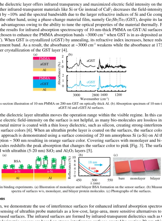
enhancement by ~10% and the spectral bandwidth due to the larger refractive indices of Si and Ge compared to that of CaF2. On the other hand, using a phase-change material film, namely Ge2Sb2Te5 (GST), despite its large refractive index can be advantageous owing to the ability to tune the optical properties of the material thermally. Fig. 2 summarizes the results for infrared absorption spectroscopy of 10-nm-thick PMMA on GST/Al surfaces. The GST thickness is chosen to enhance the PMMA absorption bands ~3000 cm-1 when GST is in as-deposited amorphous phase (aGST). When GST is crystallized (cGST) by annealing, its refractive index increases, hence redshifts the field-enhancement band. As a result, the absorbance at ~3000 cm-1 weakens while the absorbance at 1732 cm-1 is enhanced after crystallization of the GST layer [4].
-1 0 1 2 -1 0 1 2 -1 0 1 2 -1 0 1 2 Wavenumber (cm-1) (b) aGST cGST aGST PMMA Al 3000 2800 1600 1200 cGST PMMA Al A b so rb an ce (% ) (a)
Fig. 2. (a) Cross-section illustration of 10-nm PMMA on 200-nm GST on optically thick Al. (b) Absorption spectrum of 10-nm-thick PMMA on aGST/Al and cGST/Al surfaces.
Keeping the dielectric layer ultrathin moves the operation range within the visible regime. In this case,
enhancing the electric field-intensity on the surface is not helpful, as many bio-molecules are lossless in the visible. Instead, the metal mirror is coated with a thin lossy dielectric, such as silicon, creating strong-interference surfaces with distinct surface colors [6]. When an ultrathin probe layer is coated on the surfaces, the surface colors noticeably change. This approach is demonstrated using a surface consisting of 20 nm amorphous Si (a-Si) on Al that exhibits a strong absorption ~ 500 nm resulting in orange surface color. Covering surfaces with monolayer and bi-layer BSA protein molecules redshifts the peak absorption that changes the surface color to pink [Fig. 3]. The surfaces are further tested with ultrathin (5-20 nm) SiO2 and Al2O3 layers [5].
0 2 4 6 8 10 450 500 550 a-Si a-Si λ (nm) (a) BSA R ef le ct an ce ( %) 8 nm 18 nm
Al Al bare monolayer bilayer
(b) (c)
Fig. 3. Protein binding experiments. (a) Illustration of monolayer and bilayer BSA formation on the sensor surface. (b) Measured reflection spectra of surfaces w/o, monolayer, and bilayer protein molecules. (c) Photographs of the surfaces.
3. Conclusions
In conclusion, we demonstrate the use of interference surfaces for enhanced infrared absorption spectroscopy and colorimetric sensing of ultrathin probe materials as a low-cost, large-area, more sensitive alternatives to the
plasmonics-based surfaces. The infrared surfaces are formed by infrared-transparent dielectrics such as CaF2 or GST on Al. The colorimetric sensors are based on strong-interference surfaces created by coating 20-nm-tthick a-Si on Al. The surface color noticeably changes when an ultrathin probe material is coated on the surface.
4. References
[1] B. Liedberg, C. Nylander, and I. Lunström, “Surface plasmon resonance for gas detection and biosensing,” Sensors and Actuators. 4, 299–304 (1983).
[2] R. Adato and H. Altug, “In-situ ultra-sensitive infrared absorption spectroscopy of biomolecule interactions in real time with plasmonic nanoantennas.,” Nat. Commun. 4, 2154 (2013).
[3] S. Ayas, G. Bakan, E. Ozgur, K. Celebi, and A. Dana, “Universal Infrared Absorption Spectroscopy Using Uniform Electromagnetic Enhancement,” ACS Photonics 3, 337–342 (2016).
[4] G. Bakan, S. Ayas, E. Ozgur, K. Celebi, and A. Dana, “Thermally Tunable Ultrasensitive Infrared Absorption Spectroscopy Platforms Based on Thin Phase-Change Films,” ACS Sensors 1, 1403–1407 (2016).
[5] S. Ayas, G. Bakan, E. Ozgur, K. Celebi, G. Torunoglu, and A. Dana, “Colorimetric Detection of Ultrathin Dielectrics on Strong-Interference Coatings,” Opt. Lett. (in press).
[6] M. A. Kats, R. Blanchard, P. Genevet, and F. Capasso, “Nanometre optical coatings based on strong interference effects in highly absorbing media,” Nat. Mater. 12, 20–24 (2013).

