IN VITRO AND IN VIVO IMMUNOMODULATORY
EFFECTS OF EXTRACELLULAR VESICLES
a thesis
submitted to the department of molecular biology and
genetics
and the graduate school of engineering and science
of bilkent university
in partial fulfillment of the requirements
for the degree of
master of science
By
Mehmet S
¸ahin
December, 2013
I certify that I have read this thesis and that in my opinion it is fully adequate, in scope and in quality, as a thesis for the degree of Master of Science.
Assoc. Prof. ˙Ihsan G¨ursel(Advisor)
I certify that I have read this thesis and that in my opinion it is fully adequate, in scope and in quality, as a thesis for the degree of Master of Science.
Assoc. Prof. Reng¨ul C¸ etin-Atalay
I certify that I have read this thesis and that in my opinion it is fully adequate, in scope and in quality, as a thesis for the degree of Master of Science.
Prof. Dr. Kamil Can Ak¸calı
Approved for the Graduate School of Engineering and Science:
Prof. Dr. Levent Onural Director of the Graduate School
ABSTRACT
IN VITRO AND IN VIVO IMMUNOMODULATORY
EFFECTS OF EXTRACELLULAR VESICLES
Mehmet S¸ahin
M.S. in Molecular Biology and Genetics Supervisor: Assoc. Prof. ˙Ihsan G¨ursel
December, 2013
The major theme of this thesis was to characterize and understand the im-munomodulatory potential of extracellular vesicles (EVs) isolated from different mouse and human cell lines on immune cells. To this end, we first purified and char-acterized the particle nature of exosomes and microparticles (MPs) and exosomes by AFM and DLS analyses. Next, we documented their in vitro or in vivo differential uptake/internalization kinetics by immune cells. Finally, we tested the potential ap-plication of EVs as a drug delivery system. The physicochemical characterization studies confirmed that EVs were in vesicular form and upon reconstitution followed by lyophilization they retained their original sizes (i.e. size ranges were 100-150 nm for exosomes, and 250-500 nm for MPs, respectively). MPs derived from mouse cell lines (macrophage, T-cell and fibroblast) were stained and administered in vitro and in vivo to track their internalization by immune cells. When incubated in culture, origins of MPs greatly affected uptake rate and ratio by different immune cells. This differential internalization pattern of distinct MPs was reproduced when they were injected i.p. to mice. When incubated in culture B-cells took up the most MPs from fibroblast origin cells. Macrophages of peritoneal exudate cells took up RAW264.7 derived MPs at the highest level upon 24h post-ip injection. We tested whether MPs have a role in transportation or presentation of bacterial products during an ongo-ing infection by adherongo-ing circulatongo-ing pathogenic by-products and carryongo-ing them to distant immune cells. Our data suggest that MPs could contribute to the severity of ongoing infection in vivo, since MPs from different cells can adhere distinct ligands such as LPS or DNA or even RNA and transmit these ligands to naive innate im-mune cells augmenting the immunostimulatory response raised against these ligands. Lastly, the immunotherapeutic potential of EVs harboring immunosuppressive syn-thetic oligodeoxynucleotide sequence namely, A151 was tested following dehydration-rehydration method on mouse splenocytes. EV-associated A151 displayed improved inhibition of immune activation triggered by TLR7/8 and TLR9 ligands on spleen cells. When taken together, this study established that various types of EVs derived from different cells induces plethora of activities on immune cells. Furthermore, EVs are potential drug carrier systems when loaded externally with suitable agents and can be harnessed in immunotherapy of diseases.
Keywords: Exosome, microparticle, TLR, immunosuppressive DNA, immunomodu-lation .
¨
OZET
H ¨
UCRE DIS
¸I KESEC˙IKLER˙IN ˙IN V˙ITRO VE ˙IN V˙IVO
˙IMM ¨
UNMOD ¨
ULAT ¨
OR ETK˙ILER˙I
Mehmet S¸ahin
Molek¨uler Biyoloji ve Genetik, Y¨uksek Lisans Tez Y¨oneticisi: Do¸c. Dr. ˙Ihsan G¨ursel
Aralık, 2013
Bu tezin ana konusu, de˘gi¸sik fare ve insan h¨ucre hatlarından elde edilen, h¨ucreler arası ileti¸simi sa˘glayan partik¨ullerin ba˘gı¸sıklık sistemi h¨ucreleri ¨uzerindeki olası etkilerini an-lamak ve ara¸stırmaktır. Bu ama¸cla ¨oncelikle h¨ucre dı¸sı kesecikleri izole edip partik¨uler yapılarını atomik g¨u¸c mikroskopu (AFM) ve dinamik ı¸sık sa¸cılması (DLS) metodları kul-lanarak analiz ettik. Ek olarak bu partik¨ullerin in vitro ve in vivo olarak ba˘gı¸sık sistemi h¨ucreleri tarafından h¨ucre i¸cine alınma kineti˘gini de belgelemeye ¸calı¸stık. Son olarak da ila¸c ta¸sıma sistemi olarak olası kullanımlarını tanımladık. Keseciklerin fiziko-kimyasal karak-terizasyonları DLS ve AFM ile yapılarak, bunların liyofilizasyon sonucu kesecik yapılarını korudukları ve boyutlarının de˘gi¸smedi˘gi g¨osterildi (Ekzosomlar 100-150 nm boyutlarında, mikropartik¨uller ise 250-500 nm boyutlarında). Fare h¨ucre hatlarından elde edilen mikropar-tik¨uller (Makrofaj, T h¨ucresi ve Fibroblast) boyanarak in vitro ve in vivo olarak ba˘gı¸sıklık sistemi h¨ucrelerince alımları takip edildi.H¨ucre k¨ult¨ur¨u ¸calı¸smalarında mikropartik¨ullerin kayna˘gının de˘gi¸sik ba˘gı¸sıklık sistemi h¨ucrelerince alım oranını ¨onemli derecede etkiledi˘gi g¨ozlemlendi. Intraperitonal olarak da farelere enjekte edilen mikropartik¨ullerin h¨ucre i¸cine alım farkları g¨osterilmi¸stir. K¨ult¨urde dalak h¨ucreleri ile ink¨ube edildi˘ginde fibroblastlar tarafından ¨uretilmi¸s vesik¨ullerin ¸co˘gunlu˘gu B h¨ucreleri tarafından alınırken, Ip enjeksiyonun-dan 24 saat sonra peritonal sıvı h¨ucrelerinin RAW 264.7 tarafından sentezlenen mikropar-tik¨ulleri en y¨uksek d¨uzeyde aldı˘gı g¨ozlemlenmi¸stir. Daha sonra mikropartik¨ullerin enfek-siyon sırasında patojenlere ba˘glanarak bakteri ¨ur¨unlerinin ta¸sınmasında ve uzak yerlerdeki ba˘gı¸sıklık sistemi h¨ucrelerine sunulmasında rol alıp almadı˘gı sorgulandı. Farklı mikropar-tik¨ullerin LPS, DNA ya da RNA gibi farklı ligandlara ba˘glanarak bunları uyarılmamı¸s ba˘gı¸sıklık sistemi h¨uclerine iletti˘gini ve imm¨un aktivasyonu artırdı˘gı g¨ozlenlenmi¸stir. Bu bulgular mikropartik¨ullerin in vivo enfeksiyonun d¨uzeyine katkı sa˘gladı˘gını i¸saret etmek-tedir. Son olarak, bu keseciklerin potansiyel terap¨otik ¨ozellikleri sentetik ba˘gı¸sık sistemi baskılayıcı bir oligodeoksin¨ukleotid olan A151 dehidrasyon-rehidrasyon y¨ontemi ile dalak h¨uclerinde test edilmi¸stir. A151 i¸ceren kesecikler TLR7/8 ve TLR9 ligandları ile uyarılan dalak h¨ucreleri ¨uzerinde daha etkin bir baskılama g¨ostermi¸stir. Sonu¸c olarak bu ¸calı¸sma de˘gi¸sik h¨ucreler tarafından ¨uretilen keseciklerin ba˘gı¸sık sistemi h¨ucreleri ¨uzerinde bir ¸cok etkisi oldu˘gunı g¨ostermi¸stir. Ek olarak keseciklerin potansiyel bir ila¸c ta¸sıma sistemi olarak dı¸sarıdan y¨uklenebilen uygun ajanlar oldu˘gu ve hastalıkların tedavisinde kullanılabilece˘gi anla¸sılmı¸stır.
Acknowledgement
First of all, I would like to express my deepest gratitude to my spectacular supervisor ˙Ihsan G¨ursel for his endless support and patience. Thank you for accepting me as a M.Sc. student and for allowing me to pursue diverse and borderless ideas. You were not only a supervisor but also a decent parent for me. Thank you very much for the all fun and instructing experience we have lived together.
I would like to thank to my laboratory mates, Gizem, ˙Ihsan, Fuat, Tamer, Banu, G¨ozde, Defne, Beg¨um, Merve, Umur and Yusuf and dearest interns Ece, Sevgican and ˙Idil for their help, precious friendship and support in the lab and in my experiments. I should also mention all senior project students past two years, I am grateful for their support. It was a privilege for me to be part of IG family.
I also thank to veterinarians Emre Bu˘gdaycı and Gamze Aykut, and Turan amca for their help, support and patience during animal experiments and Abdullah amca for solving every technical problem I have encountered.
I would also like to thank to The Scientific and Technological Research Council of Turkey (T ¨UB˙ITAK) for their financial support throughout my master studies.
My sincere thanks to Mayda G¨ursel for her assistance and support during my studies. Due to her exceptional personality she has a special place in hearts of all IG group members. I also would like to express my special thanks to MG Group members, Bilgi, Esin, Mine and Soner for their friendliness and support.
Moreover, I would like to thank all my instructors and friends in MBG graduate school for their guidance, companionship and assistance. I have learned a piece of science and friendship from every each of you. Yet, in particular I want to express my deepest gratitude to B¨u¸sra, G¨okhan abi, Derya, Nil¨ufer, Gurbet, Emre the French and the dangerous duo, Engin-Ender for their support, help and warm friendship. Also, I would like to thank my past advisor Uygar Tazebay for his support and assistance.
Very special thanks to my uncle Murat Ko¸c for trusting me and accepting to be my guarantor for my T ¨UB˙ITAK fellowship.
Finally, I want to thank my parents, Semavil and Medine, my brother Emre and my beloved fianc´ee Berna for supporting me and being there whenever I need them. Without them, none of the exceptional things in my life would have been possible. Their love, care and support is the most significant motivation for me, which makes my accomplishments meaningful.
Contents
1 Introduction 1
1.1 Immune System . . . 1
1.2 Innate Immunity . . . 2
1.3 Adaptive Immunity . . . 4
1.4 Cells of the Immune System . . . 4
1.4.1 Dendritic cells . . . 5
1.4.2 T lymphocytes . . . 6
1.4.3 B lymphocytes . . . 7
1.4.4 Macrophages . . . 8
1.5 Cytokines . . . 8
1.6 Pattern Recognition Receptors . . . 10
1.6.1 Toll-like receptors . . . 10
1.7 Immunmodulatory synthetic DNA motifs . . . 13
1.7.1 Immune stimulatory synthetic DNA motifs . . . 13
CONTENTS vii 1.8 Extracellular Vesicles . . . 16 1.9 Microparticles . . . 17 1.9.1 Microparticle formation . . . 18 1.9.2 Microparticle composition . . . 19 1.9.3 Microparticle function . . . 21 1.9.4 Microparticle immunotherapy . . . 24 1.10 Exosomes . . . 25 1.10.1 Exosome formation . . . 26 1.10.2 Exosome composition . . . 27 1.10.3 Exosome function . . . 30 1.10.4 Exosome immunotherapy . . . 33
2 Aim of the Study 35 3 Materials and Methods 36 3.1 Materials . . . 36
3.1.1 Immunomodulatory Agents . . . 36
3.1.2 ELISA Reagents . . . 37
3.2 Methods . . . 37
3.2.1 Cytokine ELISA . . . 37
3.2.2 NO detection by Griess Method . . . 38
CONTENTS viii
3.2.4 Preparation of Single Cell Suspensions of Mouse Spleen Cells . . . 39
3.2.5 PBMC Isolation . . . 40
3.2.6 Maintenance of Cell Lines . . . 40
3.2.7 Microvesicle Isolation . . . 42
3.2.8 SP-DiOC Staining of Extracellular Vesicles . . . 45
3.2.9 Atomic Force Microscopy(AFM) . . . 45
3.2.10 Binding & Uptake and Internalization Assays . . . 45
3.2.11 Association of TLR ligands with Microparticles . . . 46
3.2.12 Cell Counting . . . 46
3.2.13 Cargo Loading to Extracellular Vesicles . . . 47
3.2.14 Micro BCA Protein Assay . . . 47
4 Results & Discussion 49 4.1 Physicochemical analyses of the extracellular vesicles . . . 49
4.2 Uptake and Internalization studies with EVs . . . 52
4.2.1 Influence of cell source of EVs on binding and internalization by spleen cells . . . 55
4.2.2 Studies on differential internalization of EVs by immune cells . . . 57
4.2.3 In vivo distribution of MPs upon intrperitoneal administration to mice 65 4.3 EVs transmit TLR ligands to distant cells and induce immune activation . . 67
4.3.1 Effects of EVs on murine immune system cells . . . 68
CONTENTS ix
4.4 Studies with A151 ODN loaded EVs . . . 74 4.4.1 CD81 Expression on EVs before and after reconstitution . . . 75 4.4.2 Nucleic Acid detection from EVs either unloaded or loaded with A151
ODN . . . 78 4.4.3 Immunosuppressive effects of EVs associated with A151 . . . 80
5 Conclusion 86
A Buffers, Solutions, Culture Media 118
List of Figures
1.1 Mechanisms of Innate Immunity . . . 3
1.2 Translation from Innate to Adaptive Immunity . . . 5
1.3 G-tetrad Structure of A151 . . . 15
1.4 Release of MPs and exosomes. . . 17
1.5 Microparticles in Pathology . . . 19
1.6 Microparticle Composition . . . 20
1.7 Potential mechanisms of EV-mdiated intercellular communication. . . 21
1.8 MP-evoked Physiological Actions . . . 23
1.9 Electron microscopy image of exosomes released by exocytosis of MVBs . . 27
1.10 Exosome biogenesis and secretion . . . 28
1.11 Exosome Composition . . . 29
1.12 Extracellular Vesicles and Immune System . . . 31
3.1 CFSE Stained Microvesicle Quantification . . . 44
3.2 Determination of microparticle amount by serial dilutions . . . 44
LIST OF FIGURES xi
4.1 3D Topography images of EVs by AFM . . . 50
4.2 Phase 2D topography images of EVs by AFM . . . 50
4.3 Size and zeta potential distribution of EVs . . . 51
4.4 Uptake of different RAW derived EVs by mouse spleen cells. . . 53
4.5 Internalization of various RAW derived EVs by mouse spleen cell. . . 54
4.6 Uptake and Internalization kinetics of RAW derived normal and apoptotic vesicles. . . 55
4.7 Uptake and internalization kinetics of RAW and EG-7 derived normal and apoptotic vesicles. . . 56
4.8 Internalization of CFSE labeled RAW, EG-7 and NIH-3T3 MPs to mice spleen cells. . . 57
4.9 Analysis of internalization of MPs to B cells specifically. . . 58
4.10 Analysis of internalization of MPs to T cells specifically. . . 59
4.11 Analysis of internalization of MPs to DCs specifically. . . 60
4.12 Analysis of internalization of MPs to macrophages specifically. . . 61
4.13 Internalization of CFSE-MPs to CD54 surface protein presenting spleen cells. 62 4.14 Dot plots of different splenic cells positive for CFSE-labeled MPs at 8h . . . 63
4.15 Dot plots of different splenic cells positive for CFSE-labeled MPs at 16h . . . 64
4.16 Immune cell ratios among MP treated CFSE positive cells. . . 65
4.17 CFSE positivity of PEC dissected from MP treated mice. . . 66
4.18 Mean fluorescence intensities (MFI) of PEC, Spleen and LN cells dissected from MP treated mice. . . 66
LIST OF FIGURES xii
4.20 Immunostimulatory properties of MPs incubated with TLR ligands . . . 70
4.21 Immunostimulatory properties of mouse and human cell line MPs incubated with TLR ligands . . . 71
4.22 MEG-01, Jurkat, CAL-1 MPs stimulate immune activation on PBMCs after incubation with TLR ligands. . . 73
4.23 Effects of lyophilization-reconstitution and A151 association on presentation of surface markers . . . 76
4.24 Effect of lyophilization and A151 loading on surface marker CD81. . . 77
4.25 Analysis of CD81 presentation among various RAW derived vesicles. . . 77
4.26 Nucleic acid content of MPs and A.Blebs derived from RAW cell lines. . . . 78
4.27 Nucleic acid content of exosomes derived from naive and apoptotic RAW cells. 79 4.28 Reduced IL-6 and IL-12 secretion upon simultaneous administration of A151 loaded EVs with K23. . . 81
4.29 Reduced IL-6 and IL-12 secretion upon simultaneous administration of A151 loaded EVs with R848 . . . 82
4.30 IL-6 and IL-12 secretion by mouse splenocytes treated with K23 and RAW derived EVs simultaneously. . . 83
4.31 IL-6 and IL-12 secretion by mouse splenocytes treated with R848 and EVs simultaneously. . . 84
4.32 Immunomodulatory effect of RAW derived EVs on mouse spleen cells. . . 85
B.1 MP uptake by B cells in PEC. . . 121
B.2 MP uptake by T cells in PEC. . . 122
B.3 MP uptake by macrophage cells in PEC . . . 123
List of Tables
1.1 TLRs . . . 11
3.1 Synthetic ODNs . . . 36
3.2 TLR Ligands . . . 36
3.3 Mouse Elisa Reagents . . . 37
3.4 Human Elisa Reagents . . . 37
3.5 Standard Preparation for Micro BCA Protein Assay . . . 47
4.1 CD11b, CD11c, CD3, B220 cell population percentages in CFSE positive cells 67 B.1 CD11b, CD11c, CD3, B220 cell population percentages in PEC . . . 122
ABBREVIATIONS
A.Bleb Apoptotic Bleb A.Exo Apoptotic Exosome
Ab Antibody
AFM Atomic force microscopy APC Antigen presenting cell
AIDS acquired immunodeficiency syndrome AO Acridine orange
bp Base pairs BCR B-cell receptor
Cag Cytotoxin associated gene CCR C-C chemokine receptor CD Cluster of differentiation
cDNA Complementary Deoxyribonucleic Acid
CpG Unmethylated cytosine-phosphate-guaniosine motifs CFSE Carboxyfluorescein succinimidyl ester
CXCL CXC-chemokine ligand DC Dendritic cell
DSL Dynamic light scattering DTP Delayed type hypersensitivity dsRNA Double-stranded RNA
ELISA Enzyme Linked-Immunosorbent Assay
ESCRT Endosomal sorting complex responsible for transport EV Extracellular vesicle
Exo Exosome
FasL Fas ligand
FBS Fetal Bovine Serum
GPI Glycosylphophatidylinositol HIV Human Immunodeficiency Virus HSC Heat shock cognate
HSP Heat shock protein
ICAM Intercellular Adhesin Molecule Ig Immunoglobulin
IκK Inhibitor kappa B kinase IL Interleukin
ILV Intraluminal vesicle IFN Interferon
IP Interferon induced protein IRAK IL-1 receptor-associated kinase IRF3 Interferon-regulatory factor 3 LBP LPS-binding protein
LN Lymph Node LPS Lipopolysaccharide LRR Leucine-rich repeats LTA Lipotheicoic Acid
MALP Mycoplasmal lipopeptide MAP Mitogen-activated protein
MCP Monocyte Chemoattractant Protein MFI Mean fluorescence intensity
MHC Major Histocompatibility Complex MIP Macrophage Inflammatory Protein miRNA Micro RNA
MP Microparticle
MSR Macrophage scavenger receptor mtDNA Mitochondrial DNA
MV Microvesicle
MVE Multivesicular Body MSC Mesenchymal stem cell
MyD88 Myeloid Differentiation Primary Response gene 88 NF-κK Nuclear factor-kappa B
NK Natural killer
NLR Nucleotide-binding oligomerization domain like recep-tors
NO Nitric oxide
NOD Nucleotide-binding oligomerization domain ODN Oligodeoxynucleotide
LIST OF TABLES xvi
PBS Phosphate buffered saline PEC Peritoneal exudate cells PCR Polymerase chain reaction pDC Plasmacytoid dendritic cells PGN Peptidoglycan
poly(I:C) polyinosinic acid:cytidylic acid PNPP Para-nitrophenyl pyro phosphate PRR Pattern recognition receptors PS Phosphatidylserine
RIG Retinoic acid inducible gene RIP Receptor-interacting protein R848 Resiquimod
RT Reverse transcriptase
SA-AKP Streptavidin Alkaline-phosphatase SP-DiOC
3,3’-Dioctadecyl-5,5’-Di(4-Sulfophenyl)Oxacarbocyanine
SSCL Sterically stabilized cationic liposomes ssRNA Single-stranded RNA
STAT Signal Transducer and Activator of Transcription TB Trypan Blue
TCR T-cell receptor TF Transcription factor
Th T-helper
TIR Toll/IL-1 receptor
TIRAP Toll/IL1 receptor-associated protein TLR Toll-like Receptor
TNF Tumor Necrosis Factor TRAF TNF-associated factor
TRAM TRIF-related adaptor molecules Treg Regulatory T-cell
Chapter 1
Introduction
1.1
Immune System
The immune system is an organization of cells and molecules which protects the body against infections. Because pathogens can rapidly evolve they can parry the host defense by develop-ing resistance or bedevelop-ing not detected. However, various immune mechanism have also evolved in order to recognize and eliminate the new pathogens. Even very simple organisms such as bacteria have a defense system consists of enzymes that protects against phage infections [1]. Yet, the mammalians and other higher organism have a much more complicated mechanism called immune system which involves various cells, compounds and interactions. In mam-mals, the immune system protects the body from surface to interior as layers of protection. The first layer, skin forms a physical barrier to prevent bacteria and viruses from entering the body. The mucosal immunity is also belonged to first barrier of immune system. Mucus covers the largest area of the epithelial surface of body and eliminates gut or respiratory pathogens [2]. If pathogens survive the first barrier, the innate immune system detects and forms a quick response in order to activate mechanism to kill and digest pathogens. In-nate immunity is evolutionarily one of the oldest defense mechanism found in all classes of animal and plant life. Innate immune system can distinguish self and non-self cells by recognizing specific pathogenic compounds. A baby’s and an old man’s innate immunity can recognize same range of foreign molecules. [3]. When pathogen is not eliminated by innate immune system adaptive immunity step in and activates various mechanism for recogni-tion and clearance of pathogens. Only vertebrates have adaptive immune system. Unlike
innate immunity, adaptive immunity cannot form a quick response. It takes 2-3 days for immune cells to produce specific antibodies against pathogens. Adaptive immunity is also called as acquired immune system. Immune system gains ability to detect new pathogens by encountering with them. When, body is infected with the same pathogen again, immune system forms a quick response because it remembers the pathogen specific antigens. A new-born baby acquires adaptive immunity months after birth. [4] However, breast milk contains ready-made antibodies which provide a passive immunity for infant. Passive immunity can also occur when maternal antibodies are transferred to the fetus through placenta during pregnancy [5, 6]. For many years, innate and adaptive immunity has been considered as two distinct mechanisms but recent studies of host defense against pathogens have demonstrated that the generated adaptive response is dependent on innate responses. Especially, Toll-like receptors(TLRs), the key elements of innate immunity have been shown to be have an essential role in activation of adaptive immunity [7–10].
1.2
Innate Immunity
Innate immunity is the first line of defense mechanism that protect the host from infections. It is also known as non-specific immune system because it does not develop immunity against specific pathogens. Unlike adaptive immune system it does not provide long-lasting immunity to the host and provides immediate response to the infection [10].
Innate immunity in vertebrates consist of anatomical barriers, secretory molecular and cellular components. The epithelial surfaces, mucus and saliva form a physical barrier against microorganism. Fluids secreted from our body generally contains antimicrobial chemicals. For instance, fatty acids in sweat inhibit the bacterial growth. In tears, saliva and nasal se-cretions there are lysozyme and phospholipase which can disrupt the cell wall and membrane of the bacteria. Defensins and surfactants found in the lung track also have antimicrobial activity. If microorganisms pass the anatomical barriers infection occurs and humoral factors step in. Humoral factors play important role in the recognition of pathogen, inflammation, migration of immune cells, killing pathogen and clearing pathogenic waste. Complement system is one the major humoral factors. It may lead to increased vascular permeability for recruitment of phagocytic cells, and lysis and opsonization of bacteria. Inflammation in the infection site also activates coagulation system which can trigger clotting, increased vascular
Figure 1.1: Overview of the diverse mechanisms activated during innate immune response to pathogens. Image adapted from [11].
permeability, secretion of chemotactic agents for phagocytic cells and antimicrobial molecules. Lactoferrin, transferrin, lysozyme are other antimicrobial humoral factors se-creted by immune cells. The most important factors during infections are cytokines sese-creted by immune cells, such as interferons and interleukins which help signaling between immune cells. Also, interferons can limit virus replication by increasing p53 activity. Cellular barrier in the innate immune system consist of neutrophils, monocytes, macrophages, mast cells, natural killer (NK) cells and eosinophils. Neutrophils and macrophages can phagocytose invading organisms and kill them intracellularly. NK cells kills pathogens by secreting cyto-toxic molecules. Eosinophils secrete chemical mediators which are effective in killing certain parasites. Multiple mechanisms used by innate immune system are depicted in Figure 1.1. Inflammation on the infection site is very important for the migration and activation of im-mune cells. It is initiated by pro-inflammatory cytokines released from macrophages after recognition of pathogen. These cells present certain receptors termed pattern recognition receptors (PRRs) on their surface, which can recognize common pathogenic molecules that are distinguishable from host molecules. Those non-self molecules are known as pathogen-associated molecular patterns (PAMPs). When PRRs on the cell surfaces interact with PAMPs inflammatory pathways are activated [1, 10].
1.3
Adaptive Immunity
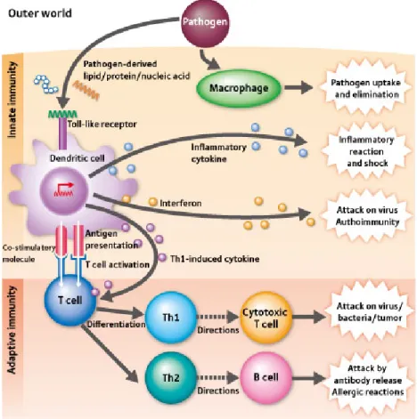
Adaptive immunity a is a highly sophisticated defensive system against pathogens and found in vertebrates. It is also known as acquired immune system because it develops new strategies during infections. Unlike innate immune system, adaptive immune response forms very slow (4-7 days), but it provides very long protection. Adaptive immunity is composed of highly specialized processes initiated with the activation of innate immune cells by pathogen recognition and presentation of digested pathogenic antigens by APCs. Three main classes of APCs are DCs, macrophages and B-cells. APCs begin processing pathogenic antigen by either phagocytosis or receptor mediated internalization of pathogen. After degradation of foreign structures, small fragments of antigens presented on cell surface as complexed with MHC class I and II molecules. Only professional APCs and certain activated epithelial cells have express MHC molecules but many infected cells can present pathogenic antigens. Secreted cytokines from innate immune cells and interaction from APCs initiate proliferation of naive T-cells (see Figure 1.2). Activated T-cells either kill recognized pathogens or help other cells to proliferate or activate them. T helper cells activates B-cells with matching antigen by interaction and IFN-γ secretion. Activated B-cells multiply and mature into antibody producing plasma cells. Then, released antibodies attach to the pathogenic antigens and mark them. Last, those marked pathogens or infected cells are cleared killer cells such as cytotoxic T-cell, macrophages and NK-cells. Activated B-cells during primary immune response, can form memory B cells which are able to live for a long time and respond quickly in the case of infection with same pathogen [10, 11].
1.4
Cells of the Immune System
Immune response against pathogens is prevailed complex interactions and activities of the diverse cells types. Primary lymphoid organs like the bone marrow and the thymus are the sites of lymphocyte production and maturation. Immune cells circulate through the blood and move to secondary lymphoid organs like the spleen and lymph nodes, where APCs present antigens to lymphocytes [10].
Figure 1.2: Innate response triggered by PRR-PAMP interactions induces and instruct adap-tive immunity through proinflammatory cytokines and dendritic cell mediated antigen pre-sentation to T-cells. Image adapted from [12].
1.4.1
Dendritic cells
Dendritic cells (DCs) are the most specialized antigen presenting cells, initiating both pri-mary and secondary immune responses. The main function of DCs is to process antigens and transmit the information to the T cells of the immune system, acting as a messenger which links innate and adaptive immunity [10]. DCs are in an immature state with high phagocytic capacity when they originate from hematopoietic bone marrow progenitor cells. Once activated DCs migrate to lymph nodes were they interact with T-cells and B-cells to initiate adaptive immune response. DCs have PRRs such as TLRs which makes them detect danger signals [13] and become activated. During activation DCs leave their immature state; becoming less phagocytic, producing cytokines and upregulating co-stimulatory molecules such as CD86 which is required for T-cell activation [14].
DCs are mainly present in tissues but rarely found in the blood circulation where they constitute approximately 1 percent of the peripheral blood mononuclear cells (PBMC). Tis-sues in contact with external environment are more likely to present DCs, such as skin and the inner lining of the nose, lungs, stomachs and intestines. Humans have two subsets of DCs in peripheral blood called myeloid (m)DC and plasmacytoid (p)DC. Phenotypes and functions of mDCs and pDCs are incompletely described but some of their common and contrary characteristics have been determined. mDCs are CD11c+ and major stimulators of T-cells whereas pDCs are DC11c- and can produce large amounts of type I interferon upon activation [15].
1.4.2
T lymphocytes
T cells are the critical components of the adaptive immunity. T-cell are originated from haematopoietic stem cells in bone marrow but they complete their maturation in thymus. The T cell receptor (TCR) is a highly specific molecule found on the surface of T cells and responsible for recognizing antigen bound to MHC class proteins. TCR diversity is carried by a series of gene rearrangement events in the thymus. T cells expressing a wide repertoire of TCR undergo positive and negative thymic selection before completing their maturation [10]. In the positive selection, thymic stroma cells express MHC molecules and T-cells with TCR that are able to interact with those MHC molecules properly selected. MHC interaction provides surival signal during this step and T-cells with no survival signals die. Survived T-cells migrate from cortex to the medulla of the thymus. In medulla (negative selection), stroma cells express MHC/self-peptide complexes and T-cells which strongly interact with those molecules receive apoptotic signals and die. Survived ones leave thymus as matured naive T cells. When a naive T cell interact with an activated APC it differentiates into effector cell of which there are several types. After a pathogen infection is cleared most of the effector T-cells undergo apoptosis, but some of them survive as long-lived memory cells which can form rapid immune response against same pathogen [10].
1.4.2.1 Cytotoxic T cells
The cytotoxic T-cells (also known as killer T cells) harbor pathogens and cancer cells. They have the CD8 co-receptor in association of TCR on their surface which can bind to MHC
class I:peptide complex. Upon activation they interact with target cells and release cytotoxic effector molecules like perforin and granzymes [10].
1.4.2.2 T helper cells
T helper cells express CD4 co-receptor which can recognize MHC class II:peptide complexes together with TCR. Upon TCR triggered activation, T helper cells differentiate into two major subtypes of cells known as Th1 and Th2 [16]. Each subtype produce different cytokines have distinct functions. Th1 helper cell facilitate eradication of intracellular bacteria and protozoa through activation of natural killer cells and macrophages by IFN- γ secretion whereas Th2 helper cells activate B-cells to produce immunoglobulins [17].
1.4.2.3 Regulatory T cells
During negative selection of in thymus, auto-reactive T-cells are not completely excluded and move into circulation. Regulatory T cells (T regs) suppress the effector functions of those auto-reactive T-cells through release of IL-10, and anti-inflammatory cytokine and TGF-β [18]. T regs divide into two subtypes, naturally occurring and adaptive [19]. Naturally occurring T regs are maturated in the thymus with immunosuppressive properties whereas adaptive T regs differentiate from the naive peripheral T cells [18].
1.4.3
B lymphocytes
B cells are vital components of adaptive immune system. In mammals, they are formed in bone marrow and complete their maturation here. B cells which leave bone marrow re-circulate and populate in the spleen and lymph nodes. They present the B cell receptors (BRC) on the surface, which can bind antigens. Self-reactive BCR carrying B cells are also eliminated in the bone marrow. BCR is a membrane bound antibody composed of a conserved shaft attached to cell membrane and a unique, randomly determined antigen-binding site (immunoglobulin). Each B cell express unique BCR which can bind to only one particular antigen. B cells become activated when two BCR are cross-linked by this particular antigen. Then, as an APC, B cell process antigen and present the peptides as attached to MHC class II molecules which attract a matching T helper cell. Cytokines secreted by T cell
triggers complete activation of B cells. Activated B cells begin to multiply and mature into soluble immunoglobulin (antibody) producing plasma cells. Released antibodies attach to the matching antigens. Cells or pathogens marked with antibodies then cleared by complement cascade or killer immune cells. TLR signaling or repetitive bacterial peptides may also trigger B cell activation without T cell interaction [10, 20, 21].
1.4.4
Macrophages
Macrophages are produced by differentiation of monocytes in tissues. They are the first cells which recognize pathogens and produce immune response. Macrophages are specialized phagocytic cells which can ingest pathogens or attack with killer molecules. Macrophages have role in both innate and adaptive immunity. When pathogenic compounds are recog-nized through PRRs in a non-specific manner, macrophages become activated, increasing surface area and becoming highly phagocytic. Macrophages present antigens of ingested pathogens on their surface and can activate T cells. Also, upon activation macrophages secrete chemokines and cytokines which can attract and activate other immune cells [10, 22].
1.5
Cytokines
Cytokines are the main contributers of communication between immune cells. Various se-creted cytokines have roles in triggering inflammation, activation and differentiation of im-mune cells as well as suppressing imim-mune response. Some of the cytokines are proinflam-matory which are necessary for the initiation of immune response to recruit granulocytes and lymphocytes. Other cytokines are anti-inflammatory and function in the inhibition of inflammation and promotion of healing once the infection is secured [23]. Over-exuberant immune responses may cause autoimmune and auto-inflammatory diseases and serious in-jury. Therefore, anti-inflammatory cytokines are essential for the recovery of the body after infections and injuries.
Tumor necrosis factor alpha (TNF-α) is mostly secreted by activated macrophages and triggers local containment of infection. TNF-α secretion by macrophages on the infection site induces increased release of plasma proteins into tissue, enhanced blood vessel permeability, increased phagocyte and lymphocyte migration into tissue and improved platelet adhesion
to blood vessel wall. Although TNF-α secretion is essential local containment of infection, in certain bacterial infections in bloodstream, systemic release of TNF-α into the circulation causes loss a significant volume of plasma and eventually to shock, known as septic shock [24]. Interleukins (ILs) are subgroup of cytokines which mediates activation, differentiation and development of immune cells. IL-1β is mainly produced by blood monocytes and macrophages as a potent mediator to immune response to infections and injuries. IL-1β is one of the early cytokines released during infection and induces expression of other flammatory cytokines and activation of T-cells and macrophages [24]. IL-6 is another proin-flammatory cytokine mostly released by T-cells, macrophages and endothelial cells. It has a role in growth and differentiation of B-cells, acute phase protein production and fever [24]. Increased expression and secretion of IL-6 induces autoimmune and autoinflammatory con-ditions in various diseases such as atherosclerosis [25], diabetes [26], Alzheimer’s Disease [27], systemic lupus erythematosus [28], multiple myeloma [29], Beh¸cet’s disease [30], rheumatoid arthritis [31] and prostate cancer [32].
IL-12 (known as NK-cell stimulatory factor) is chiefly secreted by macrophages and DCs. It activates NK-cells and induces differentiation of T helper cells into Th1-like cells. Disrupted IL-12 production or functioning causes impaired IFN-γ production and Th1 re-sponses [24]. Interferons (IFN) are large group of proteins which have various functions in metabolism, such as anti-viral activity, modulating immune responses against viruses, bacteria, parasites and tumor cells and promoting activation and differentiation of cells. In-terferons (IFNs) are proteins made and released by host cells in response to the presence of pathogens such as viruses, bacteria, parasites or tumor cells. They allow communication be-tween cells to trigger the protective defenses of the immune system that eradicate pathogens or tumors. They are mainly divided into two subclasses according to the their roles: Type I IFN, Type II IFN. Type I interferons (IFN-α and IFN-β) are essential for protection against viral infections , whereas type II interferon (IFN-γ) has roles against bacterial and parasitic infections. IFN-α and IFN-β are secreted by many cell types especially pDCs and lympho-cytes and stimulate macrophages and NK cells to elicit antiviral and anti-tumor responses. IFN-γ is secreted by activated T-cells and NK cells, and function in the regulation of the inflammatory responses in both innate and adaptive immunity. T helper cells produces IFn-g to help macrophages kill the bacteria and to induce proliferation of B-cells and class switching [24]. Extensive production of IFN-γ is associated with various autoinflammatory and autoimmune diseases [33].
1.6
Pattern Recognition Receptors
PRRs are primitive part of the immune system, which are essential for detecting invading pathogens and initiating innate and adaptive immunity. PRRs are activated by PAMPs present in microbial structures or by damage-associated molecular patterns (DAMPs) ex-posed or released by damaged cells. PRRs are comex-posed of multiple membrane-associated and cytosolic receptor families. Toll-like receptors (TLRs) and C-type lectin receptors (CLRs) are membrane associated PRR families whereas NOD-like receptors (NLRs), RIG-I-like receptors (RLRs) and AIM2-like receptors (ALRs) are cytosolic ones. Recognition of PAMPs by PRRs usually triggers the intracellular signal transduction cascades which initiate expression of pro-inflammatory cytokines, chemokines and antiviral molecules [34]. On the other hand, ligand binding to some NLRs or ALRs may trigger clustering of re-ceptor monomers, inducing formation of inflammasome complexes. Inflammasomes activate Caspase-1 which induces expression of pro-inflammatory cytokines IL-1β and IL-18 [35]. Be-cause a pathogen contains various PAMPs such as lipopolysaccharides (LPS), ssRNA and ssDNA multiple PRRs can be activated simultaneously, causing crosstalk between different receptors and cascades which can also affect the direction of the immune response [36].
1.6.1
Toll-like receptors
TLRs are PRRs that have been conserved from insects to mammals and essential molecules that alerts the immune system against microbial infections. They are named for their sim-ilarity to Toll which was firstly discovered as in the fruit fly Drosophila melanogaster as a receptor required for dorsoventral axis formation in early development [37]. Later, Toll was shown to have essential roles in fly’s immunity against fungal functions [38]. TLRs, the mammalian homologue of Toll was identified in 1997, which was shown to actively induce cytokine production and immune activation of T-cells [39]. Role of TLRs in recognition of PAMPs discovered in genetic experiments which demonstrated that a mouse strain with a missense mutation in the Tlr4 gene was resistant to LPS induced endotoxic shock [40]. Subsequently, many other TLRs recognized which can recognize diverse variety of PAMPs derived from bacteria, viruses, protozoa and fungi. To date, 13 TLRs identified in mam-malians, each recognizing unique PAMPs- table. Each TLR is composed of three regions: a leucine-rich repeat (LRR) region mediating ligand-binding specificity, a transmembrane
T able 1.1: Ligands, adapter proteins and lo calization of TLRs. T able is mo dified from [11, 41–63].
domain and a Toll/Interleukin-1 receptor (TIR) domain initiating intracellular signaling pathways [64]. Most TLRs can recognize specific PAMPs by itself but there are certain co-receptors augmenting receptor-ligand affinity, such as CD14 which cooperates with TLR4 for detection of LPS in the presence of Lipopolysaccharide-binding protein (LBP), a soluble acute-phase protein that binds LPS [65].
TLRs are localized either on cell surface or on the membrane of cellular compartments. TLRs are differentially expressed most cell types, especially immune cells. The cell types which each TLR is expressed, and the cellular localization of them are summarized in Ta-ble 1.1.
Upon activation, the TIR domain of TLRs can interact different adapter molecules in cytoplasm such as Myleoid differentiation factor-88 (MyD88) and TIR-domain-containing adapter-inducing interferon-β (TRIF), which may induce various signal pathways. TLR signaling is divided into two distinct adapter dependent pathways, the MyD88-dependent and TRIF-dependent (or MyD88-independent) pathway.
The MyD88-dependent signal pathway induced by ligand triggered dimerization of TLR receptors, except TLR3. Activation of NFκB (nuclear factor kappa-light-chain-enhancer of activated B cells) and mitogen-activated protein (MAP) kinase pathways is primarily induced by MyD88. NFB is a very important transcription factor involved in control of diverse variety of cellular actions such as stress induced and immune responses, developmental processes, growth and apoptosis.
Activated MyD88 recruits kinases to trigger the signal pathway which eventually leads phosphorylation and degradation of IκB (inhibitor of NFκB), allowing NFκB to diffuse into cell nucleus and induce transcription of inflammatory cytokines [66].TRIF-dependent path-way is utilized by TLR3 and TLR4 upon recognition of dsRNA and LPS respectively. TRIF can activate certain kinases which initiate a branch of signal pathways which eventually lead activation of NFB or interferon regulatory factor (IRF3) [66,67]. IRF3 is a transcription fac-tor which enables production of type I interferons [68]. Depending on the circumstances and the ligands, TLR activation can trigger signal pathways through different adapter proteins but TLR 4 is the only one which uses all known four adapters. TIR mediated simultaneous activation of different adapter signal pathways is necessary in certain conditions for produc-tion of inflammatory cytokines [66]. The known adapters used by TLRs are represented in Table 1.1 .
As the importance of innate immunity for generation of adaptive immune responses was indicated before [7–9], the key elements of innate immunity, TLRs are very important for activation adaptive immunity. Therefore, using TLR ligands as adjuvants within human vaccines may provide stronger and prolonged adaptive immunity [69]. Especially TLR7/8 and TLR9 ligands are very promising adjuvants due their presentation on immune-adaptive immunity linking pDC surfaces [70]. Although, TLR7/8 ligands demonstrated great success as vaccine adjuvantas against pathogens [71, 72], TLR9 ligands (CpG ODNs) were reported to be more effective for augmenting both humoral and cell mediated immune response [73]. Also, extensive variety of CpG ODNs with diverse and potent effects on immune responses offers therapies against pathogens, autoinflammatory diseases and even cancer [74].
1.7
Immunmodulatory synthetic DNA motifs
TLR9 can discriminate self and non-self DNA and trigger immune response according to the circumstance. Even though the stimulatory effect of bacterial DNA has been reported, the DNA motifs in mammalian and adenoviral genomes have been shown to be immunosup-pressive. Both stimulatory and suppressive synthetic DNA motifs are promising agents for vaccine development and treatment of inflammatory conditions, respectively.
1.7.1
Immune stimulatory synthetic DNA motifs
In 1995, the immune stimulatory effects of unmethylated CpG motifs in bacterial DNA on B-cells was demonstrated [75]. Later, it was discovered the recognition of bacterial CpG motifs was mediated by TLR9 [57]. Although, the recognition mechanism of CpG motifs by TLR9 is unclear, synthetic CpG ODN induced various responses depending on their sequence, length and backbone. The immunostimulatory strength of different CpG ODN may differ but they have been divided into three subclasses according to their structural characteristics and impact [76].
Type D ODNs (also known as class A) are characterized by a central palindromic phosphodiester (PO) CpG sequence and PS-modified 3’ end poly(G) track. The poly
G favors forms G-tetrad formation which provides higher molecular weight and subse-quently high stability and increased endosomal uptake. D ODNs activates pDCs and triggers IFN-α expression. They weakly stimulate TLR9-dependent NF-B signaling and pro-inflammatory cytokine production [77–79].
Type K ODNs (also known as class B) are 18-28mer(much larger than D-ODNs) mo-tifs with a linear structure. They contain one or more 6mer CpG momo-tifs with full phosphorothioate backbone. K ODNs strongly activate B-cells and TLR9-dependent NF-B signaling but weakly promote IFN-α expression. They are promising vaccine adjuvants [75, 79, 80].
Class C ODNs have characteristics of both K and D ODNs. They are composed of a CpG-containing palindromic motif with full PS backbone. They can activate both pDC and B-cells, also stimulate production IFN-α and pro-inflammatory cytokine [81].
1.7.2
Immunosuppressive synthetic DNA motifs
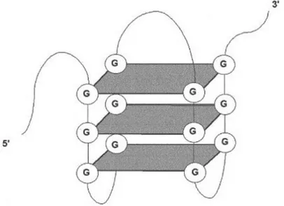
There are also synthetic DNA motifs which have immunosuppressive effects on immune system in a TLR9-dependent or independent way. Inhibitory sequence motifs were firstly described in DNA of adenovirus serotype [82]. Later, G-rich or methylated mammalian DNA motifs suppressing CpG induced immune activation through NF-B and AP-1 induc-tion were also reported [83–85]. Suppressive motifs were also determined in mammalian telomeric sequences (TTAGGG) [86] and DNA of commensal bacteria [87]. After the discov-ery of suppressive sequences, synthetic various ODNs were produced to reveal mechanism and therapeutic potential. When immunosuppressive motifs were initially identified, they were known to have ability to block CpG-induced immune activation. However, suppressive ODNs were not inhibitors of CpG uptake [85] and they were shown to hamper multiple forms of immune stimulation [88–91]. One of the most effective and studied suppressive ODN is A151 which express multiple TTAGGG motifs, mimicking the immunosuppressive activity of telomeric DNA [86]. The immunosuppressive effect of the A151 is connected to its G-tetrad structure formed by Hoogstein binding of poly-G runs (Depicted in Figure 1.3). The action mechanism of A151 is still unclear but it can down-regulate the production of proin-flammatory and Th1 cytokines by inhibiting phosphorylation of STAT1/STAT4 [86, 92]. In vivo administration of A151 demonstrated promising progresses against various pathogenic conditions like autoimmune and inflammatory diseases in murine. [92–95].
Disorders in immune system cause abnormal immune responses which give rise lethal autoimmune diseases. The immune system attacks the body’s own cells and tissues, ending up with tissue, organ damage. There is no accurate treatment for autoimmune diseases but immunosuppressive are used to down-regulate immune response and avoid tissue damage. Systemic lupus erythematosus (SLE) is an autoimmune disease which mostly harms the heart, joints, lungs, blood vessels, liver, kidneys, skin and nervous system [96]. Intraperi-toneal (IP) injection of A151 to female NZB/W mice (a useful model to study pathogenesis and treatment of human SLE [97]) significantly decreased the severity of the disease and prolonged the survival [95, 98]. Septic arthritis is another autoimmune disease caused by joint infections by certain bacteria. It was reported that, both IP and intra-articular ad-ministration of A151 significantly reduced inflammation and the swelling on the joint of the CpG-induced septic arthritis murine models [99, 100]. Uveitis is a autoimmune disease characterized by inflammation of uvea and IP or local administration of A151 successfully reduced ocular inflammation in three differently uveitis induced murine models [95, 101]. Protective effect of A151 against inflammatory conditions such as lethal endotoxic shock and acute slicosis was also demonstrated in mice [92, 95].
As a result, A151 has a great potential as a immunosuppressive drug against diverse pathological conditions , but its delivery in biological milieu is prone to digestion and serum adsorption, leading
Figure 1.3: Representation of the G-tetrad structure of A151, formed through Hoogsteen hydrogen bonding among G bases.
to limited bioavailability, hampering its in vivo performance. Surely, phosphorothioate chemistry makes A151 more stable for in vivo use in experimental animal models of auto/inflammatory and auto-immune diseases, better delivery systems are required to unveil its full potential.
1.8
Extracellular Vesicles
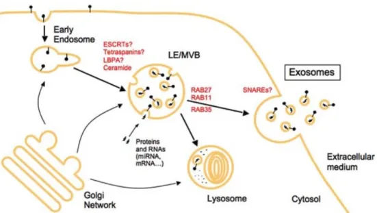
Intercellular communication is essential for all life forms, especially multicellular organism. Communication between cells can be achieved by cell-to-cell contact, and transmission of specific molecules. In last two decades, a new mechanism for intercellular communication has been discovered that covers transfer of plasma membrane derived extracellular vesicles (EVs). The release of apoptotic blebs during apoptosis was known long ago but the secretion of membrane vesicles from healthy cells has recently demonstrated. Since then, EVs have been isolated from nearly all types of mammalian cells, including stem cells, immune cells, nerve cells and tumor cells. Intercellular communication through EVs were identified in not only mammalian cells but in lower eukaryotes and prokaryotes. Until 1996 these vesicles were initially regarded as membrane debris or waste containers with no real biological ef-fect. However, Raposa et al. showed that extracellular vesicles could stimulate adaptive immune responses [102]. Since then, many studies have demonstrated the significance of EVs in intercellular communication via transfer of nucleic acids, lipids, cytokines and pro-teins. As EVs have been isolated from cell culture medium of cell lines and primary cells they have also been purified from most bodily fluids and the studies indicate that these vesicles have a key role in not only the maintenance of physiological processes stem cells but immune surveillance, tissue repair, blood coagulation, but also in the pathology causing several diseases [103]. When the cell derived vesicles were identified, they were known to be originated from cell membrane by direct shedding. Although, the term exosome was firstly used for vesicles ranging from 40 to 1,000 nm the subcellular origin of these vesicles was remained as secret until 1984 [104]. Then it has been determined that the release of many nanovesicles during reticulocyte differentiation were outcome of multivesicular endo-some (MVE) fusion with the plasma membrane [105, 106]. Currenty, these multivesicular endosome derived nanovesicles(40-100 nm) are referred as exosomes, whereas the directly shedded vesicles from plasma membrane (40 to 1,000 nm) are referred as microvesicles, ec-tosomes, shedding vesicles or microparticles( [104, 107]). Distinct mechanisms of exosome
and MP secretion are simply represented in Figure 1.4. The secretory mechanisms, function and therapeutic applications of exosomes and microparticles (MP) are growing research area in the field of physiology, immunology and genetics. However, there are controversial issues about distanciation of those vesicles from each other. Current methods for exosome and MP isolation (ultracentrifugation, filtering, sucrose gradient) are not able to separate MPs from exosomes completely. MPs with the sizes in the range between 50-100 nm are also isolated as mixed up with exosomes. Therefore, the function and actions of exosomes may not be accurate. On the other hand, most of the previous studies did not deal with exosomes and MPs separately, but experimented on mixed populations, spawning controversial data.
Figure 1.4: MPs are formed by directly shedding from the plasma membrane whereas, ex-osomes are constituted in MVE and released through fusion of cell membrane and MVE. Image adapted from [107].
1.9
Microparticles
Microparticles are small (40-1000 nm) vesicles shed from a variety of cells [108]. MPs were termed as platelet dust when they were first identified by Peter Wolf during an ongoing blood coagulation research in 1967 as a product of platelets [109]. Although MPs were
initially thought to be useless cellular waste, decades of research focused on the tiny vesicles have defined their roles as participators in thrombosis, cellular signaling, vascular injury, homeostasis, inflammation and messengers in the transport of bioactive lipids, cytokines, complement and immune signaling [108, 110–112]. Function of the MPs are dependent on the derived cell type. However, distinct phenotypic subsets of MPs may have different functions, even if derived from same cell type.
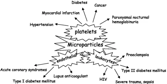
In circulation of healthy individuals, MPs were derived most abundantly from platelets followed by red blood cells, leukocyte and endothelial cells [111,113]. MP amount, phenotype in circulation altered by various diseases, such as atherosclerosis, diabetes, cancer, sepsis, pulmonary hypertension and rheumatic diseases [114, 115]. For instance, platelet MPs are increased in myocardial infarction [116], hypertension [117], diabetes [118] and cancer [119], whereas endothelial MPs abundantly present in acute coronary syndromes [116] and Type 1 diabetes mellitus [120]. Diabetic patients have leukocyte originated MPs in their circulation approximately threefold higher than healthy individuals [120]. Under different pathological conditions, cellular origins and phenotypes of circulating MPs may shift (See Figure 1.5). Since diverse phenotypes MPs are associated with various homeostatic activities, microparti-cle research is an important emerging field focused on characterization of distinct phenotypes, investigation of their formation mechanism and factors triggering their release and determi-nation of their effects on pathogenesis of cardiovascular diseases or distinct inflammatory conditions.
1.9.1
Microparticle formation
Unlike exosomes, MPs directly shed from the plasma membrane of the parent cell. The mechanism behind MP formation is unclear but studies have suggested some factors have role in MP formation, chop off mechanism.
Remodeling of the plasma membrane is essential for MP formation. Cell membrane bi-layer has a specific lipid composition in inner and outer leaflets. Aminophospholipids (PS and phospha-tidylethanolamine) are specifically found in the inner leaflet, whereas phos-phatidylcholine and sphingomyelin are enriched in outer one. Localization and composition of those factors are essential for the preserving plasma membrane asymmetry. The control of lipid distribution on the transbilayer is carried by three transmembrane lipid transporters;
Figure 1.5: The cellular origins and phenotypes of circulated MPs are affected by pathological conditions. Image adapted from [121].
flippase, floppase and lipid scramblase [122]. Cell activation or apoptotis induction cause significant and sustained increase of cytosolic Ca2+ which lead to the disruption of the mem-brane asymmetry by enhancing scramblase and floppase activities and inhibiting PS specific flippase. Especially, flippase inhibition is a very essential stage which cause exposure of the PS, followed by cytoskeleton degradation by Ca2+- dependent proteolysis and lastly MP release [123]. Because apoptotic or dying cells loose membrane asymmetry and secrete MPs it is a problematic to distinguish whether they are derived from healthy or unhealthy cells during in vitro culturing. In future studies, healthy cell MPs may be sorted out from the others and better understanding of function and composition of MP subpopulations.
1.9.2
Microparticle composition
In comparison with exosomes, less is known about the protein and lipid composition of MPs. Each MP have cytoplasmic and plasma membrane properties of the originated cell however it is not clear whether they carry enriched particular components. If their secretion mechanisms are clearly understood, more information about their content can be revealed.
Because composition of MPs is dependent on cell origin, any alteration on the cell effect the MP content. Any environmental stimuli which make cell produce different proteins,
mRNA, miRNAs also directly affect the MP composition. A general representation of MP composition is depicted in Figure 1.6.
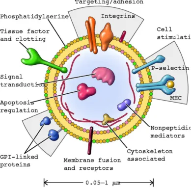
Each MP present specific surface membrane proteins based on their origin. For instance, endothelial MPs have CD31 and CD146, whereas leukocyte MPs present CD4, CD3, or CD8 at their surface membrane [116, 124]. Thus, source of the circulating MPs can be detected with cell marker Ab staining. Also, MP surface is phosphatidylserine(PS) enriched which makes them bind to labeled Annexin-V and consequently detectable by flow cytometry [125].
Figure 1.6: Representation of MP composition. Surface of a MP is consist of molecules from parental cell plasma membrane. Cellular origin and conditions affect the MP phenotype. This representation does not reflect exact and complete content of MPs. Image adapted from [123].
In addition to the cytoplasmic proteins, MPs also contains some mRNA, microRNA and proteases of the parental cell [126–129]. Also a very recent study has revealed the presence of genomic DNA fragments in MPs isolated from human plasma [130].
1.9.3
Microparticle function
MPs are secreted by normal or activated cells as a consequence of differential environmental effects. Therefore, MPs can been seen as pathways which exchange information between cells. Because MPs carry mRNA, miRNA, transcription factors of the parent cell, they can affect the transcription profile of the target cell [126, 127]. Microparticles can interact with target cell through different mechanisms in order to mediate cell-to-cell communica-tion. They can activate intracellular signaling by interacting target cell membrane receptors, release their content into cytoplasm by fusing cell membrane, internalized through phagocy-tosis, macropinocytosis or receptor-mediated endocytosis. Additionally, membrane proteins presented on surface of microparticles can be cleaved by extracellular proteases and ex-posed ligands can bind to target cell receptors to induce signal pathways [127, 131, 132](see Figure 1.7). However, the physiological and pathophysiological roles of MPs are unclear. Findings suggest that MPs have roles in inflammation, coagulation, vascular function, apop-tosis and cell proliferation or differentiation [123, 133]. Especially, circulating MPs allows membrane interactions between distant vascular cells.
Figure 1.7: Extracellular vesicles can transmit signals through their inside content or surface proteins. The proposed mechanisms behind EV-mediated signal pathways are Receptor binding, direct fusion with target cell plasma membrane, endocytosis and receptor cleavage. Image adapted from [132].
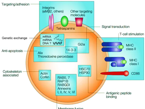
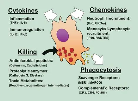
MPs can be beneficial or deleterious depending on condition. Normally, MPs are trans-ducers of biological information. MPs shed from activated macrophages can transmit inflam-matory signals to foreign cells thus accelerate and strengthen immune responses [134](also
see Fig 1.12). On the other hand, extensive secretion of MPs may cause significant proco-agulation and inflammatory effects provoking adverse clinical symptoms in several vascular diseases [133, 135]. Moreover, during some viral infections they may cause infection of viral genetic information to foreign cells easily because they are considered as ”host” by other cells [136]. Also, tumor derived MPs can suppress anti-tumor responses in favor of tumor growth [137].
Role of the MPs in inflammation is one of the most popular research field currently. The implications of MPs in inflammation has been reported by many studies. The most apparent relationship is the elevated MP levels in various inflammatory conditions such as sepsis [138, 139], HIV and Ebola virus infection [140,141] and malaria [142,143]. Although, MP secretion is enhanced during infections, stimulation with inflammatory cytokines such as TNF, IL-1, IL-6 and type 1 interferons in the absence of infection also caused elevated MP secretion [144, 145]. As expected, both in vitro and in vivo administration of bacterial endotoxins induced MP secretion in various cell types. For instance, apoptosis related MP secretion was observed in LPS stimulated epithelial cells, whereas monocytes secrete MPs upon immune activation [146–149]. Moreover, increased MP levels were reported in the circulation Plasmodium-inocuated laboratory animals [150]. Especially platelet derived MPs in circulation have significant roles in inflammation. Activated platelets secrete proinflammatory IL-1beta-rich MPs which may favor atherosclerosis plaque formation [151,152]. Also, MPs secreted from in vitro activated platelets enhanced expression of cell adhesion molecules and proinflammatory cytokines in endothelial cells and THP-1 cell line [153]. On the line, leukocyte MPs in circulation induced secretion of cytokines and expression of surface coagulation factors in endothelial cells [154].
MPs have been suggested to have roles behind certain physiological immune escape mech-anism. It was reported that, during pregnancy trophoblast release FasL bearing MPs to induce T-cell death and subsequently escape from immune system despite its semi-allograft properties [155]. Tumor cells such as ovarian cancer cells also may secrete FasL-bearing MPs into the extracellular medium and cause apoptosis of lymphoid cells. Considering the fact that normal ovarian epithelial cells do not secrete FasL bearing MPs, it is suggested that tumor cells developed a mechanism that neutralize Fas-bearing immune cells which facili-tate their escape from immune system [156, 157]. Another implication of MPs in immune escape also reported in AIDS. CCR5 is chemokine receptor localized on the surface of white blood cells and this receptor is used by many HIV versions to enter and infect the cell [158].
Figure 1.8: Physiological effects and actions of MPs from different cellular origin. Image adapted from [121].
MPs released from CCR5-positive cells rendered deficient peripheral blood mononuclear cells CCR positive and thus susceptible to HIV-1 infection [159]. Another study also confirmed the receptor transfer from CXCR4 positive platelets and megakaryocytes to CXCR4-null cells through MPs [160].
It was reported that neutrophil derived MPs, termed extosomes may have potential roles in opsonization due to their ability to efficiently bind opsonized bacteria and become adherent to monocytic and endothelial cells [161, 162].
Among countless significant physiological activities of MPs in human body, their role in immune system is very diverse and essential. MP can remotely transport bacterial or viral antigens via blood stream to APCs such as macrophages and DCs in the spleen without the need for direct interaction with infected cells. Also, they can penetrate almost all tissues and physiological barriers which cells cannot pass through and convey the signals. Such remote interactions maintain a distance between site of infection and the site of immune induction, also enhance the probability of early detection of infection. In certain circumstances MPs can embrace parent cells assignment. For instance, TNF-stimulated, endothelial cell-derived MPs can stimulate activation and maturation of pDCs [163]. Moreover, MPs secreted from
Mycobacterium-infected macrophages were able to activate nave T cells by antigen present-ing, thus rendering APC T-cell interactions unnecessary. Also, according to that study, MPs were more effective than exosomes at inducing T-cell proliferation [164, 165].
Additionally, elevated levels of MPs presenting CD4 antigen in circulation of human immunodeficiency virus (HIV)-infected patients have been determined [140]. Enhanced lev-els of MPs derived from activated leukocytes were observed in circulation in case of severe trauma [166]. Also, in preeclampsia, granulocyte and lymphocyte originated MP subpop-ulations significantly increased [167]. Almost every mammalian cell types are known to be able to secrete MPs . Even smooth muscle cells release MPs which were suggested to have potential functions in atherosclerosis and thrombus formation [121, 168, 169]. MPs may have pleiotropic effects on the vascular wall in a consequence of their variety of subpopula-tions. MPs act as vectors transmitting specific messages to other cells in regulating vascular function and dysfunction depending on the donor cell type [121].
Mouse macrophage cell line secreted significantly higher amounts of MPs when activated by various TLR ligands [170]. In the same study, a relevance between MP production and nitric oxide (NO) release was reported and NO synthase inhibitors also greatly hampered MP secretion [170]. NO is most commonly observed in monocytes and macrophages. Therefore, it is plausible that each cell may have distinct mechanisms for MP production. Moreover, same cell types may have different mechanism for producing subpopulations of MPs under distinct conditions.
1.9.4
Microparticle immunotherapy
MPs are promising therapeutic tools for diverse applications such as vaccination, drug deliv-ery, gene therapy due to their physiological structure. They can pass through every tissues and be accumulated by cells rapidly. Unlike artificial vesicles, MPs are completely natural and non-toxic. MPs from non-activated immune cells generally does not cause any im-mune response in the recipient circulation, thus minimize the side effects. There are many therapeutic agents which are effective in vitro studies but become impotent upon in vivo administration. Absorption by serum or other tissues and degradation before reaching target site hamper the functioning of those therapeutic agents. MPs are potential delivery systems for those agents because of their cell membrane derived lipid structure. Also, MPs can be
used for targeted therapies which also enhance the activity of the agents. MPs derived from different cell types generally have distinct affinities to other cells. Choosing right MP type may provide both protection and target specificity for therapeutic agents or drugs.
Because MPs conveys immunological signals between site of infection and immune in-duction, they are suggested to be promising vaccination tools. Since, antigen carrying MPs can effectively stimulate both innate and humoral responses, they can be used to increase efficiency of adjuvants.
MPs may affect transcription profile of target cell by transporting mRNA, miRNA and TFs [126]. Non-coding small RNA based therapies have shown great promise for many diseases such as cancer [171]. However, their nucleic acid structure limits their bioavailability in biological fluids. MPs are great delivery vehicles for such nucleic acid based therapeutic agents.
Altering MP composition by genetically engineering of donor cell is another promising approach in MP based therapeutics [172]. It has been reported that, MPs derived from suicide gene mRNA transfected donor cells, were administered to schwannoma and success-fully hampered tumow growth [173]. Several studies demonstrated that, mesenchymal stem cell (MSC) MPs have MSC mimicking properties such as apoptosis inhibition, induction of cell proliferation subsequent wound healing [174]. Thus MSC derived MPs are potential therapeutic tools in regenerative medicine.
MPs are potential markers for early diagnosis of cardiovascular and other diseases by their virtue of joining circulation from any site. Therefore, characterization of MPs from healthy and unhealthy cells is essential to exploit their such property in future. There are some studies carried out to use MPs as diagnostic tool for atherosclerosis vascular disease [175], cancer [176] and smoking dependent lung damage [177].
1.10
Exosomes
Exosomes are small membrane vesicles with the size 40-100 nm. It is secreted from multivesic-ular endosomes in most cell types including B cells [102], T cells, macrophages, DC [178], mast cells, epithelial cells, platelets, reticulocytes, tumor cells. Moreover, exosomes and
exosome-like vesicles have been isolated from various body fluids such as semen [179], blood serum [180], urine [181], saliva [182], breast milk [183], amniotic fluid [184], ascites fluid [185], cerebrospinal fluid [186] and bile [107, 187]. Exosomes were initially described in 1983 in the maturing reticulocytes [188, 189] and they become a significant target in immunology in 1996, when Raposo demonstrated the feature of B-cell isolated exosomes in stimulating T-cells [102]. Since the Raposo’s milestone achievement in exosome history, the exosomes have become an significant subject in antigen presentation and immune regulation. Although their biological role has not been fully revealed yet, they have shown great success in therapeutic approaches.
1.10.1
Exosome formation
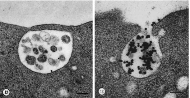
Exosomes are formed in MVBs/endosomes of the cell which is a membrane-bounded com-partment functioning as an intermediate in the degradation pathway of proteins either inter-nalized from plasma membrane or transported from Golgi network [190]. Interestingly, the secretion of exosome mechanism was demonstrated on a study about the iron-binding trans-ferrin receptor by reticulocytes during maturation into erythrocytes in 1983 by Harding et al. [188]. The down-regulation of transferrin receptors was considered as related to endocyto-sis and lysosomal degradation and the visualization of colloidal gold-conjugated transferrin accumulated by reticulocytes by electron microscopy suddenly reveled the multivesicular bodies and their fusion with plasma membrane [188](see Figure 1.9).
Exosomes are formed by inward budding of the membrane of the MVB and released to extracellular space after fusion of MVB with cell membrane [191]. Although these com-partments share many elements, MVBs either fuse with lysosomes in lysosomal pathway for degradation of their contents or fuse with plasma membrane for secretion of exosomes. These two distinct fates rely on cholesterol proportion on MVBs. When cholesterol-rich MVBs are in exosome secretory pathway, morphologically identical but cholesterol-poor ones are prone to fuse with lysosomes [192].
Generation of exosomes in MVBs involves the lateral segregation of cargo at the delimiting membrane of an endosome and inward budding and pinching of vesicles into the endosomal lumen. Two mechanisms for exosome production have been identified so far; endosomal sorting complex responsible for transport (ESCRT)-dependent and ESCRT-independent.
![Figure 1.8: Physiological effects and actions of MPs from different cellular origin. Image adapted from [121].](https://thumb-eu.123doks.com/thumbv2/9libnet/5798243.118090/39.918.173.755.119.467/figure-physiological-effects-actions-different-cellular-origin-adapted.webp)