ORIGINAL ARTICLE
Medicine Science 2020;9(1):191-6
A new approach for green synthesis and characterization of Artemisia L. (Asteraceae)
genotype extracts -Cu
2+nanocomplexes (nanoflower)
and their effecitve antimicrobial activity
Ayse Baldemir Kilic1, Cevahir Altınkaynak2, Nilay Ildiz3, Nalan Ozdemir4, Vedat Yilmaz5, Ismail Ocsoy5
1Erciyes University, Faculty of Pharmacy, Department of Pharmaceutical Botany, Kayseri, Turkey
2Nevsehir Haci Bektas Veli University, Avanos Vocational School,Department of Plant and Animal Production, Nevsehir, Turkey 3Erciyes University, Faculty of Pharmacy, Department of Pharmaceutical Microbiology, Kayseri, Turkey
4Erciyes University, Faculty of Science, Department of Chemistry, Kayseri, Turkey 5Erciyes University, Faculty of Pharmacy, Department of Analytical Chemistry, Kayseri, Turkey
Received 27 September 2019; Accepted 20 October 2019 Available online 07.03.2020 with doi: 10.5455/medscience.2019.08.9165
Abstract
In this study, we have demonstrated the fabrication of novel organic-inorganic nanobio-antimicrobial agents called “nanoflowers” (NFs) and elucidate the increase in the antimicrobial activity of NFs. This is the first report that the NFs were formed of plant extracts as the organic components and copper (II) ions (Cu2+) as the inorganic component. The Artemisia L. (Asteraceae) methanol extracts from three genotypes including A. absinthium L. (Aa), A. vulgaris L. (Av) and A. ludoviciana Nutt. (Al) were selected in the NF synthesis. The effect of the plant extract concentrations on the morphology of NFs was examined. Most regular and uniform flower-shaped morpholo-gies were observed when a concentration of 0.1 mg mL-1 plant extract was used in the synthesis of NFs. The syntesized NFs were characterized with several techniques such as scanning electron microscopy (SEM), Fourier transform infrared spectrometer (FT-IR), energy-dispersive X-ray (EDX) and X-ray diffraction analysis (XRD). The NFs exhibited much antimicrobial activity against the pathogens even at low concentrations compared to the extracts. The MICs and MBCs values for NFs were found to be range between 0.4 to 40 μg mL-1 and 40 to 400 μg mL-1 while those values for Aa, Av and Al extracts were ranged from 500-2000 μg mL-1 and 1000-4000 μg mL-1 for the studied pathogens, respectively.
Keywords: Artemisia extracts, hybrid nanoflower, inhibitory property, bacterial and fungal pathogens.
Medicine Science International Medical Journal
Introduction
The development of nanomaterials has provided many multifunctional versatile tools used in scientific and technical fields [1-3]. Various colloidal nanomaterials (NMs), such as metallic or polymeric, have been synthesized and characterized using several techniques [4-8]. Among the NMs, the biocompatible molecules (DNA, protein, enzyme and plant extract) integrated nano or micro sized materials have been commonly used in last two decades due to much biocompatibility and very less environmental toxicity [9-13].
The plant extracts have been receivied considerably much attention in NMs synthesis compared to other biomolecules owing to their quite less price, high stability, lack of contamination risk and easy preparation. Up to now, researchers have intensively used various plant extracts as reducing and capping agent in the synthesis of metallic NMs. In typical synthesis, certain amount of plant extract and metal salt are accordingly mixed in aqueous solution and incubated under stirring at various temperatures for different periods of time. The plant extracts are mainly composed of natural chemicals, such as flavonoids and polyphenols. The oxidation of those of which induces the reduction of metal ion and eventual formation of metallic nanomaterials [14-19].
Rather than conventional plant extract based metallic NMs synthesis, we were inspired from an encouraging breakthrough discovered by Zare and co-workers in 2012 for prepration of protein-inorganic nanoflowers (NFs) [20,22]. In several reported *Coresponding Author: Nilay Ildiz, Erciyes University, Faculty of
Pharmacy, Department of Pharmaceutical Microbiology, Kayseri, Turkey E-mail: [email protected]
works, protein, enzyme and amino acid were successfully utilized as organic component and some metal ions were used as inorganic component in the formation of flower-shaped nanostructures. However, to the best of our knowledge, plant extracts have not been previously involved in synthesis of organic-inorganic hybrid nanostructures [22-30]. Artemisia L. species are medicinal and aromatic herbs in the Asteraceae family, which has a long history of use in culinary traditions [31]. They have been actively used in various purposes, such as coughs and colds, chills, stomatch-ache, dry dyspepsia, purgative effect; tea, poultice, inhale (vapours from boiling leaves); insect repellent [32]; a vermifuge, in the treatment of chronic fevers and for inflammation of the liver, as an antispasmodic and antiseptic [33,34].
Herein, for the first time, a simple and rational approach is reported for the preparation of novel organic-inorganic nanobio agents called “nanoflowers” (NFs) and elucidate the increase in the antimicrobial activity of NFs. The NFs were formed of plant extracts as the organic component and copper (II) ions (Cu2+) as
the inorganic component. The extract of Artemisia from three different genotypes including Artemisia absinthium L. (Aa),
A. vulgaris L. (Av) and A. ludoviciana Nutt. (Al) were selected
in the NF synthesis. The effects of the plant extracts and Cu2+
concentrations on the morphology of NFs were systematically examined. The antimicrobial activities of Aa, Av and Al extracts and NFs synthesized from extracts were evaluated against bacterial and fungal pathogens.
Materials and Methods Chemicals and reagents
Copper sulfate pentahydrate, methanol, phosphoric acid, dimethyl sulphoxide (DMSO) and other chemicals were purchased from Sigma-Aldrich. NaCl, KCl, Na2HPO4, KH2PO4, HCl and NaOH were used to prepare phosphate buffer saline solution (PBS, pH 7.4). Coomassie brilliant blue G-250 were used for buffer solution and prepared using ultrapure water.
Preparation of extracts
Artemisia species were obtained from Zeytinburnu Medicinal
Plant Garden, Istanbul, Turkey. Herba of Artemisia spp. were washed several times with deionized water and dried at room temperature. Aa, Av and Al herbs were powdered using a blender. 100 g of the each plant powder was added into 500 mL one-necked flask containing 250 mL methanol and incubated at room temperature (RT: 25 ºC) for 1 day under stirring. After incubation, the each solution was filtered through a Whatman filter paper No. 1 to collect the extract. This step was repeated twice using the same procedure. The extracts were evoporated under vacuum at 40 ºC and stored at -20 ºC for further use.
Preparation of Aa, Av and Al-Cu2+hybrid nanoflowers
The extracts incorporated-Cu2+ NFs were prepared according to
previously reported method with some modifications [23-28]. Birefly, a volume of 0.35 mL CuSO4 solution (120 mM) was separately mixed with Aa, Av and Al extracts (concentrations increasing from 0.1 and 0.5 mg mL-1) into 50 mL of 10 mM PBS
(pH 7.4) The mixtures were vigorously shaken for 30 s to make them homogeneous and then left without disturbing at +4°C for 3 days incubation. The greenish precipitates appeared at the bottom of the solution were washed by centrifugation at 10.000 rpm for
15 min at least 3 times. Finally, the collected NFs were dried under vacuum at 50°C for overnight and stored for futher characterization and use. The supernatant of each mixture was kept for Bradford protein assay.
Characterization of plant extract incorporated nanoflowers The morphologies of the NFs were examined using ZEISS model EVO LS10 scanning electron microscope (SEM). The elemental analysis of the NFs was undertaken by energy-dispersive X-ray (EDX) (ZEISS EVO LS10) to determine weight and atomic percentage of Cu2+ in the NFs. The infrared spectra of the NFs
were recorded using a Fourier Transform Infrared Spectrometer (FT-IR) (Perkin Elmer Spectrum 400). The 20 mg of NFs was also used for X-ray Diffraction Analysis (XRD) (BRUKER AXS D8). The encapsulation yield of Aa, Av and Al NFs was determined to be ~58%, ~68% and ~48%, respectively via Bradford protein assay using an UV-vis spectrophotometer (HITACHI) [35]. Antimicrobial study
The bacterial strains (E. coli ATCC 35218, S. typhi ATCC 14028
P. aeruginosa ATCC 27853 C. albicans ATCC 10231 and S. aureus ATCC 25923) were obtained from Medical Microbiology
Laboratory, Faculty of Medicine, Inonu University culture collection.
The minimum inhibitory concentration (MIC) and mimimum bactericidal concentration (MBC) values of both the extracts (Aa, Av and Al) and NFs (Aa, Av and Al NFs) were determined via broth microdilution method based on Clinical Laboratory Standards Institute (CLSI) [36,37] guidelines protocole modified by Bazargani and coworkers [38] The MIC assays were carried out in 96-well microtitre plates in triplicate at a two fold serial dilution of the tested materials from 500 µg mL-1 to 4000 µg mL-1
for the extracts and from 0.4 µg mL-1 to 400 µg mL-1 for the NFs.
The inoculum concentration for the MIC and MBC tests was standardized by using the optical density of the bacterial suspension to a turbidity according to spectrophotometric absorbance. The bacterial suspensions (5×10⁵ CFU mL-1) were added in each well.
Appropriate antibacterial agent and Mueller Hinton Broth (MHB) + bacterial suspension were used as positive control while MHB served as negative control. The plates were then incubated at 37 0C for 16 to 20 h. After incubation, the plates were visually examined for bacterial growth. No visible growth was observed in each well, then the samples were subcultured on sterile Mueller hinton agar (MHA) plates to determine the MBC value. The plates were then incubated at 37 0C for 24 h. The well containing the lowest concentration with no visible bacterial growth was taken as the MIC value. This is further validated by addition of 3-(4,5-dimethyl-2-thiazolyl)-2,5-diphenyl-2H tetrazolium bromide (MTT) to the wells. 0.2 mg/mL of 4 μL MTT was added to wells and they were incubated at 25 0C for 15 minutes. The pink appeared in the wells was considered as a positive meaning active bacterial growth, while wells with colourless solution were reprepsentetive of negative for bacterial growth. The well containing the lowest concentration of the colourless solution was interpreted as the MIC. The MBC was interpreted as lowest concentration showing no visible growth on agar subculture [38].
Both the extracts and the NFs were also used to determine their minimum fungicidal concentration (MFC) values via the broth microdilution method according to CLSI, 2008 guidelines [36]. A
mixture of L-glutamine and sodium bicarbonate-free RPMI 1640 broth containing 0.2 % (m/v) glucose was used as media. The pH of the media was adjusted to pH 7.0 by using a solution of 0.165 mol L-1 morpholinepropanesulfonic acid (MOPS). Mc Farland 0.5
inoculum was prepared in 0.85% (m/v) NaCl aqueous solution. Amphotericin B and dimethyl sulfoxide (DMSO) were used as a positive control and a negative control, respectively. Each experiment was performed in duplicate and repeated three times. The plates were examined visually. Sabaraud dextrose agar (SDA) was used to determine the MFC values.
Result
Characterization of Aa, Av and Al-Cu2+hybrid nanoflowers
Images of Aa, Av and Al NFs were generated by the SEM. The elemental composition of each NF was analyzed by EDX. The crystal structure and chemical structure of NFs were characterized using XRD and FT-IR spectroscopy, respectively.
The NFs were formed by the combination of each extract with Cu2+ ions in PBS buffer. Although all NFs were quite spherical, Aa
and Av NFs were much uniform and monodispersed compared to Al NFs. The potential reason can be attributed to contents of each extract. Interestingly, the NFs were formed using 0.1 mg mL-1 of
each extract (Figure 1a, 1c and 1e). However, no NFs were formed when 0.5 mg mL-1 extracts were used (Figure 1b, 1d and 1f). The
sizes of Aa and Av NFs were around 11±1 μm and 8±1 μm (Figure 1a and 1a), while the size of Al NF was determined in the range of 2 μm and 10 μm (Figure 1e). These differences showed that the content and concentrations of the extracts can be the key point for formation and controlling the size of the NFs.
Figure 1. a-b) Aa 0.1 mg and 0.5 mg, c-d) Av 0.1 mg and 0.5 mg, e-f) Al 0.1 mg and
0.5 mg Aa: A. absinthium; Av: A. vulgaris; Al: A.ludoviciana
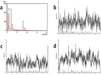
The presence of Cu metal in NF was analyzed by the EDX as shown in Figure 2a, The diffraction peaks in the NFs corresponding to Cu3(PO4)2 nanocrystals were almost consistent with the JCPDS card (00-022-0548) as presented in Figure 2b, 2c and 2d. The FT-IR spectrums of the only extract and the NFs are illustrated in Fig. 3a and 3b, respectively. The absorption bands (in Figure 3b) at 559, 1032 and 1150 cm−1 in the the NFs were attributed to P–O and P=O
vibrations, indicating the existence of phosphate groups (PO43- )
[39]. It is worthy to mention that molecules in the extracts were packed in the NFs with different conformations, which increase localized molecules concentration in the extract and may lead to strong and shifted vibration peaks. The vibration bands of –NH2 groups were at 1595 cm−1 and 1627 cm−1, for extracts and the NFs,
respectively. The stretching bands of –CH2 and –CH3 groups in the extract Al were at 2850 and 2918 cm−1. The adsorption bands
at 1675 cm−1 and 3232 cm−1 were assigned to carbonyl (>C=O)
and hydroxyl (–OH) groups from the compounds in the extract, respectively.
Figure 2. a) EDX analysis of Cu metal in the NFs, b-d) XRD of pattern of AI NFs,
Aa NFs and Av NFs. Aa: A. absinthium; Av: A. vulgaris; AI: A. ludoviciana, NFs: Nanoflowers
Figure 3. FT-IR spectra of extract AI (a) and the extract AI-Cu2+ hybrid nanoflower (b). AI: A. ludoviciana
Evaluation of antimicrobial activity of Aa, Av and Al-Cu2+hybrid nanoflowers
According to literature, Artemisia spp. containing various active molecules (phenolic compounds, essential oil) have high antibacterial and antifungal activity. Previous report revealed that Artemisia spp. did not exhibited any antimicrobial effect to Gram negative bacteria but showed moderate and strong activity against Gram posititive bacteria and fungi [40-44]. Park et al. [45] used the A. capillaris Thunb. extract as reducing agent to synthesis the silver nanoparticles (Ag NPs) and to improve the antimicrobial activity against several Gram negative and Gram positive bacterial strains. Some studies demonstrated that the green synthesis of AgNPs using Artemisia extracts and found them as effective antimicrobial agents towards pathogenic bacteria, such as Staphylococcus aureus, Bacillus cereus,
Acinetobacter baumannii, and Pseudomonas aeruginosa [46].
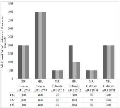
In this study, antimicrobial activities of the NFs and extracts of three Artemisia genotype (Aa, Av, Al) and were tested against standard pathogens using the broth microdilution method. Subsequently MIC, MBC and MFC values were determined. The NFs showed excellent antimicrobial acitivity against bacteria and fungi even at very low concentrations (Figure 4,5). The MIC and MBC values for NFs were between 0.4 to 40 μg mL-1 and 40 to 400 μg mL-1 while those values for Aa,
Av and Al extracts were 500-2000 and 1000-4000 μg mL-1
for bacterial pathogens, respectively. The extracts did not show any antibacterial activity towards E. coli, S. typhi and
P. aeruginosa at 4000 μg mL-1. According to these results, the
NFs have very promising antimicrobial properties compared to the extracts.
Figure 4. MIC and MBC values (μg/ml) of Artemisia sp. extracts (Aa, Al, Av).
Aa: A. absinthium; Av: A. vulgaris; Al: A. ludoviciana MIC: Minimum inhibitory concentration, MBC: Minimum bactericidal concentration
Figure 5. MIC and MBC values (µg/ml) of nanoflowers (NFs). Aa: A. absinthium;
Av: A. vulgaris; AI: A. ludoviciana MIC: Minimum inhibitory concentration: MBC: Minimum bactericidal concentration
Discussion
Several plant extracts were used for extracellular synthesis of silver nanoparticles and they can selectively inhibit growth of the Gram negative and Gram positive and they can be considered an interesting versatile biotechnological resource due to their antimicrobial activity [47-50]. The resistance of Gram negative bacteria to plant extract and essential oil have been associated to hydrophilic outer membrane that may block the penetration of hydrophobic compounds into target bacterial cell membrane [51]. The NFs as amphipathic hybrid nanomaterials can attach Gram positive and Gram negative bacteria outer membrane and they may show antibacterial activity. In addition, the NFs exhibited much higher antibacterial activity against Gram positive bacteria than Gram negative.
Conclusion
In this work, we have successfully firstly produced to synthesize the three Artemisia genotypes (Aa, Av, Al) extracts-metal ion NFs with very narrow size distribution, high production yield, and also demonstrated their excellent inhibitory property. Differences of the antimicrobial activities of the NFs and extracts may be related to due to the varied amounts of plant extracts of main molecules (proteins, flavonoids, polyphenol) contained in the Aa, Av and Al extracts. Importantly, the NFs exhibited dramatically enhanced antimicrobial activities towards standard bacterial (E. coli, S. typhi,
P. aeruginosa and S. aureus) and fungal pathogens (C. albicans)
compared to extracts. The results suggest that green sytnthesis of the NFs present promising a great potential for development of eco-friendly antimicrobial agents for especially nasocomial human pathogens.
Acknowledgements
The authors would like to thank Zeytinburnu Medicinal Plant Garden due to provide to plant materials. This manuscript was partially presented as an oral presentation at the DRD-2019 symposium.
Competing interests
The authors declare that they have no competing interest. Financial Disclosure
This work was supported by the Scientific Research Projects Coordination Unit of Erciyes University [Project number: THD-2016-6590].
Ethical approval
No ethic approvell is needed to this research. Ayse Baldemir Kilic ORCID: 0000-0003-2473-4837 Cevahir Altinkaynak ORCID: 0000-0003-0082-8521 Nilay Ildiz ORCID: 0000-0002-3799-856X Nalan Ozdemir ORCID: 0000-0002-8930-5198 Vedat Yilmaz ORCID: 0000-0001-6194-6527 Ismail Ocsoy ORCID: 0000-0002-5991-3934
References
1. Lee JH, Yigit MV, Mazumdari D, et al. Molecular diagnostic and drug delivery agents based on aptamer-nanomaterial conjugates. Adv Drug Deliv Rev. 2010;62:592–605.
2. Yigit MV, Moore A, Medarova Z. Magnetic nanoparticles for cancer diagnosis and therapy. Pharm Res. 2012;29:1180–8.
3. Öçsoy I, Yasun E, Ocsoy MA, Tan W. Nucleic acid-functionalized nanomaterials. Nano LIFE. 2013;3:1340004.
4. Leung KCF, Xuan S, Zhu X, et al. Gold and iron oxide hybrid nanocomposite materials. Chem Soc Rev. 2012;41:1911–28.
5. Kharisov BI. A Review for synthesis of nanoflowers. Recent Pat Nanotechnol. 2008;2:190-200.
6. Nakayama Y, Puzauskie PJ, Radenovic A, et al. Tunable nanowire nonlinear optical probe. Nature. 2007;447:1098–101.
7. Balazs AC, Emrick T, Russell TP. Nanoparticle polymer composites: Where two small worlds meet. Science. 2006;314:1107–10.
8. Ocsoy I, Gulbakan B, Shukoor MI, et al.Aptamer conjugated multifunctional nanoflowers as a platform for targeting, capture, and detection in laser desorption ionization mass spectrometry. ACS Nano. 2013;7:417–27. 9. Zhu G, Hu R, Zhao Z, et al.Noncanonical self-assembly of multifunctional
DNA nanoflowers for biomedical applications. ACS. 2013;135:16438–445. 10. Zhu G, Zhang S, Song E, et al.Angew, Building fluorescent DNA nanodevices
on target living cell surfaces. Chem Int Ed. 2013;52:5490–96.
11. Ocsoy I, Gulbakan B, Chen T,et al. DNA-guided metal-nanoparticle formation on graphene oxide surface. Adv Mater. 2013;25:2319–25.
12. Ocsoy I, Paret ML, Ocsoy MA, et al. Nanotechnology in Plant Disease Management: DNA-directed silver nanoparticles on graphene oxide as an antibacterial against Xanthomonas perforans. ACS Nano. 2013;7:8972–80. 13. Rica R, Stevens MM. Plasmonic ELISA for the ultrasensitive detection of
disease biomarkers with the naked eye. Angew Chem Int Ed. 2008;47:5415– 17.
14. Kim JS, Kuk E, Yu KN, et al. Antimicrobial effects of silvernanoparticles. Nanomed Nanotechnol Biol Med. 2007;3:95-101.
15. Sun S, Murray C, Weller D, et al. Monodisperse FePt nanoparticles and ferromagnetic FePt nanocrystal superlattices. Science 2000;287:1989–92. Thakkar KN, Mhatre SS, Parikh RY. Biological synthesis of metallic nanoparticles. Nanomed Nanotechnol Biol Med. 2010;6:257–62.
16. Mukherjee P, Ahmad A, Mandal D, et al. Fungus-Mediated Synthesis of Silver Nanoparticles and Their Immobilization in the Mycelial Matrix: A Novel Biological Approach to Nanoparticle Synthesis. Nano Lett. 2001;1:515–9. 17. Rica De La R, Matsui H. Urease as a nanoreactor for growing crystalline ZnO
nanoshells at room temperature. Angew Chem Int Edit. 2008;47:5415–17. 18. Duman F, Ocsoy I, Kup FO. Chamomile flower extract-directed CuO
nanoparticle formation for its antioxidant and DNA cleavage properties. Mat Sci Eng C. 2016;60:333–8.
19. Demirbas A, Welt BA, Ocsoy I. Biosynthesis of red cabbage extract directed Ag NPs and their effect on the loss of antioxidant activity. Mater Lett. 2016;179:20–3.
20. Schröfel A, Kratošová G, Šafařík I, et al. Applications of biosynthesized metallic nanoparticles – a review. Acta Biomater. 2014;10:4023–42. 21. Ge J, Lei J, Zare RN. Protein–inorganic hybrid nanoflowers. Nature
Nanotechnol. 2012;7:428–32.
22. Zhu L, Gong L, Zhang Y, et al. Rapid detection of phenol using a membrane containing laccase nanoflowers. Chem–Asian J. 2013;8:2358–60.
23. Yilmaz E, Ocsoy I, Ozdemir N, et al. Bovine serum albumin-Cu(II) hybrid nanoflowers: An effective adsorbent for solid phase extraction and slurry sampling flame atomic absorption spectrometric analysis of cadmium and lead in water, hair, food and cigarette samples. Anal Chim Acta. 2016;906:110–7. 24. Somturk B, Hancer M, Ocsoy I, et al. Synthesis of copper ion incorporated horseradish peroxidase-based hybrid nanoflowers for enhanced catalytic activity and stability. Dalton Trans. 2015;44:13845–52.
25. Ocsoy I, Dogru E, Usta S. A new generation of flowerlike horseradish peroxides as a nanobiocatalyst for superior enzymatic activity. Enzyme Microbiol Technol. 2015;75:25–9.
26. Shi J, Zhang S, Wang X, et al. Preparation and enzymatic application of flower like hybrid microcapsules through a biomimetic mineralization approach. J Mater Chem B. 2014;2:4289–96.
27. Somturk B, Yilmaz I, Altinkaynak C, et al.Synthesis of urease hybrid nanoflowers and their enhanced catalytic properties. Enzyme Microbiol Technol. 2016;86:134–42.
28. Altinkaynak C, Yilmaz I, Koksal Z, et al. Preparation of lactoperoxidase incorporated hybrid nanoflower and its excellent activity and stability. Int J Biol Macromol. 2016;84:402–9.
29. Wu ZF, Wang Z, Zhang Y, et al. Zircon U–Pb dating and in-situ Hf isotopic analysis of Permian peraluminous granite in the Lhasa terrane, southern Tibet: Implications for Permian collisional orogeny and paleogeography. Sci Rep. 2016;1:1–7.
30. Altinkaynak C, Tavlasogluc S, Özdemir N, et al. A new generation approach in enzyme immobilization: Organic-inorganic hybrid nanoflowers with enhanced catalytic activity and stability. Enzyme Microbiol Technol. 2016;93:105–12.
31. Obolsky D, Pischel I, Feistel B, et al. Artemisia dracunculus L. (tarragon): a critical review of its traditional use, chemical composition, pharmacology, and safety. J Agric Food Chem. 2011;59:11367–84.
32. Graven EH, Dean SG, Svoboda KP, et al. Antioxidant activity of newly discovered lineage of marine actinobacteria. Flavour Frag J. 1992;7:121–3. 33. Weyerstahl P, Marschall H, Schröder M, Wahlburg H-C, et al.Biosynthesis
and chemical synthesis of presilphiperfolanol natural products. Flavour Frag J. 1997;12:315–25.
34. Baytop T. Therapy with Medicinal Plants in Turkey (Past and Today). 2nd ed. İstanbul University, İstanbul;1999.
35. Bradford MM. A Rapid and sensitive method for the quantitation of microgram quantities of protein utilizing the principle of protein-dye binding. Anal Biochem. 1976;72:248–54.
36. Clinical Laboratory Standards Institute. Reference method for broth dilution antifungal susceptibility testing of yeasts; approved standard-third ed. CLSI document M27-A3. CLSI: Wayne;2008
37. Clinical Laboratory Standards Institute. Performance standards for antimicrobial susceptibility testing. Twenty-second informational supplement ed. CLSI document M100-S22. CLSI: Wayne;2012.
38. Bazargani MM, Rohloff J. Antibiofilm activity of essential oils and plant extracts against Staphylococcus aureus and Escherichia coli biofilms. Food Control. 2016;61:156–64.
39. Lin Z, Xiao Y, Yin Y, et al. Correction to facile synthesis of enzyme inorganic hybrid nanoflowers and ıts application as a colorimetric platform for visual detection of hydrogen peroxide and phenol ACS Appl Mater Interfaces. 2014;6:10775−82.
40. Bora KS, Sharma A. The genus Artemisia: a comprehensive review. Pharm Biol. 2011;49:101–9.
41. Juteau F, Jerkovic I, Masotti V, et al. Composition and antimicrobial activity of the essential oil of Artemisia absinthium from Croatia and France.Planta Med. 2003;69:158–61.
42. Kordali S, Kotan R, Mavi A, et al. Determination of the chemical composition and antioxidant activity of the essential oil of Artemisia dracunculus and of the antifungal and antibacterial activities of Turkish Artemisia absinthium, A. dracunculus,Artemisia santonicum, and Artemisia spicigera essential oils. J Agr Food Chem. 2005;53:9452–8.
43. Badillo LM, Espinosa-Madrigal RM, Martinez-Muñoz RE, et al. The Mexican medical plants with antifungal properties are an economic and health opportunity area. Pharmacology Online. 2008;3:61–77.
44. Fiamegos YC, Kastritis PL, Exarchou V, Han H, Bonvin AM, Vervoort J, et al. Antimicrobial and efflux pump inhibitory activity of caffeoylquinic acids from Artemisia absinthium against gram-positive pathogenic bacteria. PLoSOne. 2011;6:e18127.
45. Park Y, Noh HJ, Han L, et al. Artemisia capillaris extracts as a green factory for the synthesis of silver nanoparticles with antibacterial activities. J Nanosci Nanotechnol. 2012;12:7087–95.
46. Salehi S, Shandiz SAS, Ghanbar F, et al. Phytosynthesis of silver nanoparticles using Artemisia marschalliana Sprengel aerial part extract and assessment of their antioxidant, anticancer, and antibacterial properties. Int J Nanomedicine. 2016;11:1835–46.
47. Kim BS, Salunke B, Sawant S, et al. Biological synthesis of silver nanoparticles using plant leaf extracts and their specific antimicrobial activity. N biotechnol. 2014;31:S173.
48. Stabili L, Acquaviva MI, Biandolino F, et al. Biotechnological potential of the seaweed Cladophora rupestris (Chlorophyta, Cladophorales) lipidic extract. N biotechnol. 2014;31:436–44.
49. MubarakAli D, Thajuddin N, Jeganathan K, et al. Plant extract mediated synthesis of silver and gold nanoparticles and its antibacterial activity against clinically isolated pathogens. Coll Surf B: Biointerfaces. 2011;85:360–5. 50. Krishnaraj C, Jagan EG, Rajasekar S, et al. Synthesis of silver nanoparticles
using Acalypha indica leaf extracts and its antibacterial activity against water borne pathogens. Coll Surf B: Biointerfaces, 2010;76:50–6.
51. Inouye S, Yamaguchi H, Takizawa T. Screening of the antibacterial effects of a variety of essential oils on respiratory tract pathogens, using a modified dilution assay method. J Infect Chemother. 2001;7:251–4.