Aesthetic Surgery Journal 2014, Vol. 34(4) 538 –544 © 2014 The American Society for Aesthetic Plastic Surgery, Inc. Reprints and permission: http://www .sagepub.com/ journalsPermissions.nav DOI: 10.1177/1090820X14526598 www.aestheticsurgeryjournal.com Breast Surgery
Gynecomastia is a benign enlargement of the male breast. In gynecomastia surgery, which is highly successful in improving this aesthetic problem, the primary concerns are to avoid contour irregularities, infection, hematoma, seroma, skin injuries, and extensive scarring. The aim of gynecomastia surgery is to restore a normal chest contour with minimal signs of breast surgery.
Suction drains traditionally have been placed after gyneco-mastia surgery. They are thought to aid the removal of unde-sirable postoperative matter and minimize tissue dead space. These drains are costly not only in economic terms but also in terms of recordkeeping, nursing time, and postoperative care. Patients often report discomfort associated with drains, and drain sites can leave conspicuous scars and may represent potential sources of tract formation or infection. Outpatient management of drains can present a source of concern for patients who have shorter hospital stays and therefore must care for the drain site at home.1 The effectiveness of surgical drains in reducing postoperative hematomas also has been questioned in other breast-related surgeries.
In this study, we retrospectively analyzed the results of surgeries performed for different grades of gynecomastia without drains to determine whether this practice had any adverse effect on patient outcomes.
MethodS
We retrospectively analyzed the charts of 138 consecutive male patients who underwent bilateral gynecomastia
From the Plastic and Aesthetic Surgery Department, Istanbul Medipol University, Istanbul, Turkey.
Corresponding Author:
Dr Mustafa Keskin, Istanbul Medipol Hastanesi Kosuyolu, Kadikoy, 34718 Istanbul, Turkey.
E-mail: [email protected]
Necessity of Suction Drains in
Gynecomastia Surgery
Mustafa Keskin, MD; Mustafa Sutcu, MD; Bulent Cigsar, MD;
and Naci Karacaoglan, MD
Abstract
Background: The aim of gynecomastia surgery is to restore a normal chest contour with minimal signs of breast surgery. Objective: The authors examine the rate of complications in gynecomastia surgery when no closed-suction drains are placed.
Methods: One hundred thirty-eight consecutive male patients who underwent gynecomastia surgery without drains were retrospectively analyzed to determine whether the absence of drains adversely affected patient outcomes. Patients were managed by ultrasonic-assisted liposuction both with and without the pull-through technique.
Results: The mean age of the patients was 29 years, and the mean volume of breast tissue aspirated was 350 mL per beast. Pull-through was needed in 23 cases. There was only 1 postoperative hematoma.
Conclusions: These results are comparable with previously published data for gynecomastia surgery in which drains were placed, suggesting that the absence of drains does not adversely affect postoperative recovery. Routine closed-suction drainage after gynecomastia surgery is unnecessary, and it may be appropriate to omit drains after gynecomastia surgery.
Level of Evidence: 4
Keywords
breast surgery, gynecomastia, drains, suction, hematoma, complications Accepted for publication July 30, 2013.
IN TE RNAT IONAL CONTR IBU TIO N
Scan this code with your smartphone to see the operative video. Need help? Visit www.aestheticsurgeryjournal.com.
Keskin et al 539 surgery without the use of drains between January 2006
and September 2012. Patient age, grade of gynecomastia, volume of liposuction, and hematoma rate were recorded.
Surgical Technique
The preoperative preparation for gynecomastia included a physical examination and laboratory work appropriate to the age of the patient. None of the patients in the study had any coagulation disorder, and none were taking medi-cation that could affect bleeding. However, prothrombin time (PT) and partial thromboplastin time (PTT) tests were conducted. Patients were reminded preoperatively to stop taking aspirin, ibuprofen, alcohol, and vitamin E and to stop smoking 10 days prior and 10 days after the surgery.
All patients received general anesthesia. An antibiotic was administered intravenously immediately before the sur-gery. Depending on breast size and the extent of the fatty component, between 500 and 1000 mL of a warm tumescent solution was infused into each breast. The solution com-prised 1000 mL Ringer’s lactate, 20 mL of 2% lidocaine, and 1 ampoule of epinephrine 1:1000. A stab incision was made about 2 to 3 cm below the inferior margin of the areola. Using a standard infiltration pump and a 3-mm-wide/20-cm-long blunt cannula, the wetting solution was uniformly administered in multiple subcutaneous layers. The right breast was infused first, followed by the left breast. The infiltration of both breasts was completed in 15 to 20 min-utes, which provided sufficient time for the adrenaline to work before applying ultrasonic energy to each breast.2 To prevent friction injuries at the entrance site, a plastic skin protector was inserted. In all the patients, glandular and fat tissues were removed with ultrasonic-assisted liposuction (VASER Shape; Sound Surgical, Denver, Colorado), with or without the pull-through technique. A 3.7-mm solid probe with 2 grooves was used at 80% of total power in continu-ous mode. The duration of the cavitation was approximately 1 minute per each 100 mL of infiltration. The cannula, with ultrasound assistance, was passed through multiple times in a fanlike pattern to separate the more fibrous breast tissue. After the application of the ultrasound energy, the emulsi-fied fat was evaluated using a standard procedure with a 3.7-mm triple-hole cannula. Final contouring of the thorax was not just limited to the breast but extended to the lateral axillary component and below the inframammary line.
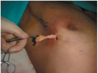
The residual glandular tissue, located mostly in the sub-areolar area, was pinched after completing the liposuction to evaluate volume; it was then removed by the pull-through technique using the same incision employed for the liposuc-tion.3 To surgically excise the gland, it first was pinched between the thumb and the index finger, grasped using a Mosquito forceps, and then passed through the skin incision made for the liposuction. When the forceps was pulled, the edge of the glandular tissue was exposed. The gland was freed from the surrounding tissue by cutting with scissors in different directions (Figure 1). This maneuver was repeated
until the gland was completely separated. It was then removed, and any remaining gland was palpated after each removal. Leaving a small amount of gland behind the areola prevented skin depression. The incision sites were closed in layers subcutaneously and intracutaneously with a continu-ous Monocryl 5-0 suture (Ethicon, Inc, Somerville, New Jersey). A video of the procedure is available online at www.aestheticsurgeryjournal.com. You may also scan the code on the first page of this article with any smartphone to be taken directly to the video at www.YouTube.com.
Postoperatively, a compression garment was applied to each patient’s torso. The garment was composed of a 9-inch waist binder (VOE, Barcelona, Spain) with 2 independent bands and an adjustable Velcro closure system. Special care was taken to apply the binder smoothly to the skin. The sur-gery was performed on an ambulatory or same-day basis, and the patients were examined within the first 24 postoperative hours to determine potential abnormalities. Patients were instructed to wear the binder continuously, except while showering, for the subsequent 2 weeks. They were also asked to refrain from any strenuous activity for 3 weeks, after which they could resume activity if they felt comfortable.
ReSultS
Gynecomastia was deemed to be idiopathic in all cases. A total of 276 breasts of 138 patients were operated on during the study period. The mean age of our patients was 29 years Figure 1. Removal of the glandular tissue with the pull-through technique via a liposuction incision.
Table 1. Volume of Breast Tissue Aspirated
Volume of Tissue Aspirated, mL Number of Breasts
<250 119
250-500 130
>500 27
(range, 17-55 years), and their mean body weight was 64 kg (range, 58-88 kg). The distributed severity of gynecomastia was as follows: 41 patients were classified as grade 1, 85 patients were classified as grade 2, and 12 patients were clas-sified as grade 3 (Table 1). In 23 patients, the glandular tissue
was removed with the pull-through technique. None of the patients in this series required skin resection or areola transpo-sition. Ecchymosis was present for 1 to 2 weeks, disappearing gradually. Some operative field swelling also persisted for sev-eral weeks postoperatively. However, in all cases, the skin Figure 2. (A) A 31-year-old man with grade II gynecomastia. (B) After 6 minutes of cavitation, total of 600 mL liposuction was carried out. One week after the surgery with compression garment. (C) Without compression garment. (D) Postoperative month 17.
Keskin et al 541 appeared to drape normally after the compression garment was removed (Figure 2). Clinical examination of all 276 breasts revealed only 1 postoperative hematoma. This was a hema-toma on a single breast, and only liposuction was performed. All patients were tracked postoperatively, with an average fol-low-up time of 14 months.
diScuSSion
Early reports described gynecomastia surgery techniques with placement of closed-suction drains,4 which were con-ventionally used to minimize the amount of fluid at the operative site and the dead space between tissues. However, the reasons for drain use have not been studied, and the criteria for using them are not clear. While closed-suction drains are generally believed to help prevent possible com-plications, there is no evidence that the drainage is benefi-cial in all cases or that closed-suction drainage has particular benefits after gynecomastia surgery (Figure 3). Figure 3. Postoperative hematoma after removal of the
breast tissue through the subareolar incision was not prevented with a closed-suction drain.
Table 2. A Review of the Current Literature Concerning Gynecomastia Surgery
Series Year Number of Patients Technique Hematoma (Patients) Drain vs Compression Garment
Varma and Henderson8 1990 20 OE 2 breast Closed-suction drain
Persichetti et al9 2001 29 OE 1 seroma Closed-suction drain
Fruhstorfer and Malata10 2003 29 L ± OE None Compression garment
Hammond et al11 2003 15 PT + UAL None Compression garment
Bracaglia et al12 2004 45 L + PT 2 Compression garment
Tashkandi et al13 2004 24 OE 0 Drains with compression garment
Wiesman et al14 2004 174 L ± OE 48 (with seroma) Not reported
Walden et al15 2004 34 L ± OE 1 Compression garment
Aslan et al16 2005 15 OE + L 2 Drains with compression garment
Ramon et al17 2005 17 PAL + PT (endoscopic assisted) 0 Compression garment
Filho et al18 2006 12 OE + L 1 hematoma 1 seroma Compression garment
Mentz et al19 2007 200 PT 2 Compression garment
Cordova and Moschella20 2008 121 L ± OE 6 Not defined
Lista and Ahmad21 2008 99 PAL + PT 1 (seroma) Compression garment
Benito-Ruiz et al22 2009 40 Liposuction with arthroscopic shaver 3 Drains with compression garment
Ratnam23 2009 42 L ± OE 2 Compression garment
Al-Allak et al24 2011 224 OE 26 Not defined
Cigna et al25 2011 37 OE + PAL 1 Drains with compression garment
Jose and Thomas26 2011 73 PAL ± OE 5 Compression garment
Petty et al27 2011 227 L ± OE (arthroscopic shaver) 4 Compression garment
Morselli and Morellini28 2012 260 L + PT %1 Suction drain
Abbreviations: L, liposuction; PAL, power-assisted liposuction; OE, open excision; UAL, ultrasound-assisted liposuction.
A review of the literature does not support the use of drains, nor does it indicate that drains prevent hematomas. The use of adrenaline in local tissue infiltration to produce preoperative vasoconstriction has been shown to reduce blood loss significantly in operations on breast tissue.4
Ultrasound evaluation of surgical wounds has shown that hematomas can occur at least 10 days postoperatively.5 Theoretically, prolonged drainage throughout the postop-erative period may reduce the incidence of hematoma development, but it can increase significantly the risk of Figure 4. (A, C) Preoperative image of a 16-year-old boy with grade 3 gynecomastia. (B, D) Postoperative images of the same patient 6 months after treatment with liposuction and the pull-through technique without a drain.
Keskin et al 543 infection. Bacteria, especially skin commensals, migrate
along the drain tube and have been cultured from drain tips as early as 48 hours after surgery.6
Closed-suction drains present other potential disadvan-tages in addition to possible infection. The percutaneous draining tubes often present a source of pain and discom-fort for patients. Drains may be inserted through a liposuc-tion-access incision, but sometimes an additional incision may be required. Additional cuts carry the possibility of additional scarring.
Also, ambulatory, nonhospital admissions for gyneco-mastia surgery are increasingly common. These patients need additional care when they are discharged with drains, and this increases the costs of the treatment and places a further burden on the patients.1 Finally, removal of the drainage tubes is often associated with a great deal of patient anxiety.7 A review of the current literature suggests omitting closed-suction drainage in gynecomastia surgery does not increase postoperative complications (Table 2).8-28 Eliminating drainage tubes may be beneficial, given that the literature review does not either confirm their value or suggest they contribute positively to patient outcomes.
Observation suggests that ultrasound-assisted liposuc-tion is an insufficient treatment for glandular gynecomas-tia. The glandular tissue is too dense and fibrous to be amenable to suction alone. Therefore, liposuction should be combined with some kind of surgical excision if the breast is purely fatty. The pull-through technique, which allows glandular tissue to be removed through the liposuc-tion incision, is our suggested method. Even when the pull-through technique is used, a closed-suction drain is unnecessary. Drains are rarely used in bodybuilding-related gynecomastia, which is considerably more prone to hematoma complications.29 In certain situations, drainage may be necessary; however, usage is determined by an individual patient’s case, rather than on the breasts’ glan-dular nature. For example, if the surgeon determines intra-operatively that the surgical site is oozing blood uncontrollably, then a drain might be considered.
In this particular series, none of the patients needed nip-ple-areola complex (NAC) repositioning, not even the 12 patients who were classed as having grade 3 gynecomastia (Figure 4). NAC repositioning should be considered a last resort and, if needed, performed in a second surgery. Patients who have lost extensive weight present a special situation in which NAC repositioning and skin excision are usually needed. However, even in NAC repositioning cases, closed-suction drains are unnecessary because—as in a comparable example— breast reduction surgeries performed on women omit closed drains. Reduction mammaplasties in which NAC repositioning and skin excision are done with-out drains have been demonstrated to be safe, withwith-out increased complications.30-33 Additionally, the pain and dis-comfort associated with wound drainage are avoided, and nursing time involved with drain care is minimized.
concluSionS
When intraoperative tumescent fluid and a postoperative compression garment are used, omitting the placement of closed-suction drainage increases patient comfort without the risk of increasing complications from gynecomastia surgery. Drainage is not needed when combining the pull-through technique with suction-assisted lipectomy. The findings of the current study and of a literature review demonstrate the advantage of omitting drains following gynecomastia surgery.
disclosures
The authors declared no potential conflicts of interest with respect to the research, authorship, and publication of this article.
Funding
The authors received no financial support for the research, authorship, and publication of this article.
ReFeRenceS
1. Matarasso A, Wallach SG, Rankin M. Reevaluating the need for routine drainage in reduction mammaplasty. Plast Reconstr Surg. 1998;102:1917-1921.
2. McKee DE, Lalonde DH, Thoma A, Glennie DL, Hayward JE. Optimal time delay between epinephrine injection and incision to minimize bleeding. Plast Reconstr Surg. 2013;131:811-814.
3. Karamese M, Keskin M, Sutcu M, et al. Pull-through tech-nique combined with liposuction in gynecomastia. Turk Plast Surg. 2011;19:78-81.
4. Teimourian B, Perlman R. Surgery for gynecomastia. Aesthetic Plast Surg. 1983;7:155-157.
5. Samdal F, Serra M, Skolleborg KC. The effects of infil-tration with adrenaline on blood loss during reduction mammaplasty. Scand J Plast Reconstr Surg Hand Surg. 1992;26:211-215.
6. Varley GW, Milner S, Turner GM, Crisp AJ, Szypryt EP. Ultrasound assessment of the efficacy of wound drains. J R Coll Surg Edinb. 1994;39:97-99.
7. Watson JD, Smith G, Forrest H, Quin RO. Contamination of suction drainage systems in vascular surgery. J R Coll Surg Edinb. 1988;33:130-131.
8. Varma SK, Henderson HP. A prospective trial of adrena-line infiltration for controlling bleeding during surgery for gynaecomastia. Br J Plast Surg. 1990;43:590-593.
9. Persichetti P, Berloco M, Casadei RM, et al. Gynecomastia and the complete circumareolar approach in the surgi-cal management of skin redundancy. Plast Reconstr Surg. 2001;107:948-954.
10. Fruhstorfer BH, Malata CM. A systematic approach to the surgical treatment of gynaecomastia. Br J Plast Surg. 2003;56,237-246.
11. Hammond DC, Arnold JF, Simon AM, Capraro PA. Combined use of ultrasonic liposuction with the pull-through technique for the treatment of gynecomastia. Plast Reconstr Surg. 2003;112:891-895.
12. Bracaglia R, Fortunato R, Gentileschi S, Seccia A, Farallo E. Our experience with the so-called pull-through tech-nique combined with liposuction for management of gynecomastia. Ann Plast Surg. 2004;53:22-26.
13. Tashkandi M, Al-Qattan MM, Hassanain JM, Hawary MB, Sultan M. The surgical management of high-grade gyne-comastia. Ann Plast Surg. 2004;53:17-20.
14. Wiesman IM, Lehman JA Jr, Parker MG, et al. Gynecomastia: an outcome analysis. Ann Plast Surg. 2004;53:97-101.
15. Walden JL, Schmid RP, Blackwell SJ. Cross-chest lipo-plasty and surgical excision for gynecomastia: a 10-year experience. Aesthetic Surg J. 2004;24:216-223.
16. Aslan G, Tuncali D, Terzioglu A, Bingul F. Periareolar-transareolar-perithelial incision for the surgical treatment of gynecomastia. Ann Plast Surg. 2005;54:130-134. 17. Ramon Y, Fodor L, Peled IJ, et al. Multimodality
gyne-comastia repair by cross-chest power-assisted superficial liposuction combined with endoscopic-assisted pull-through excision. Ann Plast Surg. 2005;55:591-594. 18. Filho DH, Arruda RG, Alonso N. Treatment of severe
gynecomastia (grade III) by resection of periareolar skin. Aesthetic Surg J. 2006;26:669-673.
19. Mentz HA, Ruiz-Razura A, Newall G, Patronella CK, Miniel LA. Correction of gynecomastia through a single puncture incision. Aesthetic Plast Surg. 2007;31:244-249. 20. Cordova A, Moschella F. Algorithm for clinical evaluation
and surgical treatment of gynaecomastia. J Plast Reconstr Aesthetic Surg. 2008;61:41-49.
21. Lista F, Ahmad J. Power-assisted liposuction and the pull-through technique for the treatment of gynecomastia. Plast Reconstr Surg. 2008;121:740-747.
22. Benito-Ruiz J, Raigosa M, Manzano M, Salvador L Assessment of a suction-assisted cartilage shaver plus
liposuction for the treatment of gynecomastia. Aesthetic Surg J. 2009;29:302-309.
23. Ratnam BV. A new classification and treatment protocol for gynecomastia. Aesthetic Surg J. 2009;29:26-31. 24. Al-Allak A, Govindarajulu S, Shere M, et al. Gynaecomastia:
a decade of experience. Surgeon. 2011;9:255-258.
25. Cigna E, Tarallo M, Fino P, De Santo L, Scuderi N. Surgical correction of gynecomastia in thin patients. Aesthetic Plast Surg. 2011;35:439-445.
26. Jose RM, Thomas S. Gynaecomastia correction—the role of power-assisted liposuction. Eur J Plast Surg. 2011;34:187-191.
27. Petty PM, Solomon M, Buchel EW, Tran NV. Gynecomastia: evolving paradigm of management and comparison of techniques. Plast Reconstr Surg. 2011;125:1301-1308. 28. Morselli PG, Morellini A. Breast reshaping in
gynecomas-tia by the “pull-through technique”: considerations after 15 years. Eur J Plast Surg. 2012;35:365-371.
29. Babigian A, Silverman RT. Management of gynecomas-tia due to use of anabolic steroids in bodybuilders. Plast Reconstr Surg. 2001;107:240-242.
30. Wrye SW, Banducci DR, Mackay D, Graham WP, Hall WW. Routine drainage is not required in reduction mam-maplasty. Plast Reconstr Surg. 2003;111:113-117.
31. Arrowsmith J, Eltigani E, Krarup K, Varma S. An audit of breast reduction without drains. Br J Plast Surg. 1999;52:586-588.
32. Corion LU, Smeulders MJ, van Zuijlen PP, van der Horst CM. Draining after breast reduction: a randomised con-trolled inter-patient study. J Plast Reconstr Aesthetic Surg. 2009;62:865-868.
33. Ngan PG, Iqbal HJ, Jayagopal S, et al. When to use drains in breast reduction surgery? Ann Plast Surg. 2009;63:135-137.