Systemic Proteasome Inhibition Induces Sustained Post-stroke
Neurological Recovery and Neuroprotection via Mechanisms
Involving Reversal of Peripheral Immunosuppression
and Preservation of Blood
–Brain–Barrier Integrity
Thorsten R. Doeppner1,2&Britta Kaltwasser1&Ulrike Kuckelkorn3&
Petra Henkelein3&Eva Bretschneider4&Ertugrul Kilic2&Dirk M. Hermann1
Received: 13 May 2015 / Accepted: 8 November 2015 / Published online: 16 November 2015 # Springer Science+Business Media New York 2015
Abstract In view of its profound effect on cell survival and function, the modulation of the ubiquitin-proteasome-system has recently been shown to promote neurological recovery and brain remodeling after focal cerebral ischemia. Hitherto, local intracerebral delivery strategies were used, which can hardly be translated to human patients. We herein analyzed effects of systemic intraperitoneal delivery of the proteasome inhibitor BSc2118 on neurological recovery, brain injury, pe-ripheral and cerebral immune responses, neurovascular integ-rity, as well as cerebral neurogenesis and angiogenesis in a mouse model of transient intraluminal middle cerebral artery occlusion. Systemic delivery of BSc2118 induced acute neu-roprotection reflected by reduced infarct volume when deliv-ered up to 9 h post-stroke. The latter was associated with reduced brain edema and stabilization of blood–brain–barrier integrity, albeit cerebral proteasome activity was only mildly reduced. Neuronal survival persisted in the post-acute stroke
phase up to 28 days post-stroke and was associated with im-proved neurological recovery when the proteasome inhibitor was continuously delivered over 7 days. Systemic proteasome inhibition prevented stroke-induced acute leukocytosis in pe-ripheral blood and reversed the subsequent immunosuppres-sion, namely, the reduction of blood lymphocyte and granulo-cyte counts. On the contrary, post-ischemic brain inflamma-tion, cerebral HIF-1α abundance, cell proliferainflamma-tion, neurogenesis, and angiogenesis were not influenced by the proteasome inhibitor. The modulation of peripheral immune responses might thus represent an attractive target for the clin-ical translation of proteasome inhibitors.
Keywords Cerebral ischemia . Stroke . Proteasome . Neuroprotection . Neuroregeneration . Inflammation
Introduction
The proteasome is a multicatalytic protease complex, which is a major site of protein turnover in eukaryotic cells. Proteasomes containing the catalytic 20S core are ubiquitous-ly expressed in mammalian tissues [1,2], controlling protein abundance under physiological conditions and degrading misfolded proteins under pathophysiological conditions. In view of its profound influence on cell cycle and signaling, inhibition of the proteasome has been used as therapeutic strategy in malignant tumors and vascular diseases as well [3–8].
Over short observation periods, the neuroprotective effects of proteasome inhibitors have repeatedly been reported in an-imal models of ischemic stroke [9–16]. These studies were hampered by severe side effects and drug resistance, which precluded restorative studies in the post-acute stroke phase Electronic supplementary material The online version of this article
(doi:10.1007/s12035-015-9533-3) contains supplementary material, which is available to authorized users.
* Thorsten R. Doeppner [email protected]
1 Department of Neurology, University of Duisburg-Essen,
Essen, Germany
2
Regenerative and Restorative Medical Research Center, Istanbul Medipol University, Istanbul, Turkey
3
Department of Biochemistry, Charité-Universitätsmedizin, Berlin, Germany
4 Department of Otorhinolaryngology, Johannes Wesling Klinikum,
with repeated drug delivery [17]. More recently, the protea-some inhibitor BSc2118 has become available, which is well tolerated and exerts both anti-melanoma and anti-myeloma activities [18–20]. Using a mouse model of cerebral ischemia, we have previously shown that the intracerebral delivery of BSc2118 induces sustained neuroprotection, cerebral neurogenesis, and angiogenesis via mechanisms that involve stabilization of the transcription factor HIF-1α [21].
In view of its invasive route of application, the local intra-cerebral delivery of proteasome inhibitors can hardly be trans-lated to human patients. Since BSc2118 poorly penetrates the blood–brain–barrier (BBB), the efficacy of systemically de-livered proteasome inhibitors was hitherto unknown. In the present study, we evaluated the effects of intraperitoneal BSc2118 delivery on post-stroke brain injury, neurological deficits, immune responses, and neurovascular remodeling, demonstrating that systemic proteasome inhibition induces sustained neuroprotection via mechanisms involving reversal of post-ischemic immunosuppression and stabilization of BBB integrity.
Materials and Methods
Study Design and Animal Procedures
All studies were performed according to local government authorities. Animals had free access to food and water and were kept under circadian rhythm. In vivo studies were per-formed on male C57BL6 mice (Harlan, Germany; 22–27 g). Animals were strictly randomized to experimental groups, and both experimenters and analysts were blinded at all stages of the study. Induction of stroke was performed as stated below, and mice were allowed to survive for 1 h (analysis of protea-some activity), 1 day (Evans blue extravasation analysis, zymography, Western blots, flow cytometry, and proteasome activity studies), 2 days (analysis of proteasome activity), 4 days (analysis of infarct volume and brain edema), 7 days (flow cytometry), or 28 days (behavioral tests and immuno-histochemistry). The number of animals used for statistical analyses is given in the figure legends.
Focal cerebral ischemia was induced as previously de-scribed [21]. Briefly, the left common carotid artery (CCA) was prepared and a silicon-coated nylon filament (Doccol, USA) was inserted into the CCA. The filament was carefully moved forward until the proximal branch of the left middle cerebral artery (MCA). Under constant laser Doppler flow (LDF) control, the filament stayed in situ for 45 min in order to induce transient focal cerebral ischemia in the left MCA territory. After filament removal, LDF control was continued for an additional 15 min in order to ensure adequate reperfu-sion. Sham animals underwent the same surgical procedure
but without inserting the filament. Application of the protea-some inhibitor BSc2118 (synthesized at the Department of Biochemistry, Charité-Universitätsmedizin, Berlin, Germany; 30 mg/kg solved in DMSO) was done via intraperitoneal (i.p.) injection (injection volume 50μl). The proteasome inhibitor was delivered as single injection (for the majority of studies 9 h post-stroke) or as chronic delivery, i.e., first injection was given 9 h post-stroke with additional daily injections until day 7. Control animals received 50μl of DMSO only. The poten-tial clinical relevance of a systemic BSc2118 treatment against stroke was further evaluated using an intravenous injection of rt-PA (10 mg/kg body weight) or NaCl during the beginning of the reperfusion followed by treatment with BSc2118 (or DMSO as control) 9 h post-stroke and infarct volume analysis on day 4. For analysis of proteasome activity in brain lysates, a volume of 5μl of BSc2118 (30 mg/kg) was stereotactically injected into the left striatum as previously described [21]. Post-stroke neurogenesis and angiogenesis were assessed by means of daily i.p. injections of bromodeoxyuridine (BrdU, 50 mg/kg; Sigma-Aldrich, Germany) on days 8–18 with sub-sequent histochemical analysis as stated below.
Behavioral Tests
Neurological recovery was evaluated using the rotarod, tight rope, corner turn, and balance beam tests, as described before by our group (e.g., see [22]). Prior to stroke induction, all mice were trained in order to ensure adequate performance, i.e., rotarod test performance >300 s, tight rope test score >19, corn turn test performance <0.6, and balance beam test per-formance <20 s. In the rotarod test, which evaluates motor coordination deficits, the time until the animal dropped off the accelerating rotating drum (velocity 4–40 rpm with max-imal testing time of 300 s) was measured. In the tight rope test, which evaluates coordinated climbing abilities as a means to assess motor coordination as well, a validated score from 0 (min) to 20 (max) was obtained. In the corner turn test, in which the mice were placed into an apparatus consisting of two vertical boards with an angle of 30°, the laterality index, defined as number of right turns/ten measurements, was eval-uated. A score of 1 indicated severe movement preference as a result of stroke-induced neurological impairment, whereas a score of 0.5 indicated no neurological impairment. In the bal-ance beam test, the time until the mice reached the opposite platform was analyzed. All tests were performed twice on occasion of each time point. Mean values were calculated for both measurements.
Analysis of Infarct Volume and Brain Edema
For analysis of infarct volume and brain edema, 2-mm-thick coronal brain slices obtained from animals sacrificed at 4 days post-stroke were stained with 2 % 2,3,5-triphenyltetrazolium
chloride (TTC). Healthy and injured tissues in both hemi-spheres were outlined using ImageJ software. For analysis of infarct volume, edema-corrected infarct areas were deter-mined by subtraction of healthy tissue in both hemispheres. Brain edema was calculated as relative increase of ipsilateral volume compared with contralateral hemispheric volume. Evaluation of BBB Permeability
As previously described [21,23], BBB permeability was eval-uated in mice receiving Evans blue injections at 22 h post-stroke, followed by transcardiac perfusion with 0.1 M phosphate-buffered saline (PBS) 2 h later. Extravasated Evans blue was measured with a luminescence spectrophotometer withλexc.at 620 nm andλem.at 680 nm.
Immunohistochemistry
Immunohistochemical stainings were performed on 2- μm-thick coronal paraffin sections obtained from mice sacrificed 28 days post-stroke by transcardial perfusion with 4 % para-formaldehyde in 0.1 M PBS. The following primary antibod-ies were used: monoclonal mouse anti-BrdU (1:400; Roche, Switzerland), monoclonal rat anti-BrdU (1:400; Abcam, UK), polyclonal goat anti-doublecortin (1:50; Santa Cruz Biotech-nology, Germany), monoclonal mouse anti-NeuN (1:1000; Millipore, UK), and monoclonal rat anti-CD31 (1:200; BD Biosciences, Germany). Secondary antibodies included goat anti-mouse Cy-3 (1:400; Dianova, Germany), goat anti-rat Alexa-594 (1:400; Dianova), donkey anti-goat Alexa-488 (1:250; Invitrogen, Germany), goat anti-mouse Alexa-488 (1:100; Jackson ImmunoResearch, Germany), and goat anti-rat Alexa-488 (1:250; Invitrogen). Sections were evaluated in three regions of interest (ROIs) of the striatum at coordinates AP +0.14 mm, ML ±1.5–2.25 mm, and DV −2.5–3.25 mm in four sections per animal. Mean values were calculated for all ROIs.
Zymography of Matrix Metalloprotease 9
As described [24], matrix metalloprotease 9 (MMP-9) activity was evaluated by zymography in brain homogenates obtained from left (ischemic) hemispheres of mice sacrificed at 1 day post-stroke. For homogenization, a lysis buffer (referred to as basic buffer below) containing 50 mmol/l Tris–HCl (pH 7.6), 150 mmol/l NaCl, 5 mmol/l CaCl2, 0.05 % BRIJ-35, 0.02 % NaN3, and 1 % Triton X-100 was used. After centrifugation, pellets were resuspended in elution buffer (10 % DMSO and 20 % volume of basic lysis buffer). Samples were incubated in a non-reducing sample buffer (0.4 mol/l Tris, pH 6.8; 5 % SDS, 20 % glycerol, 0.05 % bromophenol blue) and loaded onto commercially available 10 % sodium dodecyl sulfate polyacrylamide gel electrophoresis (SDS-PAGE) gels
containing 0.1 % gelatin (Novex Zymogram Gels; Invitrogen, USA). After electrophoresis, samples were further processed in 2.5 % Triton X-100, equilibrated with developing buffer (Novex), and incubated over 18 h at 37 °C. Coomassie blue was used for protein staining. As standards, 0.1 ng of human pro-MMP-9 and 0.01 ng of activated MMP-9 (both from Merck Biosciences, Germany) were used. Gels were scanned and densitometrically analyzed.
Western Blot Analysis of HIF-1α Abundance
As previously reported [21], HIF-1α was detected by Western
blotting in brain homogenates obtained from left (ischemic) hemispheres of mice sacrificed at 1 day post-stroke. For ho-mogenization, a lysis buffer containing 50 mmol/l Tris, pH 8.0; 150 mmol/l NaCl; 1 % Triton X-100; and protease inhib-itors was used. Equal amounts of protein (40μg) were plotted for SDS-PAGE. After transfer onto PVDF membranes, pro-teins were incubated with a polyclonal rabbit anti-HIF-1α antibody (1:1000; Abcam, UK) that was detected with a peroxidase-coupled goat anti-rabbit antibody (Santa Cruz Bio-technology). Blots were scanned and densitometrically analyzed.
Analysis of Proteasome Activity
Proteasome activity was determined in brain homogenates obtained from left (ischemic) hemispheres at 1, 24, and 48 h post-stroke or at corresponding time points after sham surgery [20, 21]. For homogenization, a lysis buffer containing 100 mM Tris–HCl, 145 mM NaCl, 10 mM EDTA, and 0.5 % Triton X-100 at pH 7.5 was used. Chymotrypsin-like activity was evaluated using Suc-Leu-Leu-Val-Tyr-AMC (Sigma-Aldrich; 50 μM) in a reaction buffer consisting of 50 mM Tris, 20 mM KCl, 1 mM magnesium acetate, 2 mM dithiothreitol, 1 mM leupeptin (Sigma-Aldrich), and 1 mM phenylmethylsulfonyl fluoride (PMSF; Merck). Protease ac-tivity was fluorimetrically measured atλexc.355 nm andλem. 460 nm and was evaluated as relative activity in comparison to corresponding control tissues set as 100 %. Protein concentra-tions were determined using the Bradford assay.
Flow Cytometry
Absolute numbers of leukocytes (CD45+high), relative amounts of lymphocytes (CD45+high, SSClow, CD11b−, CD11c−) and relative amounts of granulocytes (CD45high, SSChigh) were analyzed in ischemic hemispheres and periph-eral blood by flow cytometry 1 or 7 days post-stroke with slight modifications to a protocol previously described by our group [25,26]. For lysis of erythrocytes, blood samples were processed with lysis buffer (155 mM NH4Cl, 10 mM KHCO3, and 3 mM EDTA). Brain tissue samples were
mechanically homogenized using a buffer containing collage-nase type XI (125 U/ml), hyaluronidase (60 U/ml), and colla-genase (450 U/ml) in Ca2+/Mg2+-supplemented 0.1 M PBS. After centrifugation and resuspension in 30 % Percoll (GE Healthcare, Sweden), cells were stained with rat anti-CD45 (BioLegend, Fell, Germany), rat anti-mouse CD11b FITC (eBiosciences, Germany), and hamster anti-mouse CD11c APC (BD Biosciences, Germany) antibody and sorted. Statistics
Data is presented as mean±standard deviation (SD) values. For comparisons between two groups, Student t tests and, for comparisons between multiple groups, one-way ANOVAs followed by the Tukey’s post hoc tests were used. p values <0.05 were considered statistically significant.
Results
Systemic Delivery of BSc2118 Induces Post-ischemic Neuroprotection
To evaluate neuroprotective effects of BSc2118, we per-formed a detailed analysis of its therapeutic window using infarct volumetry. Analysis at 4 days post-stroke revealed a significant reduction of infarct volume after single administra-tion of BSc2118 when the proteasome inhibitor was delivered up to 9 h after intraluminal MCA occlusion (Fig.1a). Of note, a single systemic BSc2118 delivery was also effective in com-bination with rt-PA treatment when BSc2118 was again given at 9 h post-stroke. Infarct volumes on day 4 were 59.7 ± 6.2 mm3 (control+NaCl), 83.9±11.0 mm3(control+rt-PA), 33.5 ± 9.7 mm3 (BSc2118 + NaCl), and 35.8 ± 7.0 mm3 (BSc2118+rt-PA). However, analysis of surviving neurons in the ischemic striatum revealed that this survival-promoting effect was lost in the post-acute ischemic phase at 28 days post-stroke, as long as BSc2118 was administered as single bolus injection (Fig.1b). On the other hand, sustained promotion of neuronal survival was noted at 28 days post-stroke in animals receiving repeated BSc2118 delivery over 7 days with the first injection 9 h after MCA occlusion (Fig.1b). Under no conditions did the authors observe any clinical signs of toxicity under BSc2118 treatment, as has been reported before [21].
Systemic BSc2118 Delivery Stabilizes BBB Integrity but Does Not Influence Cerebral HIF-1α Abundance As previously reported following intracerebral BSc2118 de-livery [21], neuroprotection by BSc2118 was associated with reduced brain edema at 4 days after MCA occlusion (Fig.2a). Likewise, systemic BSc2118 delivery reduced BBB
permeability assessed by Evans blue extravasation analysis (Fig.2b) and reduced MMP-9 activity (Fig.2c). The latter is critically involved in post-ischemic BBB breakdown [27] at 24 h post-stroke. Contrary to intracerebral BSc2118 delivery [21], HIF-1α abundance was not influenced by systemic
BSc2118 delivery (Fig.2d).
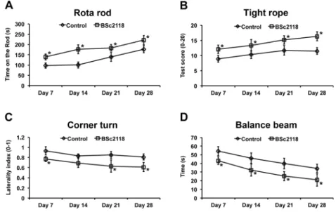
Systemic BSc2118 Delivery Enhances Post-ischemic Neurological Recovery
Rotarod, tight rope, corner turn, and balance beam tests re-vealed that repeated BSc2118 delivery over 7 days induced sustained improvement of motor and coordination recovery that persisted up to 28 days post-stroke (Fig.3). Conversely, single BSc2118 delivery 9 h after stroke did not result in sustained motor coordination improvement (data not shown). Fig. 1 Systemic delivery of proteasome inhibitor BSc2118 protects against focal cerebral ischemia. a Infarct volume in mice receiving single intraperitoneal (i.p.) injections of vehicle (control) or BSc2118 (30 mg/kg) at various time points after intraluminal middle cerebral artery (MCA) occlusion followed by animal sacrifice at 4 days post-stroke. Representative 2,3,5-triphenyltetrazolium chloride (TTC) stainings are shown from mice receiving single vehicle or BSc2118 injections 9 h after reperfusion. b Neuronal survival in the striatum evaluated by NeuN im-munohistochemistry in mice receiving single i.p. injections of vehicle or BSc2118 (30 mg/kg) 9 h after MCA occlusion (single delivery) or daily i.p. injections of vehicle or BSc2118 over 7 days starting 9 h after MCA occlusion (repeated delivery). Representative microphotographs of NeuN+ cells are shown from mice receiving repeated vehicle or
BSc2118 injections. Scale bars=50 μm. *p<0.05 compared with corre-sponding control mice (n=7 mice per group (in a)/n=12–14 mice per group (in b))
Systemic BSc2118 Delivery Does Not Influence Cerebral Proteasome Activity to a Major Extent
Since BBB permeability of BSc2118 is low [20,21], we next compared cerebral proteasome activity after intracerebral (i.e., intrastriatal) and systemic (i.e., i.p.) BSc2118 delivery. While a single intracerebral bolus injection of BSc2118 resulted in near-complete proteasome inhibition in sham-operated and ischemic mice that persisted over up to 48 h (Fig. 4a),
systemic BSc2118 delivery only mildly affected cerebral pro-teasome activity (Fig.4b).
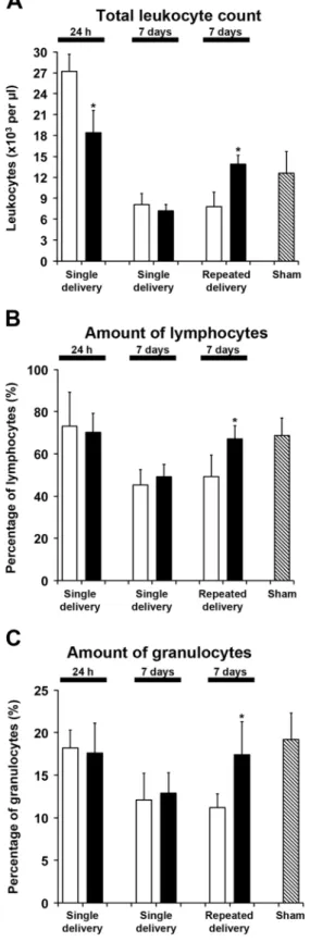
Systemic BSc2118 Delivery Attenuates Acute
Post-ischemic Leukocytosis and Reverses Subsequent Peripheral Immunosuppression
Considering that focal cerebral ischemia induces inflammato-ry responses in the brain [28,29] and peripheral blood, name-ly, acute leukocytosis followed by subsequent immunosup-pression [30,31], we next evaluated immune responses in-duced by BSc2118. Interestingly, systemic BSc2118 delivery did not affect cerebral post-stroke leukocyte infiltration, nei-ther 24 h nor 7 days post-stroke (Fig. 5), but attenuated the acute stroke-induced peripheral leukocytosis 24 h post-stroke (Fig.6a). Furthermore, repeated but not single BSc2118 de-livery reversed the stroke-induced immunosuppression, re-storing total leukocyte counts, relative lymphocyte amounts and relative granulocyte amounts in the blood at 7 days post-stroke (Fig.6a–c).
Systemic BSc2118 Delivery Does Not Influence Post-ischemic Angioneurogenesis
Since intracerebral BSc2118 delivery enhances post-ischemic neurogenesis and angiogenesis [21], we finally examined whether systemic BSc2118 administration induced post-stroke neuroregeneration despite limited brain bioavailability. Analysis of proliferating cells by BrdU immunohistochemis-try within the ischemic lesion site did not reveal any difference between vehicle-treated control mice and mice receiving BSc2118 over 7 days (Fig.7a). Likewise, differentiation anal-ysis of BrdU+cells did not detect any differences in the co-expression of the neuronal markers Dcx and NeuN and the endothelial marker CD31 (Fig. 7b–d). Of note, analysis of BrdU+cells (47.1±12.5 in controls vs. 43.3±9.4 per mm2in BSc2118-treated mice), determination of co-expression be-tween BrdU and Dcx (8.8±4.3 % in controls vs. 7.9±6.2 % in BSc2118-treated mice), as well as co-expression between BrdU and NeuN (no co-expression observed in both groups) within the ipsilateral subventricular zone did not show any significant difference, either.
Discussion
The present study demonstrates for the first time that repeated, systemic delivery of the proteasome inhibitor BSc2118 in-duces sustained neuroprotection after focal cerebral ischemia, characterized by reduced infarct volume, decreased brain ede-ma, and enhanced BBB integrity, resulting in neurological recovery that persists in the post-acute stroke phase. Albeit intracerebral proteasome activity was only slightly reduced Fig. 2 Systemic delivery of BSc2118 reduces post-ischemic brain edema
and blood-brain-barrier permeability without affecting HIF-1α abun-dance. a Brain edema as evaluated by TTC staining in mice receiving single intraperitoneal (i.p.) injections of vehicle (control) or BSc2118 (30 mg/kg) 9 h after intraluminal MCA occlusion followed by animal sacrifice at 4 days post-stroke. b Blood-brain-barrier (BBB) permeability, revealed by extravasation of Evans blue in mice receiving i.p. injections of vehicle (control) or BSc2118 (30 mg/kg) 9 h after MCA occlusion followed by animal sacrifice at 24 h post-stroke. c Matrix metalloprotease-9 (MMP-9) activity assessed by gelatinase zymography in ischemic brain tissue of mice receiving i.p. injections of vehicle (control) or BSc2118 (30 mg/kg) 9 h after MCA occlusion followed by animal sacrifice at 24 h post-stroke. d HIF-1α abundance analyzed by Western blots in ischemic brain tissue of mice receiving i.p. injections of vehicle (control) or BSc2118 (30 mg/kg) 9 h after MCA occlusion follow-ed by animal sacrifice at 24 h post-stroke. *p<0.05 comparfollow-ed with cor-responding control group (n=7 mice per group (in a)/n=4 mice per group (in b–d))
by systemic BSc2118 delivery, distinct changes of peripheral immune responses were noted after systemic BSc2118 deliv-ery, namely, the prevention of stroke-induced acute leukocy-tosis and reversal of subsequent immunosuppression in pe-ripheral blood. Cerebral neurogenesis and angiogenesis were not influenced by BSc2118 delivery, arguing in favor of a neuroprotective rather than true restorative action. Our study provides a new mechanism of action of proteasome inhibitors in models of ischemic stroke.
Although proteasome inhibition is a double-edged sword [32], neuroprotection induced by proteasome inhibitors has previously been described in experimental models of focal cerebral ischemia [9–16]. The clinical relevance of these find-ings, however, was limited. Due to severe side effects, phar-macological inhibitors could not repeatedly be delivered, and as such, observation periods of these studies were limited to a maximum of 7 days only. More recently, we locally adminis-tered the later generation proteasome inhibitor BSc2118 that lacks such side effects using an intracerebral delivery ap-proach, demonstrating that proteasome inhibition protected against stroke when administered up to 12 h after focal cere-bral ischemia [21]. These experiments extended therapeutic windows of proteasome inhibitors in earlier studies, which had been in the range of a maximum of 4–6 h. The local intracerebral BSc2118 delivery yielded almost total protea-some inhibition, resulting in stabilization of HIF-1α followed by increased neurogenesis and angiogenesis that translated into neurological recovery which still persisted after as long as 3 months post-stroke.
Since intracerebral application is not feasible in the clinical setting, we now evaluated the effects of systemic BSc2118 delivery. Previous data from our group showed that BSc2118 only has limited bioavailability in the brain after systemic application [20]. In line with this, cerebral protea-some activity was only mildly inhibited in the present study Fig. 3 Systemic BSc2118
delivery induces sustained motor coordination recovery after focal cerebral ischemia. a Rotarod, b tight rope, c corner turn, and d balance beam test in mice receiving daily intraperitoneal injections of vehicle (control) or BSc2118 (30 mg/kg) over 7 days starting at 9 h after intraluminal MCA occlusion. *p<0.05 compared with corresponding control group (n=12–14 mice per group)
Fig. 4 Systemic BSc2118 delivery does not influence cerebral proteasome activity to a major extent. Proteasome activity in brain lysates of sham operated (open columns) and ischemic (closed columns) mice 1 to 48 h after single a intracerebral (i.e., intrastriatal) or b intraperitoneal (i.p.) BSc2118 (30 mg/kg) delivery. Note the near-complete inhibition of cerebral proteasome activity after local intracere-bral but not systemic i.p. delivery. *p<0.05 compared with baseline (n=4 mice per group)
following i.p. delivery with a maximum inhibition 24 h after stroke. This is in line with in vitro data, demonstrating a de-cline of BSc2118 inhibition potency in microsomal fractions after 24 h by approximately 75 % despite stability in aqueous solution [20,21]. The latter observations prompted us to use once daily injections for repeated BSc2118 delivery. We were
surprised to see that systemic BSc2118 delivery induced sustained neuroprotection when delivered up to 9 h post-stroke. Yet, sustained neuroprotection associated with neuro-logical recovery was observed only after repeated and not after single BSc2118 delivery. Since systemic BSc2118 delivery only slightly reduced cerebral proteasome activity, it is not surprising that cerebral HIF-1α abundance was not affected in this study. Our data indicate that systemic BSc2118 delivery acts via a different mode of action than intracerebral applica-tion. Notably, brain edema, BBB permeability, and cerebral MMP-9 activity were reduced by systemic BSc2118 adminis-tration. These findings argue in favor of a stabilization of the neurovascular unit that accompanies the BSc2118-induced promotion of neuronal survival.
Furthermore, distinct alterations of peripheral immune re-sponses were noted after systemic BSc2118 delivery, namely, a prevention of acute post-stroke leukocytosis and reversal of subsequent immunosuppression in the peripheral blood. Pe-ripheral immune responses are a well-established component in the progression of ischemic injury [28,29]. Rapid activa-tion of the immune response associated with leukocytosis in peripheral blood is followed by a transient phase of immuno-suppression [30,31,33]. Although proteasome inhibition is known to modulate inflammation under various pathological conditions [34], the here-reported prevention of acute post-stroke leukocytosis and reversal of subsequent immunosup-pression, i.e., the normalization of total leukocyte counts and more specifically lymphocyte and granulocyte amounts, is new. It is noteworthy that unlike in other proteasome inhibi-tion studies [9,13,14,21,35], alterations in the brain inflam-matory response were not noticed after systemic BSc2118 delivery. Evaluating precise immune mechanisms underlying BSc2118-induced neuroprotection was beyond the scope of this work. Thus, we did not analyze subsets of immune cells in the brain. It is conceivable that BSc2118 induced changes in the composition of brain immune cell infiltrates, which might explain its neuroprotective action. Further studies are needed on this issue. Moreover, the present study was done on ado-lescent and healthy animals not precisely reflecting the clinical situation. As a matter of fact, age and/or diseases critically affect post-ischemic immune responses, which has been shown for hypercholesterolemic mice by our group before
Fig. 5 Systemic BSc2118 delivery does not influence post-ischemic cerebral inflammatory response. Open columns indicate controls and closed columns indicate treatment with BSc2118. Number of brain a leukocytes, b lymphocytes, and c granulocytes, evaluated by flow cytom-etry in ischemic hemispheres of mice receiving single i.p. injections of vehicle (control) or BSc2118 (30 mg/kg) at 9 h after MCA occlusion (single delivery) or daily i.p. injections of vehicle or BSc2118 (30 mg/kg) over 7 days starting at 9 h after MCA occlusion (repeated delivery), followed by animal sacrifice at 24 h (single delivery only) or 7 days post-stroke (single or repeated delivery). No differences were detected between groups (n=6 animals per group)[26]. Studies using the latter mice strain were, however, be-yond the scope of the present work and might have to be performed in the future.
In line with an increased HIF-1α abundance due to inhibi-tion of cerebral proteasome activity, the local intracerebral
delivery of BSc2118 was previously shown to stimulate cere-bral cell proliferation, neurogenesis, and angiogenesis after focal cerebral ischemia [21]. In contrast to local intracerebral BSc2118 delivery, systemic BSc2118 administration did not
Fig. 6 Systemic BSc2118 delivery attenuates acute post-ischemic leu-kocytosis and reverses subsequent peripheral immunosuppression in the blood. Open columns indicate controls and closed columns indicate treat-ment with BSc2118. a Total leukocyte count, b relative amount of lym-phocytes, and c relative amount of granulocytes in peripheral blood of mice receiving single i.p. injections of vehicle (control) or BSc2118 (30 mg/kg) at 9 h after MCA occlusion (single delivery) or daily i.p. injections of vehicle or BSc2118 (30 mg/kg) over 7 days starting at 9 h after MCA occlusion (repeated delivery), followed by animal sacrifice at 24 h (single delivery only) or 7 days post-stroke (single or repeated delivery). Cell counts and percentages determined in sham operated mice are also shown. *p<0.05 compared with corresponding control group (n=6 animals per group)Fig. 7 Systemic BSc2118 delivery does not influence post-ischemic angioneurogenesis. a Cell proliferation evaluated by BrdU immunohisto-chemistry, b neurogenesis assessed by BrdU co-expression with the im-mature neuronal marker doublecortin (Dcx), c neurogenesis analyzed by BrdU co-expression with the mature neuronal marker NeuN, and d angiogenesis examined by BrdU co-expression with the endothelial marker CD31 in the ischemic striatum of mice receiving daily i.p. injec-tions of vehicle (control) or BSc2118 (30 mg/kg) over 7 days starting at 9 h after MCA occlusion, followed by animal sacrifice at 28 days post-stroke. No differences were detected between groups (n=12–14 mice per group)
stimulate post-ischemic cell proliferation, neurogenesis, and angiogenesis, most likely as a consequence of lack of HIF-1α stabilization. HIF-1α is known to stimulate post-ischemic neurogenesis and angiogenesis in otherwise hostile cellular microenvironments [36–39]. Although neurogenesis and an-giogenesis are stimulated by ischemia [40–43], their signifi-cance for neurological recovery is still a matter of debate due to low survival and differentiation rates of newborn cells [44]. Considering that systemic BSc2118 delivery promoted neuro-nal survival without affecting neurogenesis and angiogenesis, this delivery paradigm appears to represent a neuroprotective rather than true neurorestorative strategy. The modulation of peripheral immune responses might represent an attractive target for the clinical translation of proteasome inhibitors.
Acknowledgments This study was supported by grants from the Ger-man Research Council (DFG, No. HE3173/2-2 and No. HE3173/3-1 to DMH) and a grant from the Scientific and Technological Research Coun-cil of Turkey (TUBITAK, No. 2221 to TRD).
Compliance with Ethical Standards All studies were performed ac-cording to local government authorities.
Conflict of Interest The authors declare that they have no competing interests.
References
1. Inobe T, Matouschek A (2014) Paradigms of protein degradation by the proteasome. Curr Opin Struct Biol 24:156–164. doi:10.1016/j. sbi.2014.02.002
2. Kniepert A, Groettrup M (2014) The unique functions of tissue-specific proteasomes. Trends Biochem Sci 39(1):17–24. doi:10. 1016/j.tibs.2013.10.004
3. Buac D, Shen M, Schmitt S, Kona FR, Deshmukh R, Zhang Z, Neslund-Dudas C, Mitra B et al (2013) From bortezomib to other inhibitors of the proteasome and beyond. Curr Pharm Des 19(22): 4025–4038
4. Xolalpa W, Perez-Galan P, Rodriguez MS, Roue G (2013) Targeting the ubiquitin proteasome system: beyond proteasome inhibition. Curr Pharm Des 19(22):4053–4093
5. Jankowska E, Stoj J, Karpowicz P, Osmulski PA, Gaczynska M (2013) The proteasome in health and disease. Curr Pharm Des 19(6):1010–1028
6. Elliott PJ, Ross JS (2001) The proteasome: a new target for novel drug therapies. Am J Clin Pathol 116(5):637–646. doi:10.1309/ 44HW-5YCJ-FLLP-3R56
7. Kukan M (2004) Emerging roles of proteasomes in ischemia-reperfusion injury of organs. J Physiol Pharmacol 55(1 Pt 1):3–15 8. Di Napoli M, McLaughlin B (2005) The ubiquitin-proteasome sys-tem as a drug target in cerebrovascular disease: therapeutic potential of proteasome inhibitors. Curr Opin Investig Drugs 6(7):686–699 9. Berti R, Williams AJ, Velarde LC, Moffett JR, Elliott PJ, Adams J,
Yao C, Dave JR et al (2003) Effect of the proteasome inhibitor MLN519 on the expression of inflammatory molecules following middle cerebral artery occlusion and reperfusion in the rat. Neurotox Res 5(7):505–514
10. Buchan AM, Li H, Blackburn B (2000) Neuroprotection achieved with a novel proteasome inhibitor which blocks NF-kappaB activa-tion. Neuroreport 11(2):427–430
11. Henninger N, Sicard KM, Bouley J, Fisher M, Stagliano NE (2006) The proteasome inhibitor VELCADE reduces infarction in rat models of focal cerebral ischemia. Neurosci Lett 398(3):300–305. doi:10.1016/j.neulet.2006.01.015
12. Phillips JB, Williams AJ, Adams J, Elliott PJ, Tortella FC (2000) Proteasome inhibitor PS519 reduces infarction and attenuates leu-kocyte infiltration in a rat model of focal cerebral ischemia. Stroke 31(7):1686–1693
13. Williams AJ, Hale SL, Moffett JR, Dave JR, Elliott PJ, Adams J, Tortella FC (2003) Delayed treatment with MLN519 reduces in-farction and associated neurologic deficit caused by focal ischemic brain injury in rats via antiinflammatory mechanisms involving nuclear factor-kappaB activation, gliosis, and leukocyte infiltration. J Cereb Blood Flow Metab 23(1):75–87
14. Zhang L, Zhang ZG, Liu X, Hozeska A, Stagliano N, Riordan W, Lu M, Chopp M (2006) Treatment of embolic stroke in rats with bortezomib and recombinant human tissue plasminogen activator. Thromb Haemost 95(1):166–173
15. Zhang L, Zhang ZG, Zhang RL, Lu M, Adams J, Elliott PJ, Chopp M (2001) Postischemic (6-hour) treatment with recombinant human tissue plasminogen activator and proteasome inhibitor PS-519 re-duces infarction in a rat model of embolic focal cerebral ischemia. Stroke 32(12):2926–2931
16. Zhang Y, Xiong M, Yan RQ, Sun FY (2010) Mutant ubiquitin-mediated beta-secretase stability via activation of caspase-3 is relat-ed to beta-amyloid accumulation in ischemic striatum in rats. J Cereb Blood Flow Metab 30(3):566–575. doi:10.1038/jcbfm. 2009.228
17. Ruschak AM, Slassi M, Kay LE, Schimmer AD (2011) Novel proteasome inhibitors to overcome bortezomib resistance. J Natl Cancer Inst 103(13):1007–1017. doi:10.1093/jnci/djr160
18. Braun HA, Umbreen S, Groll M, Kuckelkorn U, Mlynarczuk I, Wigand ME, Drung I, Kloetzel PM et al (2005) Tripeptide mimetics inhibit the 20 S proteasome by covalent bonding to the active thre-onines. J Biol Chem 280(31):28394–28401. doi:10.1074/jbc. M502453200
19. Sterz J, Jakob C, Kuckelkorn U, Heider U, Mieth M, Kleeberg L, Kaiser M, Kloetzel PM et al (2010) BSc2118 is a novel proteasome inhibitor with activity against multiple myeloma. Eur J Haematol 85(2):99–107. doi:10.1111/j.1600-0609.2010.01450.x
20. Mlynarczuk-Bialy I, Doeppner TR, Golab J, Nowis D, Wilczynski GM, Parobczak K, Wigand ME, Hajdamowicz M et al (2014) Biodistribution and efficacy studies of the proteasome inhibitor BSc2118 in a mouse melanoma model. Transl Oncol 7(5):570– 579. doi:10.1016/j.tranon.2014.07.002
21. Doeppner TR, Mlynarczuk-Bialy I, Kuckelkorn U, Kaltwasser B, Herz J, Hasan MR, Hermann DM, Bahr M (2012) The novel pro-teasome inhibitor BSc2118 protects against cerebral ischaemia through HIF1A accumulation and enhanced angioneurogenesis. Brain 135(Pt 11):3282–3297. doi:10.1093/brain/aws269
22. Doeppner TR, Kaltwasser B, Bahr M, Hermann DM (2014) Effects of neural progenitor cells on post-stroke neurological impairment— a detailed and comprehensive analysis of behavioral tests. Front Cell Neurosci 8:338. doi:10.3389/fncel.2014.00338
23. Chiba Y, Sasayama T, Miyake S, Koyama J, Kondoh T, Hosoda K, Kohmura E (2008) Anti-VEGF receptor antagonist (VGA1155) reduces infarction in rat permanent focal brain ischemia. Kobe J Med Sci 54(2):E136–146
24. Doeppner TR, Kaltwasser B, ElAli A, Zechariah A, Hermann DM, Bahr M (2011) Acute hepatocyte growth factor treatment induces long-term neuroprotection and stroke recovery via mechanisms in-volving neural precursor cell proliferation and differentiation. J
Cereb Blood Flow Metab 31(5):1251–1262. doi:10.1038/jcbfm. 2010.211
25. Doeppner TR, Kaltwasser B, Teli MK, Bretschneider E, Bahr M, Hermann DM (2014) Effects of acute versus post-acute systemic delivery of neural progenitor cells on neurological recovery and brain remodeling after focal cerebral ischemia in mice. Cell Death Dis 5, e1386. doi:10.1038/cddis.2014.359
26. Herz J, Hagen SI, Bergmuller E, Sabellek P, Gothert JR, Buer J, Hansen W, Hermann DM et al (2014) Exacerbation of ischemic brain injury in hypercholesterolemic mice is associated with pro-nounced changes in peripheral and cerebral immune responses. Neurobiol Dis 62:456–468. doi:10.1016/j.nbd.2013.10.022
27. Candelario-Jalil E, Yang Y, Rosenberg GA (2009) Diverse roles of matrix metalloproteinases and tissue inhibitors of metalloprotein-ases in neuroinflammation and cerebral ischemia. Neuroscience 158(3):983–994. doi:10.1016/j.neuroscience.2008.06.025
28. Macrez R, Ali C, Toutirais O, Le Mauff B, Defer G, Dirnagl U, Vivien D (2011) Stroke and the immune system: from pathophys-iology to new therapeutic strategies. Lancet Neurol 10(5):471–480. doi:10.1016/S1474-4422(11)70066-7
29. Gelderblom M, Leypoldt F, Steinbach K, Behrens D, Choe CU, Siler DA, Arumugam TV, Orthey E et al (2009) Temporal and spatial dynamics of cerebral immune cell accumulation in stroke. Stroke 40(5):1849–1857. doi:10.1161/STROKEAHA.108.534503
30. Dirnagl U, Klehmet J, Braun JS, Harms H, Meisel C, Ziemssen T, Prass K, Meisel A (2007) Stroke-induced immunodepression: ex-perimental evidence and clinical relevance. Stroke 38(2 Suppl): 770–773. doi:10.1161/01.STR.0000251441.89665.bc
31. Famakin BM (2014) The immune response to acute focal cerebral ischemia and associated post-stroke immunodepression: a focused review. Aging Dis 5(5):307–326. doi:10.14336/AD.2014.0500307
32. Ge P, Luo Y, Liu CL, Hu B (2007) Protein aggregation and protea-some dysfunction after brain ischemia. Stroke 38(12):3230–3236. doi:10.1161/STROKEAHA.107.487108
33. Offner H, Subramanian S, Parker SM, Afentoulis ME, Vandenbark AA, Hurn PD (2006) Experimental stroke induces massive, rapid activation of the peripheral immune system. J Cereb Blood Flow Metab 26(5):654–665. doi:10.1038/sj.jcbfm.9600217
34. Dahlmann B (2007) Role of proteasomes in disease. BMC Biochem 8(Suppl 1):S3. doi:10.1186/1471-2091-8-S1-S3
35. Sinn DI, Lee ST, Chu K, Jung KH, Kim EH, Kim JM, Park DK, Song EC et al (2007) Proteasomal inhibition in intracerebral hem-orrhage: neuroprotective and anti-inflammatory effects of bortezomib. Neurosci Res 58(1):12–18. doi:10.1016/j.neures. 2007.01.006
36. Cunningham LA, Candelario K, Li L (2012) Roles for HIF-1alpha in neural stem cell function and the regenerative response to stroke. Behav Brain Res 227(2):410–417. doi:10.1016/j.bbr.2011.08.002
37. Mazumdar J, O’Brien WT, Johnson RS, LaManna JC, Chavez JC, Klein PS, Simon MC (2010) O2 regulates stem cells through Wnt/ beta-catenin signalling. Nat Cell Biol 12(10):1007–1013. doi:10. 1038/ncb2102
38. Tsai YW, Yang YR, Wang PS, Wang RY (2011) Intermittent hyp-oxia after transient focal ischemia induces hippocampal neurogenesis and c-Fos expression and reverses spatial memory deficits in rats. PLoS ONE 6(8), e24001. doi:10.1371/journal. pone.0024001
39. Shi H (2009) Hypoxia inducible factor 1 as a therapeutic target in ischemic stroke. Curr Med Chem 16(34):4593–4600
40. Arvidsson A, Collin T, Kirik D, Kokaia Z, Lindvall O (2002) Neuronal replacement from endogenous precursors in the adult brain after stroke. Nat Med 8(9):963–970
41. Tobin MK, Bonds JA, Minshall RD, Pelligrino DA, Testai FD, Lazarov O (2014) Neurogenesis and inflammation after ischemic stroke: what is known and where we go from here. J Cereb Blood Flow Metab 34(10):1573–1584. doi:10.1038/jcbfm.2014.130
42. Hermann DM, Chopp M (2012) Promoting brain remodelling and plasticity for stroke recovery: therapeutic promise and potential pitfalls of clinical translation. Lancet Neurol 11(4):369–380. doi:
10.1016/S1474-4422(12)70039-X
43. Xiong Y, Mahmood A, Chopp M (2010) Angiogenesis, neurogenesis and brain recovery of function following injury. Curr Opin Investig Drugs 11(3):298–308
44. Yu TS, Washington PM, Kernie SG (2014) Injury-induced neurogenesis: mechanisms and relevance. Neuroscientist. doi:10. 1177/1073858414563616