EXPERIMENTAL STUDY
The effect of diosmin on pancreatic injury induced by hepatic
ischemia reperfusion in rats
Serin Kilicoglu S
1, Tanrikulu Y
2, Kismet K
3, Devrim E
4, Erel S
3, Sen Tanrikulu C
5, Aydogan A
3,
Tasova V
3, Sabuncuoglu MZ
3, Kilicoglu B
3Department of Histology and Embriology, Ufuk University Faculty of Medicine, Cankaya, Ankara, Turkey.
Abstract: Background: Hepatic ischemia-reperfusion causes histological injury to the pancreatic cells during transplantation, trauma and emergency surgery. We investigated the effects of diosmin, a phlebotrophic drug with antioxidant and antiinfl ammatory effects, on pancreatic injury in the experimental liver ischemia-reperfusion model. Materials and methods: Forty rats were divided into the four groups: sham (Group 1), control (Group 2), perop-erative diosmin (50 mg/kg) treatment (Group 3) and preopperop-erative 10-day diosmin (50 mg/kg) treatment group (Group 4). Ischemia-reperfusion model was carried out by clamping the hepatic pedicle for 60 min and then reperfusing the liver for 90 min. At the end of the procedures, blood and pancreas tissue samples were obtained for biochemical and histopathological assessment.
Results: According to the results of liver function tests, amylase and the plasma and pancreatic tissue oxidative stress parameters, there was a signifi cant difference between the control and other groups. Histopathologically; the specimens of the Group 2 showed specifi c morphological abnormalities. The groups 3 and 4 showed the pancreas histomorphology similar to the sham group. Pathological scores were signifi cantly different between the Group 2 and other groups.
Conclusions: Diosmin can be administered for a protection of destructive effects of hepatic ischemia–reperfusion injury on pancreas both emergent and elective hepatic surgical operations in which possible ischemic periods were expected (Fig. 1, Tab. 3, Ref. 33). Full Text in PDF www.elis.sk.
Key words: diosmin, liver, ischemia-reperfusion, pancreatic injury.
1Department of Histology and Embriology, Ufuk University Faculty of
Medicine, Cankaya, Ankara, Turkey, 22ndGeneral Surgery Department,
Erzurum Area Training and Research Hospital Yakutiye, Erzurum, Turkey,
34thGeneral Surgery Department, Ankara Training and Research
Hospi-tal Ulucanlar, Ankara, Turkey,4Department of Biochemistry, Ankara
Uni-versity Faculty of Medicine, Cankaya, Ankara, Turkey, and 5Emergency
Medicine Department, Erzurum Area Training and Research Hospital Ya-kutiye, Erzurum, Turkey
Address for correspondence: B. Kilicoglu, Ankara Training and Research Hospital General Surgery Department, Ulucanlar, Ankara, 06340, Turkey. Phone: +90.3122044201
Hepatic ischemia-reperfusion injury is ordinarily at the time of the repair of extensive liver trauma, shock, tumour resection and liver transplantation (1). Prolonged organ ischemia is character-ized by a lack of tissue oxygen. The several situations occur at the lack of tissue oxygen such as adenosine triphosphate (ATP) deple-tion, derangements in calcium homeostasis, conversion of cellu-lar metabolism to anaerobic pathways, loss of cellucellu-lar function and cell death. However, once the blood fl ow is re-established, reperfusion enhances the injury caused by the ischemic period, aggravating the damage caused at the cellular level. Ischemia-reperfusion injury in various organs was recognized as a complex phenomenon. The various factors play a role in the pathogenesis of ischemia reperfusion injury such as increased infl ammatory response, reactive oxygen species, cytokine release and neutro-phile activation (2, 3).
There are many techniques for liver infl ow occlusion dur-ing hepatectomy; continuous or intermittent Prdur-ingle manoeuvre is used most frequently (4). The Pringle manoeuvre induces not only a stagnation of the portal venous blood, thereby increas-ing portal venous pressure, but also an ischemic liver damage. There is an increasing evidence that the changes in the portal circulation during hepatic ischemia reperfusion also causes dam-ages in other organs, such as the intestine, lung, heart (5–7). In addition, an increased portal venous pressure may induce pan-creatitis and contribute to mortality (8). Nevertheless, the way how the pancreas responds to congestion and acute elevation of portal venous pressure by portal vein occlusion is not well under-stood (9, 10).
Furthermore, the relationship between liver resection and hy-peramylasemia are examined. Hyhy-peramylasemia is a well-docu-mented sequel of liver surgery, and has been demonstrated follow-ing other types of abdominal surgery and even extra-abdominal surgery (5, 6). In addition, hyperamylasemia occurs in the process of an acute pancreatitis and in the postoperative period of various types of surgery, including hepatic surgery (8, 11, 12).
Diosmin is an antioxidant, phlebotrophic and vascular protec-tive fl avonoid, which increases venous tone, improves lymphatic drainage and reduces capillary hyperpermeability. It also inhib-its the activation, migration and adhesion of leukocytes, which leads to a reduction in the release of infl ammatory mediators and
thereby a reduction in capillary hyperpermeability. The antioxi-dant activity of diosmin has been shown in many studies (13–15). In the present study, we investigated the effects of diosmin on pancreatic injury in the experimental liver ischemia-reperfusion model.
Materials and methods
Animals
Forty Wistar-Albino female rats, weighing 250 ± 30 g, were housed under a constant temperature (21 ± 2 °C) individually in wire cages with 12 h light-dark cycle, fed water and rodent chow ad libitum. Twelve hours before anaesthesia, animals were de-prived of food, but had free access to water 2 h before anaesthesia. No enteral or parenteral antibiotics were administered. The pro-cedures in this experimental study were performed in accordance with the National Guidelines for The Use and Care of Laboratory Animals and approved by the Animal Ethics Committee of Ankara Research and Training Hospital.
Study groups and operative procedure
Rats were randomly divided into the four groups each includ-ing 10 animals: SHAM group, Control group (ischemia-reperfu-sion) and treatment groups (peroperative group and preoperative group). Animals were anaesthetized by intramuscular injection of 80 mg/kg ketamine hydrochloride (Ketalar; Parke- Davis, Istan-bul, Turkey) and 20 mg/kg xylasine (Rompun, Bayer, IstanIstan-bul, Turkey). After the abdomen was shaved and disinfected, a mid-line incision was made and rats underwent either sham surgery or ischemia-reperfusion. Ischemia was carried out by clamping for 60 min with a microvascular “bulldog” clamp of hepatic pedicle. After the ischemic period, liver was reperfused by opening the clamp, and reperfusion was achieved for 90 min. In the perop-erative treament group, diosmin was given to the rats, just after ischemia induction at a dose of 50 mg/kg in gavage form with nasogastric tube; therefore the peak plasma levels of diosmin were achieved before reperfusion was initiated. In the preopera-tive treatment group, 50 mg/kg/day diosmin (Vendios; Bilim, Is-tanbul, Turkey) was given to the rats one dose daily for ten days before the operation, in gavage form with nasogastric tube (7 Gauge feeding tube) that was inserted daily and taken off after drug administration.
At the end of the procedures, blood and pancreas tissue sam-ples were obtained for biochemical and histopathological as-sessment.
Biochemical analyses
Plasma alanine aminotransferase (ALT), aspartate aminotrans-ferase (AST) and amylase levels were measured for evaluating the liver functions by using Olympus Au 640 autoanalyzer. To assess oxidative injury, malondialdehyde (MDA) levels, glutathione peroxidase (GSH-Px) and xanthine oxidase (XO) enzyme activi-ties were determined in the blood samples and malondialdehyde (MDA) levels and glutathione peroxidase (GSH-Px) enzyme ac-tivities were determined in the pancreas samples.
Evaluation of oxidative stress
After sacrifi cation of the animals, pancreas samples were re-moved and kept on an ice bath until homogenization. The sample of pancreas was fi rst washed with distilled water, the tissues were homogenized in (20 % w/v, approximately 1 g in 5 ml for each) physiological saline, then they were centrifuged 4000 x g for 15 min and upper clear supernatants were used in the assays. All the procedures were performed at +4 °C throughout the experiments. Protein level of the clear supernatants was studied by Lowry’s method (16), and then they were adjusted to equal concentrations before the other analyses. All the results were expressed as unit/ mg protein for those of pancreas tissues. Malondialdehyde (MDA) level (nmol/mg) and glutathione peroxidase (GSH-Px) enzyme activity (mlU/mg) were measured in the supernatants.
MDA level was measured by thiobarbituric acid reactive sub-stances method. GSH-Px activity was measured by following changes in NADPH absorbance at 340 nm.
Histological evaluation
For the light microscopic analyses, the samples obtained from the pancreas were fi xed in 10% neutral buffered formaline solu-tion for 2 days. Tissues were washed in fl owing water and were dehydrated with rising concentrations of ethanol (50 %, 75 %, 96 %, 100 %). After dehydration, specimens were put into xylene to obtain transparency and were then infi ltrated with and embed-ded in paraffi n. Embedembed-ded tissues were cut into sections of 5 μm thickness by Leica RM 2125 RT and stained with hematoxylin and eosin. Histopathologic examinations were performed by two histopathologists, blinded to the study design. Photographs were taken by Olympus BX-51 microscope with Olympus DP-71 photo attachment. The histological grading of edema was made using a scale ranging from 0 to 3 (0 = no edema, 1 = interlobular edema, 2 = interlobular and moderate intralobular edema, and 3 = inter lobular edema and severe intralobular edema). Leukocytic infi ltration was also graded from 0 to 3 (0 = absent, 1 = scarce perivascular infi l-tration, 2 = moderate perivascular and scarce diffuse infi ll-tration, 3 = abundant diffuse infi l tration). Grading of vacuolization was based on the appropriate percentage of acinar cells in volved: 0 = absent, 1 = less than 25 %, 2 = 25 –50 % and 3 = more than 50 % of acinar cells. Haemorrhage was graded as: 0 = no haemorrhage, 1 = 1 –2 haemorrhagic foci per slide, 2 = 3 –5 hae morrhagic foci per slide, 3 = more than 5 haemo rrhagic foci per slide. Necrosis was graded as: 0 = no necrosis, 1= less than 15 % of pancreatic cells involved, 2 = 15 –35 % of cells involved, 3 = more than 35 % of cells involved.
Statistical analysis
Data analysis was performed using the SPSS 15.0 package program. Data were presented as the mean ± standard deviation. Any possible differences among the groups were evaluated by the One-Way ANOVA or Kruskal Wallis variance analysis, where appropriate. When the p-value from the variance analysis was statistically signifi cant, the Mann –Whitney U multiple compari-son test was used to know which group differs from the others. Furthermore, the Student’s t-test variance analysis was used for
the evaluation of the histopathological results. All results were accepted as statistically signifi cant when p<0.05.
Results
All rats were sacrifi ced on postoperative day 10. No rat died during the experimental period.
Biochemical analyses
The results of liver function tests of the groups are summa-rized in the Table 1. There was a signifi cant difference between the SHAM group and the Control group according to the levels of AST, ALT and amylase (p < 0.001 for AST and ALT, p = 0.001 for amylase). There was a signifi cant difference between the Control group and the Peroperative treatment group according to the levels of AST, ALT and amylase (p < 0.05 for all). There was a signifi cant difference between the Control group and the Preoperative treat-ment group according to the levels of AST, ALT and amylase (p < 0.001 for all). There was a signifi cant difference between the Sham group and the Peroperative treatment group according to the levels of AST (p < 0.001). There was a signifi cant difference between the Sham group and the Peroperative treatment group according to the levels of AST and ALT (p < 0.001 for all).There was no signifi cant difference between the Sham group and the treatment groups according to the levels of amylase. There was no signifi -cant difference between the Peroperative treatment group and the Preoperative treatment group according to the levels of AST (p = 0.940). Serum ALT and amylase levels were higher in the Perop-erative group than the PreopPerop-erative treatment group and the dif-ference was signifi cant (p < 0.001 for AST, p < 0.05 for amylase).
Oxidative stress
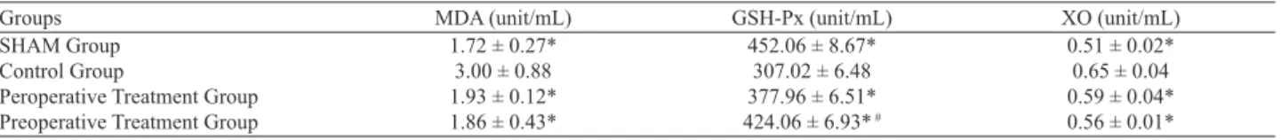
The plasma levels of MDA, GSH-Px and XO are summarized in the Table 2. There was a signifi cant difference between the Con-trol group and other groups according to the MDA, GSH-Px and XO levels (p < 0.05 for all). There was not a signifi cant difference between the Peroperative treatment group and the Preoperative treatment group according to the MDA and XO levels (p > 0.05),
GSH-Px levels were higher in the Preoperative treatment group than in the Peroperative treatment group and the difference was signifi cant (p < 0.001). There was not a signifi cant difference be-tween the Sham group and the treatments groups according to the MDA, GSH-Px and XO levels (p > 0.05 for all).
The pancreas tissue levels of MDA and GSH-Px are summa-rized in the Table 3. There was not a statistically signifi cant dif-ference between the SHAM group and the Control group accord-ing to MDA and GSH-Px levels (p > 0.05 for all). There was a statistically signifi cant difference between the Control group and the Peroperative treatment group according to MDA and GSH-Px levels (p < 0.05 for MDA, p = 0.001 for GSH-Px). There was not a statistically signifi cant difference between the Control group and the Preoperative treatment group according to MDA and XO levels (p > 0.05). GSH-Px levels were higher in the Preoperative treatment group than in the Control group and the difference was signifi cant (p < 0.05). There was a statistically signifi cant differ-ence between the Peroperative treatment group and the Preopera-tive treatment group according to MDA levels (p < 0.05). GSH-Px levels were higher in the Peroperative treatment group than in the Preoperative treatment group and the difference was not signifi cant (p = 0.596).
Histopathological results
The pancreas of the sham operated animals showed no tissue alteration on light microscopy in rats (Fig. 1A). The Control group (ischemia-reperfusion) showed interlobular edema and there was a moderate intralobular edema in four rats. We observed 1–2 hem-orrhagic foci per slide (Fig. 1B1). The leukocytic infi ltration was
Groups AST (U/L) ALT (U/L) Amylase (U/L)ψ
SHAM Group 97.60±18.15* 36.60 ± 3.13* 1879.00 ± 184.18**
Control Group 465.60 ± 43.49 201.20 ± 49.49 2119.80 ± 156.00
Peroperative Treatment Group 318.60 ± 108.32#,¥ 159.40 ± 83.59# 1830.50 ± 353.07#
Preoperative Treatment Group 282.60 ± 38.02£, μ 78.40 ± 16.04£,κ, μ 1547.50 ± 195.01£,κκ
Abbreviations: ALT – alanine aminotransferase, AST – aspartate aminotransferase, LDH – lactate dehydrogenase, (ψ) Amylase results were diluted 5 times, * p < 0.001 and
** p= 0.001 vs control, # p < 0.05 vs control, £ p < 0.001 and vs control, κ p <0.001 and κκ p < 0.05 vs perop.treatment group, (¥) p < 0.001, vs SHAM group, (μ) p < 0.001 vs
SHAM group
Tab. 1. Liver function tests.
Groups MDA (unit/mL) GSH-Px (unit/mL) XO (unit/mL)
SHAM Group 1.72 ± 0.27* 452.06 ± 8.67* 0.51 ± 0.02*
Control Group 3.00 ± 0.88 307.02 ± 6.48 0.65 ± 0.04
Peroperative Treatment Group 1.93 ± 0.12* 377.96 ± 6.51* 0.59 ± 0.04*
Preoperative Treatment Group 1.86 ± 0.43* 424.06 ± 6.93* # 0.56 ± 0.01*
Abbreviations: MDA – malondialdehyde, GSH-Px – glutathione peroxidase enzyme activity, XO – xanthine oxidase enzyme activity, * p < 0.05 vs control, # p < 0.001 vs
perop. treatment group
Tab. 2. Plasma oxidative stress activities of the groups.
Groups MDA (nmol/mg) GSH-Px (mlU/mg)
SHAM Group 0.31±0.07 36.33±17.11
Control Group 0.47±0.13 24.39±5.68
Peroperative Treatment Group 0.32±0.14* 38.21±8.89** Preoperative Treatment Group 0.31±0.07£ 33.46±9.00#
Abbreviations: MDA – malondialdehyde, GSH-Px – glutathione peroxidase enzyme activity, * p < 0.05 and ** p = 0.001 vs control, # p < 0.05 vs control, £ p < 0.05 vs
perop. treatment group
moderate in the perivascular region and a diffuse infi ltration was scarce (Fig. 1B3). In one focus per slide, the interlobular region was infi ltrated with polymorphonuclear leukocytes and lympho-cytes (Fig. 1B2). The vacuolization of cells was not remarkable in whole tissue. Only less than 25 % of the acinar cells were vacuolized. The collapsed acinar cells were present in the bound-ary of the lobules (Fig. 1B4). The Langerhans islets were quali-tatively larger in the ischemia-reperfusion group and congestion in the islets’ capillaries were remarkable (Fig. 1B5). The treated groups with diosmin (the Peroperative group and the Preoperative group) showed no histological alteration to the ordinary pancreas tissue. There was no histological evidence of leuko cyte infi ltra-tion, necrosis and/or vacuolization. We observed no perivascular infi ltration or edema in the interlobular/intralobular region of the parenchyma (Figs 1C and D).
Discussion
Ischemia reperfusion injury is a complex phenomenon where-by cellular damage in a hypoxic organ is accentuated following the
restoration of oxygen delivery. An interruption of an organ’s blood fl ow, with its subsequent lack of oxygen and nutrient supply, is an inherent phenomenon during diverse surgical procedures. In liver surgery, there are clinical situations in which the ischemic periods can be particularly long, such as during the resection of large he-patic tumours, management of hehe-patic trauma of diverse origins, vascular reconstructions, and liver procurement for transplanta-tion. Once the blood fl ow and oxygen supply are re-established, reperfusion enhances the injury caused by the ischemic period, aggravating the damage caused at the cellular level (3, 17).
Ischemia-reperfusion injury is now thought to be the main cause. In the early stages of hepatic reperfusion, endothelial cells swell, vessels constrict, leukocytes become entrapped, and plate-lets aggregate within sinusoids, resulting in microcirculatory fail-ure. There have also been reports of sinusoids failing to refi ll and infl ammatory cytokine production increasing during the reperfu-sion period (18, 19).
Hepatic ischemia-reperfusion injury involves interaction be-tween different cell types and a variety of cellular and molecular mechanisms including Kupffer cell activation, formation of reac-tive oxygen species, release of cytokines and chemokines, and neutrophile recruitment (3).
There is an increasing evidence that changes in the portal cir-culation during hepatic ischemia reperfusion also causes damages in other organs, such as the intestine, lung, heart (5 –7). Further-more, there are published studies that specifi cally examine the association between pancreatitis and liver resection. In addition, there are studies that have examined the relationship between liver resection and hyperamylasemia, a biochemical marker of pancreatitis. Hyperamylasemia is a well-documented sequel of liver surgery, and has been demonstrated following other types of abdominal surgery and even extra-abdominal surgery (5, 6). Tsuzuki et al (12) observed hyperamylasemia in 70 (39.5 %) of 177 patients who had undergone hepatectomy for hepatocellular carcinoma or metastatic lesions, but they could not elucidate the mechanism responsible. Miyagawa et al (8) found that signifi -cant hyperamylasemia following liver resection was associated with prolonged occlusion of the hepatoduodenal ligament, the Pringle manoeuvre.
Many factors, such as an increased portal venous pressure, ischemic liver damage, and reduced functional liver volume may affect amylase metabolism after hepatectomy. It has been reported that the portal venous stasis associated with liver disease may pre-dispose patients to develop pancreatitis (8, 20). An occlusion of the portal vein results in venous congestion in the pancreas and hyperamylasemia and pancreatitis have been induced in an ex-perimental setting by complete occlusion of the pancreatic veins. A prolonged portal congestion also appears to be another consid-erable problem to be dealt with. Despite some recent attempts to understand it, the contribution of acute portal congestion to hepatic ischemia-reperfusion injury has not yet been fully clarifi ed (21). In the present study, we evaluated the biochemical analyses by measuring the plasma AST, ALT and amylase. Diosmin treatment was signifi cantly ameliorates these parameters both in preopera-tive and peroperapreopera-tively treated groups.
Fig. 1. The micrographs of light microscope stained with haematoxylin and eosin. Micrograph (A) sham group illustrates the typical structure of pancreas with the aciner cells (A) and langerhans islets (L). Micro-graph (B 1, 2, 3, 4, 5) ischemia-reperfusion group hemorrhagic foci (H) and infl ammation (I) in the interlobular area, lymphocytes (L), neutrophiles (N), vessels (V), vacuoles (v), collapsed cells (C). Micro-graph C and D treated with diosmin intralobular ductus (d).
Oxidative stress most likely plays a major role in the early de-velopment of acute pancreatitis and several experimental animal models show a benefi cial effect of anti-oxidative drugs. Therapy with antioxidants (such as nitric oxide, tetrandine, L-arginin, allo-purinol) administered intravenously has been investigated in a pro-spective double-blind placebo controlled randomized trial on pa-tients with predicted severe AP but no effect on mortality could be demonstrated. The prophylactic effect on the incidence of post-ER-CP pancreatitis was tested in two randomized prospective random-ized trials with 256 and 106 patients, respectively. N-acetylcysteine (NAC) was administered before and after ERCP and both studies concluded that NAC was without any preventive effect (22, 23).
Despite advances in experimental studies on the pancreas, the mechanism involved in pancreatic changes resulting in an acute increase of portal pressure secondary to portal venous clamping has not been fully clarifi ed. Aydede et al (24) pointed out that portal bed congestion can trigger an infl ammatory response in the pancreas, with a directly proportional relationship between congestion and the changes.
Diosmin is a naturally occurring fl avonoid glycoside that can be isolated from various plant sources or derived from the fl avonoid hesperidin. Diosmin is considered to be a vascular-protecting agent used to treat chronic venous insuffi ciency, hemorrhoids, lymph-edema, and varicose veins. As a fl avonoid, diosmin also exhibits anti-infl ammatory, free-radical scavenging, and antimutagenic properties. Diosmin acts by improving venous tone, increasing lymphatic drainage, protecting capillary bed microcirculation, in-hibiting infl ammatory reactions, and reducing capillary permeabil-ity. Micronised purifi ed fl avonoid fraction inhibits the activation, migration and adhesion of leukocytes at the capillary level. This leads to a reduction in the release of infl ammatory mediators such as oxygen free radicals, prostaglandins and thromboxane, resulting in a decrease in capillary hyperpermeability (25).
Diosmin-hesperidin reported to have radical scavenging prop-erties in the previous reports (26–28). Bouskela et al (29) has found that diosmin-hesperidin prevented the reperfusion injury in hamster cheek pouch ischemia/reperfusion injury model. It was reported that ischemia/reperfusion induced an increase in the microvascular permeability and the leukocyte adhesion was inhibited by diosmin-hesperidin, which is consistent with an improvement in blood fl ow through the microvascular network. This protective effect might include a decrease in endothelial cell swelling with a consequent decrease in fl ow resistance. Another possibility for the observed effects of diosmin-hesperidin is its antioxidant effect (29–32). In another study, Pehlivan et al (33) found that diosmin-hesperidin was effective in preventing intestinal reperfusion injury after an oral administration.
In the present study, we investigated the effect of diosmin on pancreas in the hepatic ischemia-reperfusion injury. There was a signifi cant difference between the control and diosmin groups ac-cording to the results of amylase and liver function tests.
The groups treated with diosmin (the Peroperative group and the Preoperative group) showed no histological alteration to the ordinary pancreas tissue. There was no histological evidence of leuko cyte infi ltration, necrosis and/or vacuolization. We observed
no perivascular infi ltration or edema in the interlobular/intralobular region of the parenchyma.
In the present study, we evaluated the oxidative stress by measuring the plasma levels of MDA, GSH-Px, XO and pancreas tissue levels of MDA and GSH-Px. Diosmin treatment was also signifi cantly decreased the oxidative stress both in preoperative and peroperatively treated groups.
Conclusion
The present study was the fi rst study about the effect of diosmin on pancreas in the hepatic ischemia-reperfusion injury. When we compared the preoperatively and peroperatively treated groups, better results were achieved in the Preoperatively treated group; but the difference was not statistically signifi cant. According to these results, we concluded that diosmin could be administered for the protection of destructive effects of hepatic ischemia–reperfusion injury on pancreas in both emergent and elective hepatic surgical operations, in which possible ischemic periods were expected.
References
1. Fondevila C, Busuttil RW, Kupiec-Weglinski JW. Hepatic ischemia/ reperfusion injury – a fresh look. Exp Mol Pathol 2003; 74: 86 –93. 2. Suzuki S, Inaba K, Konno H. Ischemic preconditioning in hepatic ischemia and reperfusion. Curr Opin Organ Transplant 2008; 13: 142 –147. 3. Montalvo-Jave EE, Escalante-Tattersfi eld T, Ortega-Salgado JA, Piña E, Geller DA. Factors in the pathophysiology of the liver ischemia – reperfusion injury. J Surg Res 2008; 147: 153 –159.
4. van Gulik TM, de Graaf W, Dinant S, Busch OR, Gouma DJ. Vas-cular occlusion techniques during liver resection. Dig Surg 2007; 24: 274 –281.
5. Sathasivam S, Ritchie A, Brooks AJ, Morris DL. Acute pancreatitis following liver resection: report of three fatal cases and a review of the literature. ANZ J Surg 2004; 74: 643 –645.
6. Miyagawa S, Makuuchi M, Kawasaki S, Kakazu T. Changes in serum amylase level following hepatic resection in chronic liver disease. Arch Surg 1994; 129: 634 –638.
7. Tanrikulu Y, Kismet K, Serin Kilicoglu S, Devrim E, Erel S, Sen Tanrikulu C, Dinc S, Edebal OH, Erdemli E, Akkus MA. Diosmin ameliorates intestinal injury induced by hepatic ischemia reperfusion in rats. Bratisl Lek Listy 2011; 112: 545 –551.
8. Miyagawa S, Makuuchi M, Kawasaki S, Kakazu T, Hayashi K, Kasai H. Serum Amylase elevation following hepatic resection in patients with chronic liver disease. Am J Surg 1996; 171: 235 –238.
9. Pringle JH. V. Notes on the Arrest of Hepatic Hemorrhage Due to Trauma. Ann Surg 1908; 48: 541 –549.
10. Man K, Fan ST, Ng IO, Lo CM, Liu CL, Wong J. Prospective evalu-ation of Pringle maneuver in hepatectomy for liver tumors by a randomized study. Ann Surg 1997; 226: 704 –711.
11. Unalp OV, Aydin U, Yazici P, Nart D, Yenisey C, Kavak T, et al. The effects of the Pringle maneuver on the pancreas: can octreotide be protective? JOP 2009; 10: 284 –291.
12. Tsuzuki T, Shimizu S, Takahashi S, Iio H. Hyperamylasemia after hepatic resection. Am J Gastroenterol 1993; 88: 734–736.
13. Lyseng-Williamson KA, Perry CM. Micronised purifi ed fl avonoid fraction: a review of its use in chronic venous insuffi ciency, venous ulcers and haemorrhoids. Drugs 2003; 63: 71 –100.
14. Cotelle N, Bernier JL, Catteau JP, Pommery J, Wallet JC, Gay-dou EM. Antioxidant properties of hydroxy-fl avones. Free Radic Biol Med 1996; 20: 35 –43.
15. Bors W, Heller W, Michel C, Saran M. Flavonoids as antioxidants: determination of radical-scavenging effi ciencies. Methods Enzymol 1990; 186: 343 –355.
16. Lowry OH, Rosebrough NJ, Farr AL, Randall RJ. Protein mea-surement with folin phenol reagent. J Biol Chem 1951; 182: 265 –275. 17. Teoh NC, Farrell GC. Hepatic ischemia reperfusion injury: patho-genic mechanisms and basis for hepatoprotection. J Gastroenterol Hepatol 2003; 18: 891 –902.
18. Serracino-Inglott F, Habib NA, Mathie RT. Hepatic ischemia-re-perfusion injury. Am J Surg 2001; 181: 160 –166.
19. Badia JM, Ayton LC, Evans TJ, Carpenter AJ, Nawfal G, Kinder-man H et al. Systemic cytokine response to hepatic resections under total vascular exclusion. Eur J Surg 1998; 164: 185 –190.
20. Murata A, Ogawa M, Uda K, Matsuura N, Watanabe Y, Baba T et al. Release of pancreatic secretory trypsin inhibitor from human hepatoblas-toma cells on stimulation with cytokines. Life Sci 1988; 43: 1233 –1240. 21. Sjövall S, Holmin T, Evander A, Stenram U. Splenic and gastro-duodenal vein occlusion--infl uence on the pancreatic gland and on the out-come of experimental pancreatitis. Int J Pancreatol 1988; 3 (2 –3): 143 –149. 22. Bang UC, Semb S, Nojgaard C, Bendtsen F. Pharmacological ap-proach to acute pancreatitis. World J Gastroenterol 2008; 14: 2968 –2976. 23. Siriwardena AK, Mason JM, Balachandra S, Bagul A, Galloway S, Formela L et al. Randomised, double blind, placebo controlled trial of intravenous antioxidant (n-acetylcysteine, selenium, vitamin C) therapy in severe acute pancreatitis. Gut 2007; 56: 1439 –1444.
24. Aydede H, Erhan Y, Ikgül O, Cilaker S, Sakarya A, Vatansever S. Effect of portal vein occlusion on the pancreas: an experimental model. World J Surg 2006; 30: 1000 –1006.
25. De-Souza DA, Greene LJ. Intestinal permeability and systemic in-fections in critically ill patients: effect of glutamine. Crit Care Med 2005; 33: 125 –135.
26. Magnotti LJ, Deitch EA. Burns, bacterial translocation, gut barrier function, and failure. J Burn Care Rehabil 2005; 26: 383 – 391.
27. Stefanutti G, Pierro A, Parkinson EJ, Smith VV, Eaton S. Moderate hypothermia as a rescue therapy against intestinal ischemia and reperfu-sion injury in the rat. Crit Care Med 2008; 36: 1564 –1572.
28. Kirimlioglu V, Kirimlioglu H, Yilmaz S, Piskin T, Tekerekoglu S, Bayindir Y. Effect of steroid on mitochondrial oxidative stress enzymes, intestinal microfl ora, and bacterial translocation in rats subjected to tem-porary liver infl ow occlusion. Transplant Proc 2006; 38: 378 –381. 29. Bouskela E, Cyrino FZ, Lerond L. Effects of oral administration of different doses of purifi ed micronized fl avonoid fraction on microvascu-lar reactivity after ischaemia/reperfusion in the hamster cheek pouch. Br J Pharmacol 1997; 122: 1611 –1616.
30. Monograph. Diosmin. Altern Med Rev 2004; 9: 308 –311. 31. Pickelmann S, Nolte D, Leiderer R, Möllmann M, Schütze E, Mess-mer K. Effects of the phlebotropic drug Dafl on 500 mg on postischemic reperfusion injury in striated skin muscle: a histomorphologic study in the hamster. J Lab Clin Med 1999; 134: 536 –45.
32. Korthui RJ, Gute DC. Anti-infl ammatory actions of a micronized, purifi ed fl avonoid fraction in ischemia/reperfusion. Adv Exp Med Biol 2002; 505: 181 –190.
33. Pehlivan M, Hazinedaroglu SM, Kayaoglu HA, Erkek AB, Keklik T, Canbolat O, et al. The effect of diosmin hesperidin on intestinal isch-aemia – reperfusion injury. Acta Chir Belg 2004; 104: 715 –718.
Received April 13, 2012. Accepted December 17, 2012.