IDENTIFICATION OF LONG NON-CODING RNAS
OVERCOMING TAMOXIFEN RESISTANCE IN
ESTROGEN RECEPTOR ALPHA POSITIVE BREAST
CANCER
A THESIS SUBMITTED TO
THE GRADUATE SCHOOL OF ENGINEERING AND SCIENCE OF BILKENT UNIVERSITY
IN PARTIAL FULFILLMENT OF THE REQUIREMENTS FOR THE DEGREE OF
MASTER OF SCIENCE IN
MOLECULAR BIOLOGY AND GENETICS
By Hilal Bal September 2017
ii
ABSTRACT
IDENTIFICATION OF LONG NON-CODING RNAS OVERCOMING TAMOXIFEN RESISTANCE IN ESTROGEN RECEPTOR ALPHA POSITIVE BREAST CANCER
Hilal Bal
M.S. in Molecular Biology and Genetics
Advisor: Özgür Şahin September, 2017
Most of the breast cancer incidences all over the world fall into Estrogen Receptor alpha (ERα)-positive breast cancer subtype, which are treated with endocrine therapy. Tamoxifen, a selective ER modulator drug, is the most prescribed endocrine therapy option for the patients, providing a decreased mortality rate. Although patients respond to tamoxifen well initially they may lose their sensitivity to tamoxifen and develop resistance which is a major obstacle when tackling ERα-positive breast cancer. Global transcriptome analyses performed in recent years demonstrated that most parts of the genomic DNA that are transcribed into RNA are not further translated into proteins. RNA molecules that are not converted into proteins and are therefore called non-coding RNAs (ncRNA) were found to be involved in cellular processes like sequence-specific chromosome modifications, gene silencing and regulation of protein signaling pathways. While the roles of protein and microRNA (miRNA) regulators in the tamoxifen resistance have been identified, the roles of long non-coding RNAs in tamoxifen resistance are still elusive.
To elucidate the impact of the long non-coding transcripts in tamoxifen resistance, I have developed acquired tamoxifen resistant ERα-positive cell line models and examined alterations in their transcriptome with respect to long non-coding RNA expression. The results of whole genome RNA-Seq analysis showed that 330 long non-coding transcripts were differentially expressed in the tamoxifen resistant cell line compared to its parental counterpart. I filtered-out ncRNAs according to criteria based on fold change,
cancer-iii
association, and being a validated lncRNA, and I ended up with two candidate lncRNAs. Here, I continued with the upregulated candidate lncRNA and confirmed its elevated expression by qRT-PCR in both of the in vitro acquired tamoxifen resistant cell line models I used. Moreover, I showed that knockdown of the candidate lncRNA using antisense oligonucleotide (ASO) re-sensitizes resistant cells to tamoxifen. This sensitization effect of candidate lncRNA was achieved via induction of autophagy shown by increased LC3 II/LC3 I ratio followed by apoptosis evidenced by cleaved Caspase 7 when the lncRNA was targeted. Finally, analysis of tamoxifen-treated, ERα-positive breast cancer patient data sets suggested that higher expression of the candidate lncRNA was associated with poor overall, relapse-free and disease-free survival of the patients. Overall, in this thesis, I identified a novel lncRNA regulator of tamoxifen resistance and a potential biomarker of therapy response.
iv
ÖZET
ÖSTROJEN RESEPTÖRÜ ALFA POZİTİF MEME KANSERİNDE TAMOKSİFEN DİRENCİNİ KIRAN UZUN KODLANMAYAN RNA’LARIN BELİRLENMESİ
Hilal Bal
Moleküler Biyoloji ve Genetik, Yüksek Lisans Tez Danışmanı: Özgür Şahin
Eylül, 2017
Tüm dünyadaki meme kanseri vakalarının büyük çoğunluğunu, ERα pozitif meme kanseri alt tipi oluşturmaktadır. Seçici ER modülatörü bir ilaç olan tamoksifen, hastalara önerilen en yaygın endokrin tedavi seçeneğidir ve hastalığın ölüm oranını önemli ölçüde düşürmektedir. Hastalar başlangıçta tamoksifene iyi derecede yanıt verseler de, zamanla direnç geliştirebilirler. Tamoksifen direnci olarak da bilinen bu durum, endokrin tedavi ile ERα-pozitif meme kanseri mücadelesinin önünde önemli bir engel teşkil etmektedir. Son yıllarda gerçekleştirilen global transkriptom analizleri, RNA'ya çevirilen genomik DNA bilgisinin çoğunun proteinlere dönüştürülmediğini göstermiştir. Proteinlere dönüştürülmeyen ve dolayısıyla kodlanmayan RNA'lar (ncRNA) olarak isimlendirilen bu RNA moleküllerinin, dizi-spesifik kromozom modifikasyonları, gen susturma ve protein sinyal yolaklarının düzenlenmesi gibi birçok hücresel süreçlerde yer aldıkları belirtilmiştir. Tamoksifen direncinde, protein ve mikroRNA (miRNA) moleküllerinin rolleri geniş ölçüde tanımlanmış olsa da, uzun kodlamayan RNA'ların (lncRNA, long non coding RNA) rolleri ise henüz yeterince anlaşılmış değildir.
Bu tez çalışmasında, tamoksifen direncindeki uzun kodlamayan transkriptlerin etkisini anlamak amacıyla tamoksifen dirençli ERα-pozitif hücre hattı modelleri geliştirilmiş olup; duyarlı ve dirençli hücre modellerinin tüm transkriptom ebadında uzun kodlanmayan RNA ifade profilleri değerlendirilmiştir. Tüm genom RNA-Seq dizileme analizinin sonuçları, 330 tane uzun kodlamayan RNA’nın, tamoksifene dirençli hücre hattında parental hücre hattına
v
kıyasla farklı şekilde ifade edildiğini göstermektedir. İfade kat değişimi, kanserle ilişkilendirilmesi ve geçerliliği onaylanmış uzun kodlanmayan RNA kriterleri dikkate alınarak bir filtreleme uygulanmış ve iki aday uzun kodlanmayan RNA çalışma için seçilmiştir. Çalışmaya bu iki lncRNA’dan hedeflenebilme potansiyeli ile ifadesinde artma görülen ile devam edilmiş olup; ifade seviyesindeki artış eş zamanlı PCR metodu ile doğrulanmıştır Daha sonra, bir antisens oligonükleotid (ASO) ile aday lncRNA'nın ifadesinin susturulmasının dirençli hücreleri tamoksifene yeniden duyarlı hale getirdiği gösterilmiştir. Aday lncRNA'nın tamoksifen direncinin kırılması üzerindeki potansiyel etkisi lncRNA hedef alındığında önce artmış LC3 II / LC3 I oranı ile gösterilen otofaji indüksiyonu, ardından kesilmiş Kaspaz 7 ile desteklenen apoptoz ile moleküler seviyede açıklanmaya çalışılmıştır. Ayrıca, tamoksifen ile tedavi edilen ERα-pozitif meme kanseri hasta veri setlerinin analizi, aday lncRNA'nın yüksek ifadesinin hastaların genel, nükssüz ve hastalıksız sağkalımlarını olumsuz yönde etkilediğini göstermiştir. Sonuç olarak, bu tez çalışması kapsamında ER pozitif meme kanserinde tamoksifen direncini düzenleyen ve tedavi yanıtının değerlendirilmesinde potansiyel bir biyobelirteç özelliği taşıyan yeni bir aday lncRNA tanımlanmıştır.
vi
I dedicate this work to my family, friends and partner who were always there for me, lifted me up when I was down and supported me.
vii Acknowledgements
I would like to express my sincere thanks to Asst. Prof. Dr. Özgür Şahin for giving me this opportunity in his lab and sharing his valuable advices and knowledge throughout my Master’s study with such a great experience.
I would like to express my gratitude to Nevin Belder and Umar Raza for their help during transfections, PCR and protein assays and at last but not the least their valuable support and friendship. I also would like to acknowledge Erol Eyüboğlu for RNA-Seq data analysis and Rasmi Mishra for her help in cell culture.
Furthermore, I would like to thank all the present and former members of Asst. Prof. Özgür Şahin’s lab, Özge Akbulut, Suhail Ansari, Ridho Assidicky, Nevin Belder, Selvi Durmuş, Pelin Gülizar Ersan, Erol Eyüpoğlu, Rasmi Mishra, Merve Mutlu, Umar Raza, Özge Saatçi, Ünal Metin Tokat and Emre Yurdusev for their friendship, support, and the cooperation.
I am also deeply grateful to the Scientific and Technological Research Council of Turkey (TÜBİTAK) for kindly providing financial support for me during my Master’s education. This thesis was supported by TÜBİTAK with the grant number 215Z357.
Finally, I would like to express my greatest gratitude to my family and my friends and my partner for their never-ending support and love.
viii Teşekkürler
Tez Danışmanım Yard. Doç. Dr. Özgür Şahin’e, labında bana da yer verdiği için ve Master öğrenimim boyunca benimle çok değerli bilgisini ve önerilerini paylaştığı için içten teşekkürlerimi sunarım. Nevin Belder ve Umar Raza’ya transfeksiyon, PCR ve protein deneyleri sırasında sundukları yardımlar için ve en önemlisi değerli destek ve arkadaşlıkları için şükranlarımı sunarım. Ayrıca Erol Eyüboğlu’na RNA-Seq very analizi için ve Rasmi Mishra’ya hücre kültüründeki yardımları için teşekkür ederim.
Ayrıca, bütün şimdiki ve önceki Yard. Doç. Dr. Özgür Şahin labın üyelerine; Özge Akbulut, Suhail Ansari, Ridho Assidicky, Nevin Belder, Selvi Durmuş, Pelin Gülizar Ersan, Erol Eyüpoğlu, Rasmi Mishra, Merve Mutlu, Umar Raza, Özge Saatçi, Ünal Metin Tokat ve Emre Yurdusev’e arkadaşlıkları, yoldaşlıkları, destekleri ve dayanışmaları için teşekkürlerimi sunarım.
TÜBİTAK’a Master öğrenimim boyunca yaptıkları maddi destekler için teşekkür ederim. Bu tez 215Z357 numaralı TÜBİTAK projesiyle desteklenmiştir.
En son olarak, aileme, arkadaşlarıma ve hayat arkadaşıma bitmez tükenmez destekleri ve sevgileri için en derin, en içten minnetimi sunarım.
1
Contents
ABSTRACT ... ii ÖZET ... iv Acknowledgements ... vii Teşekkürler ... viii Contents ... 1 List of Figures ... 4 List of Tables ... 6 Abbreviations ... 7 INTRODUCTION ... 9 1.1. Breast Cancer ... 91.1.1 Estrogen and Estrogen Receptor ... 10
1.1.2 ERα-Positive Breast Cancer ... 11
1.1.3 First-line Treatment of ERα-Positive Breast Cancer ... 12
1.1.4 Selective Estrogen Receptor Modulators and Aromatase Inhibitors ... 12
1.2 Tamoxifen ... 13
1.2.1 Mechanism of Action of Tamoxifen ... 13
1.3 Drug Resistance ... 14
1.3.1 Therapy Options for ERα-Positive Breast Cancer after Resistance Development ... 14
1.3.2 Resistance Mechanisms Against Tamoxifen ... 15
1.4 Long Non-Coding RNAs in Human Diseases ... 18
1.4.1 Effect of Long Non-Coding RNAs in Breast Cancer ... 18
1.5 Autophagy and Cancer ... 19
1.5.1 Autophagy in Breast Cancer ... 20
1.6 Hypothesis and Aim ... 21
2
2.2 Materials ... 22
2.2.1 Buffers ... 22
2.2.2 Chemicals and Reagents ... 22
2.2.3 Media and Supplements ... 23
2.2.4 Kits ... 24
2.2.5 Equipment ... 24
2.2.6 Consumables ... 25
2.3 Methods ... 26
2.3.1 Cell Culturing ... 26
2.3.1.1 Culturing Human Breast Cancer Cell Lines MCF7 and T47D ... 26
2.3.1.1.1 Development of Tamoxifen Resistant Cell lines ... 27
2.3.1.2 Antisense oligonucleotide (ASO) Transfections ... 27
2.3.2 Cell-based Assays ... 27
2.3.2.1 In Vitro Sensitization Assay ... 27
2.3.3 Molecular Biology ... 28
2.3.3.1 Quantitative Real Time Polymerase Chain Reaction (qRT-PCR) ... 28
2.3.3.1.1 RNA isolation ... 28
2.3.3.1.2 cDNA synthesis ... 28
2.3.3.1.3 qRT-PCR for RNA expression ... 29
2.3.3.1.4 qRT-PCR data analysis ... 31
2.3.3.2 RNA Localization ... 31
2.3.3.3 Protein Biochemistry ... 32
2.3.3.3.1 Protein isolation ... 32
2.3.3.3.2 Protein quantification ... 32
2.3.3.3.3 Sodium dodecyl sulfate polyacrylamide gel electrophoresis (SDS-PAGE) . ... 32
3
2.3.4 Bioinformatics Analysis ... 35
2.3.4.1 Transcriptome Sequencing (RNA-Seq) ... 35
2.3.4.1.1 RNA-Seq analysis ... 35
2.3.4.2 Patient Data Set Analysis ... 35
2.3.4.2.1 Patient Survival Analysis (Kaplan-Meier plot) ... 35
RESULTS ... 36
3.1 Development and Characterization of Tamoxifen Resistant Cell Line Models ... 37
3.1.1 Developing Tamoxifen Resistant T47D Cell Line ... 37
3.1.2 Phenotypic Characterization of the Tamoxifen Resistance ... 39
3.2 Whole Transcriptome Characterization of the Tamoxifen Resistance ... 42
3.2.1 Validation of RNA-Seq Results ... 46
3.2.2 Cellular Localization of the LINC00152 ... 46
3.3 ASO Transfection with LINC00152 and Tamoxifen Sensitization ... 48
3.3.1 Targeted Non-Coding RNA Knockdown ... 48
3.3.2 Tamoxifen Sensitization by LINC00152 Knockdown in Both TamR Cell Line Models ... 49
3.3.3 Silencing of LINC00152 Results in Enhanced Autophagy and Apoptosis in Tamoxifen Resistant Cell Models ... 50
3.4 Clinical Relevance of Findings... 52
DISCUSSION ... 56
4.1 Tamoxifen Resistance is Characterized by ERα Downregulation ... 56
4.2 LINC00152 Contributes to Tamoxifen Resistance ... 58
4.3 LINC00152 Influences Tamoxifen Resistant Cell Survival via Regulating Autophagy and Apoptosis ... 62
CONCLUSIONS AND FUTURE PERSPECTIVES ... 65
APPENDIX ... 68
4 List of Figures
Figure 1: Representative scheme of breast cancer classification.. ... 9
Figure 2: Schematic representation of the ER action. ... 11
Figure 3: Tamoxifen does not inhibit but alters transcriptional activity of ER. ... 14
Figure 4: Schematic representation of over-simplified tamoxifen sensitiveness and resistance mechanisms.. ... 16
Figure 5: Schematic representation of modulation of autophagy in cancer. . ... 20
Figure 6. Overall workflow of the study.. ... 36
Figure 7: Dose response of the T47D cells to tamoxifen.. ... 38
Figure 8: Dose response graphs of the tamoxifen resistant T47D and MCF7 cells and their sensitive counterpart. ... 39
Figure 9: Western Blot results of tamoxifen resistance models with characteristic alterations in hormone and growth factor receptors. ... 40
Figure 10: Western Blot results of tamoxifen resistant models for survival and proliferation markers. ... 41
Figure 11: Expression of epithelial and mesenchymal markers in both tamoxifen resistant models. ... 42
Figure 12: RNA-Seq analysis flow chart. ... 43
Figure 13: Preliminary in silico analysis and representation of RNA-Seq results.. ... 44
Figure 14: Funnel approach applied to identify candidate lncRNAs to be tested in functional studies.. ... 45
Figure 15: Validation of RNA-Seq results by qRT-PCR.. ... 46
Figure 16: Nuclear retention signal (NRS) in LINC00152 sequence.. ... 47
Figure 17: Percentage representation of the localization of LINC00152 RNA in the cell.. ... 48
Figure 18: qRT-PCR results for ASO-mediated knockdown of LINC00152. ... 49
Figure 19: LINC00152 knockdown re-sensitizes tamoxifen resistant cell models to tamoxifen. ... 50
Figure 20: Main apoptosis and autophagy markers were checked by Western Blot after transfection and treatment with ASO and tamoxifen, respectively. ... 51
Figure 21: LINC00152 expression associates with lower survival in breast cancer patients in METABRIC data set ... 52
Figure 22: LINC00152 expression associates with poor prognosis is ER-positive or Luminal patients in METABRIC data set.. ... 53
5
Figure 23: LINC00152 higher expression shortens relapse-free and disease-free survival time in ERα-positive breast cancer patients received tamoxifen treatment. ... 55
6
List of Tables
Table 1. List of ASO sequences used in the experiments (* stands for PS bonds) ... 27
Table 2. Components of first step of RT reaction. ... 28
Table 3. Components for last step of RT reaction. ... 29
Table 4. Thermocycler program for cDNA synthesis. ... 29
Table 5. List of qRT-PCR primers. ... 29
Table 6. Mastermix components for qRT-PCR reaction ... 30
Table 7. qRT-PCR incubation program. ... 30
Table 8. Components of polyacrylamide gels. ... 33
7
Abbreviations
AKT1 V-akt murine thymoma viral oncogene homolog 1
AKT2 V-akt murine thymoma viral oncogene homolog 2
APS Ammonium peroxodisulfate
BCA Bicinchoninic acid
BSA Bovine Serum Albumin
CDH1 E-cadherin
CDH2 N-cadherin
DMEM Dulbecco’s Modified Eagle Medium dNTP deoxynucleotide triphosphate
ECL Enhanced chemiluminescence
EGFR Epidermal growth factor receptor 1
EMT Epithelial-mesenchymal transition
ER Estrogen receptor
ERK1 Mitogen-activated protein kinase 3 (MAPK3)
ERK2 Mitogen-activated protein kinase 1 (MAPK1)
ESR1 Estrogen receptor alpha gene
FBS Fetal bovine serum
GEO Gene Expression Omnibus
HER2 Human epidermal growth factor receptor 2
kDA Kilo Dalton
LINC00152 Long intergenic non-coding RNA 152
8
METABRIC Molecular Taxonomy of Breast Cancer International Consortium
P/S Penicilllin/Streptomycin
p27 Cyclin-dependent Kinase Inhibitor 1B (CDKN1B)
p38 Mitogen-activated protein kinase 14
p62 Sequestosome 1
PARP Poly[ADP-ribose] polymerase 1
PBS Phosphate buffered saline
PR Progesterone receptor
qRT-PCR Quantitative real time polymerase chain reaction
SDS-PAGE Sodium dodecyl sulfate polyacrylamide gel electrophoresis
Ser Serine
TAE Tris-acetate EDTA
TamR Tamoxifen resistant
TBS-T Tris buffer saline Tween20
Thr Threonine
TNBC Triple negative breast cancer
ZEB1 Zinc finger e-box binding homeobox 1
9 CHAPTER 1
INTRODUCTION 1.1. Breast Cancer
Breast cancer is the most common cancer in women, and it is the second leading cause of cancer-related death among women. Annually, about 1.7 million people are diagnosed with breast cancer (World Health Organization (WHO), Globocan 2012 data). The disease is categorized into 5 subtypes according to the gene expression profiles: Luminal A, Luminal B, HER2-enriched, Basal-like and Normal-like[1]. While both Luminal A and B subtypes have distinctive overexpression of estrogen receptor alpha (ERα), the latter one expresses elevated levels of another receptor, human epidermal growth factor receptor 2 (HER2). In this respect, Luminal B and HER2-enriched subtypes have a common trait. Normal-like and Basal-like subtypes, on the other hand, are characterized by loss of ERα and HER2 expressions (Figure
1).
Figure 1: Representative scheme of breast cancer classification. Luminal A and Luminal B
subtypes belong to positive group while HER2-enriched, Basal and Normal breast-like are ERα-negative (Taken from: Colombo et al., 2011, Breast Cancer Res [2]).
ERα-positive subtype constitutes around 70% of the all breast cancer incidences [1]. Thus, ER expression has a key role in clinical diagnosis of breast cancer. Moreover, contributions of other genes together with receptor status should be taken into account when therapy option will be decided. Patient stratification based on gene expression profile does not only help decide on the treatment, but also predicts clinical outcome of the patient and of the therapy. Thus, from the gene expression profiles prognosis and diagnosis can be evaluated[3].
10 1.1.1 Estrogen and Estrogen Receptor
Estrogen is a steroid hormone synthesized from cholesterol. It has an important role in female fertility and regulates physiological processes in the body, mainly in reproductive system and mammary glands. While estrogen can be found in several different structures, estradiol (E2), also called 17β-estradiol, is the most abundant form of estrogen in mammary glands. Main source of its production is ovaries in premenopausal women, yet it also produced by converting androgen in several other tissues including breast [4]. Estradiol executes its action via binding to its receptor, estrogen receptor.
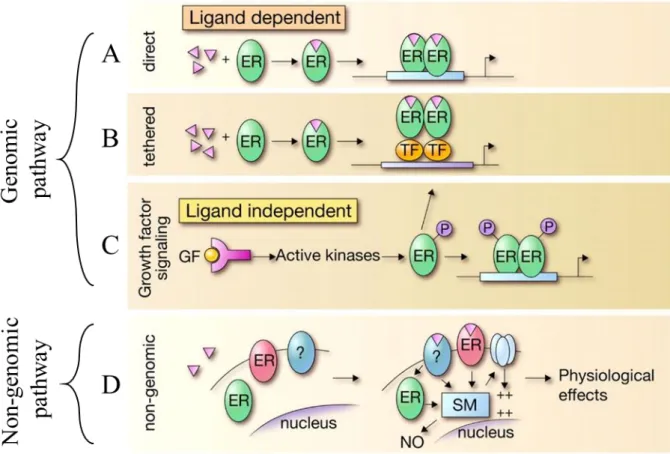
Human ER is coded by two relatively similar genes: ESR1 (ERα) and ESR2 (ERβ). They share about 80% sequence similarity. ERβ is generally overexpressed in ovaries, while ERα is mostly located in breast tissues [5]. They both can modulate gene expression in either ligand- dependent or ligand-independent manner. In ligand-dependent gene modulation, ER is required to be activated by estrogen binding. Activated ER could either directly act as a transcription factor or recruit other transcription factors to promoter of its target genes. On the other hand, ligand-independent action of ER involves its phosphorylation via kinases. Phosphorylated ER acts as if it was estrogen-bound and functions in transcription of different genes (Figure 2).
11
Figure 2: Schematic representation of the ER action. Genomic (A, B and C) and non-genomic
pathways (D) of ER is shown. Genomic pathway can be exerted through ligand-dependent (A and B) or -independent (C) actions. In A, ER is directly initiates transcription activation after ligand binding. In contrast, ligand-bound ER initiates transcription via recruiting other transcription factors in B. Furthermore, ER activation can be enabled via phosphorylation by kinases leading to the conformational change of ER (C). Lastly, membrane-bound ERs can interact with other proteins within cell and cause physiological effects (D) (Modified from: Heldring et. al., 20067, Physiol Rev [6]). See Appendix for the copyright permission.
1.1.2 ERα-Positive Breast Cancer
Luminal subtypes (Luminal A and Luminal B) of the breast cancer are the most common among all subtypes comprising roughly 70% of the all breast cancer patients. This corresponds to approximately 1.2 million new cases annually for the estrogen receptor positive (ERα-positive) breast cancer. [7]. ERα-positive breast cancer has a less aggressive, slow developing profile compared to ERα-negative subtypes, and may or may not possess progesterone receptor (PR) overexpression. However, elevated PR levels are shown to have a positive effect on patient prognosis [8] [9] [10]. Moreover, while Luminal A patients are
12
regarded having low-risk, Luminal B patients are regarded as high-risk in terms of prognosis. The difference is potentially originated from HER2 expression level that is shown to effect clinical outcome negatively [10] [11]. Because of the fact that ERα is a transcription factor and able to modify a very wide range of genes downstream, overexpressed or overactive ERα state is the main tumor-promoting factor in this subtype. Therefore, Targeting ERα and, thus, its downstream is the major approach to treat ERα-positive breast cancer.
1.1.3 First-line Treatment of ERα-Positive Metastatic Breast Cancer
In general, first-line therapy for cancer is defined as the primary choice of treatment of the disease. In case of ERα-positive metastatic breast cancer, first-line therapy is endocrine therapy. Endocrine therapy can be divided into 3 groups based on the mechanism of the action of the drugs. First group consists of selective ER modulators (SERMs) like tamoxifen and raloxifene, second group is aromatase inhibitors (AI) such as letrozole, anastrozole, and exemestane and last of the endocrine therapy groups is the selective ER down-regulators (SERDs) like fulvestrant. While raloxifene can be used in any prevention of invasive breast cancer and AIs can only be used in postmenopausal women [12] [13], tamoxifen has been used for the treatment of both premenopausal and postmenopausal, high-risk ER-positive breast cancer patients.
Both Luminal A and B patients can be assigned to anti-estrogens or aromatase inhibitors. Another crucial point which is worth considering before determining the treatment regimen is initial pathological examinations for molecular markers of breast cancer. Herein, immunohistochemistry results of the biopsy samples are reliable indicators. For instance, Luminal B patients can benefit from HER2-targeted therapies or cytotoxic chemotherapies together with endocrine therapies [14]. If patients do not show any clinical benefit, which is defined as complete or partial response or stable disease, they can be assigned to second-line therapies[15].
1.1.4 Selective Estrogen Receptor Modulators and Aromatase Inhibitors
The mechanism of the action of AI drugs, such as Letrozole, Anastrozole and Exemestane, is to cease all ER-mediated activities via blocking the main enzyme for estrogen production; aromatases. As a result, not only the ERα overactive cancer cells but the whole body deprives
13
of estradiol hormones. Estrogen is the main fertility hormone, thus patients prescribed with AI generally are post-menopausal women or have advanced stage of breast cancer [16][17]. In a meta-analysis of 30 independent studies, it was shown that aromatase inhibitors as cross-over therapy resulted in a better outcome than drug alone [18]. Apart from aromatase inhibitors, anti-estrogen modulators are used commonly in breast cancer. The most known estrogen modulator drug is tamoxifen.
1.2 Tamoxifen
For more than 3 decades, tamoxifen has been marketed as the best treatment option for patients with early-stage breast cancer with roughly 10% recurrence rate in first 5 years of the treatment [19]. Tamoxifen is an anti-estrogenic drug that was first prescribed as a contraceptive drug in 1960s [20]. However, in 1971 a study showed that, tamoxifen is a potent breast cancer drug and proven to improve progression-free survival [21]. Since then, tamoxifen has been used extensively for ERα-positive breast cancer. However, it is also well recognized that not all ERα-positive patients respond to tamoxifen well in clinics due to de novo or acquired resistance
1.2.1 Mechanism of Action of Tamoxifen
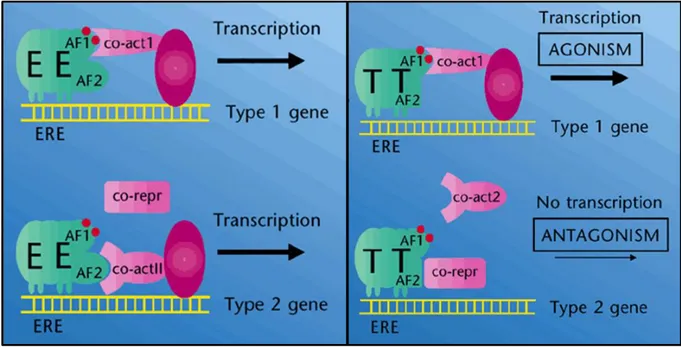
Proposed mechanism of action for tamoxifen is its binding to ER, subsequent activating its dimerization, resulting in its translocalization into nucleus and altering its downstream gene expression profile[15]. Tamoxifen could act either as an ER antagonist or as an ER agonist. ER is normally activated by binding of E2 to the ligand-binding domain of ER. This activation stimulates dimerization of ER. With the help of co-activators, active ER translocates into nucleus and binds specific sequences called estrogen responsive elements (ERE) located on DNA. ERE is a palindromic sequence, and it is found generally upstream of E2-inducible genes [22] [5]. Tamoxifen binds to ER with a lower affinity and does not impede activation of ER, but it alters its conformational dynamics and leads to co-repressor recruitment rather than co-activators. Hence, it inhibits proliferation promoting E2-indicible gene transcription. In other words, tamoxifen modifies ER transcription landscape (Figure 3) [23] [24] [25].
14
Figure 3: Tamoxifen does not inhibit but alters transcriptional activity of ER. On the left, figure
illustrates binding of estradiol (E) to ER, which subsequently synergizes with activation factor 1 and 2 (AF1 and AF2) upon dimerization to further translocate dimer structure to recognized ERE site. Co-activators attracted by AF1 or AF2 resulting in transcription of different genes. In the presence of the tamoxifen (T) conformational change is seen in activation factors, followed by recruitment of the co-repressors instead of co-activators. This conformational change led by tamoxifen results transcriptomic alteration (Modified from: Dowsett and Howell, 2002, Nat Med) [26]. See Appendix for the copyright permission.
1.3 Drug Resistance
Drug resistance is divided into two categories: First one is called intrinsic or de novo drug resistance, which is defined by pre-existing resistance factors and elements before initiation of the treatment. Because of pre-existing conditions, drug never becomes truly effective. The second one is called acquired drug resistance. Here, patients respond to treatment initially; however, response is lost over the course of treatment [27] [28]. Regardless of the type, resistance is a main issue in cancer therapies, and great number of studies is ongoing to tackle this problem.
15
Although 5 years of adjuvant tamoxifen therapy has been demonstrated to reduce mortality rate by 30-40% in the first 15 years [20] [29], unfortunately about half of patients develop intrinsic (de novo) or acquired resistance and metastasis in long-term [29] [30] [31]. After failure of tamoxifen treatment, patients with early stage of breast cancer are prescribed with other ER antagonists as well as aromatase inhibitors [18]. In advanced stages, they are treated with combination of aromatase inhibitors and ER antagonists [32]. When cancer is more aggressive and in metastatic stage, drugs with different mechanisms of action are used, such as an mTOR inhibitor Everolimus [17], a CDK4/6 inhibitor Palbociclib [33] or a VEGF modulator Bevacizumab [34]. In addition to those targeted drugs, chemotherapy is also an option for those patients [32].
Patients are generally prescribed with endocrine therapy rather than chemotherapy due to the fact that endocrine therapy is much more tolerated than the latter. On the other hand, in rapid progression and metastasis breast cancer situation, patients are given a chemotherapy regimen [33]. Lapatinib, a kinase inhibitor, showed a great potential to overcome tamoxifen resistance especially in patients with reduced or no ER expression [35] [36]. Several studies suggested that if EGFR, a commonly upregulated receptor in tamoxifen resistance, is targeted, it would benefit patients. Indeed, clinical studies showed that gefitinib, an EGFR inhibitor, combined with tamoxifen resulted in better outcome than tamoxifen alone in advanced and metastatic settings [37] [38].
1.3.2 Resistance Mechanisms Against Tamoxifen
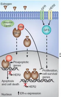
The well-known mechanisms of tamoxifen resistance are loss of ER expression lost over time and simultaneous activation of other oncogenic signaling pathways. Hence, tamoxifen cannot target ER or modify its downstream events. Drug becomes ineffective and cancer cells in this stage are considered as “tamoxifen resistant” [16]. In the tamoxifen responsive tumors, tamoxifen alters transcriptomic effect of ER by competing estradiol, subsequently alters transcription effect of it and consequently pro-apoptosis genes dominate pro-survival genes. On the other hand, in the tamoxifen irresponsive tumors, growth factor receptor-mediated pro-survival signaling pathways take place, eventually leading ER depletion. In the absence of ER, tamoxifen cannot regulate its transcriptional activity and pro-survival gene expression is restored by GFR-mediated signaling. (Figure 4)
16
Figure 4: Schematic representation of over-simplified tamoxifen sensitiveness and resistance mechanisms. On the left, tamoxifen alters transcriptomic effect of ER and switches its downstream
from proliferative gene expression to apoptosis-related genes. On the right, HER2 and GFR mediated tamoxifen resistance mechanism is shown which eventually leads to decreased ER levels. Thus, tamoxifen cannot control its transcriptional activity. (Modified from: Thomas & Gustafsson, 2011, Nat Med [39]). See Appendix for the copyright permission.
Expression levels of ER and PR decrease
Decrease in ERα expression level is a common phenomenon in tamoxifen resistance. Studies have shown that both patients and in vitro models lose expression of ERα for some extent upon tamoxifen treatment [40] [24]. Epigenetic silencing through hypermethylation in the promoter of ER is reported in tamoxifen treated patient [24] [41] and in vitro samples [42] [43] samples. Moreover, loss in ER expression was also suggested to be consequence of stimulation of MAPK/ERK and PI3K/AKT/MTOR signaling pathways through over-activated HER2 and other GFRs [40][44] [25][45] .In addition to decrease in ERα expression levels, cells could also decrease Progesterone Receptor (PR) levels as well. Since PR is a direct transcriptional target of ER, loss of PR expression clinically validated to be additive factor on tamoxifen resistance [9].
17
GFR upregulation
Not only steroid hormone regulated receptors, but also growth factor mediated receptors were shown to contribute tamoxifen resistance by both in vitro and clinical studies. EGFR and HER2 are the most frequently altered non-steroidal receptors in the breast cancer. Both of them are generally associated with basal and HER2-enriched subtypes which are considered to be rather advanced. Yet, luminal patients whose ER expression decreased or lost in the course of treatment, were known to have increased EFGR and HER2 expression which would not only give them more advanced breast cancer properties, but also compensate for ER lost and cell cycle block. Afterwards, cancer cells would have another abnormally active pathway to avoid the anti-cancer effects of tamoxifen[16] [33] [46].
Non-coding RNAs
Global transcriptome analyses performed in recent years demonstrated that most parts of the genomic DNA that are transcribed into RNA are not further translated into proteins. RNA molecules that are not converted into proteins and are therefore called non-coding RNAs (ncRNA) were found to be involved in cellular processes like sequence-specific chromosome modifications, gene silencing and regulation of protein signaling pathways. They are classified into 2 major classes: short and long non-coding RNAs. Long non-coding RNAs (lncRNAs) are further divided into several subclasses, and lincRNA or long intergenic non-coding RNA, most abundant member, is spanning a length of more than 200 nucleotides[47].
Recently, the functions of non-coding RNAs, thanks to increasing studies, are uncovered. Several of these studies proved the potential of the non-coding RNAs in disease progression. They can either take an active part in disease progression or may act as a biomarker. Short non-coding RNAs, including miRNAs, siRNAs, piwiRNAs etc., are relatively more studied compared to long non-coding RNAs. MicroRNAs (miRNAs) are 20-22 nucleotide long RNA molecules that inhibit protein synthesis by binding to mRNAs. There are few well-known miRNAs associated with cancer, such as miR-200 family and let-7. Furthermore, in terms of tamoxifen resistance in breast cancer, ectopic expression of miR-375 was shown to overcome tamoxifen resistance via modulating EMT, while another miRNA, miR-519, was proven to cause tamoxifen resistance [48] [49]. These are just few examples of many small non-coding RNAs associated with tamoxifen resistance.
18 1.4 Long Non-Coding RNAs in Human Diseases
Non-coding part of the human genome has seem shown to have functional roles in various biological processes. Genome-wide association studies (GWAS) mainly focusing on single nucleotide polymorphism (SNP) showed that about 40% of a trait or a disease- associated SNPs are found in non-coding regions of the genome [50]. Those studies played important roles to reveal functions of non-coding RNAs. The list of diseases associated by lncRNAs are long and in progress, yet it includes Prader-Willi Syndrome of neuronal system [51], fragile X syndrome [52], possibly Alzheimer’s disease [53], coronary diseases [54] [55], and lastly, cancer[56][57][58].
1.4.1 Effect of Long Non-Coding RNAs in Breast Cancer
GWAS and later on functional studies demonstrated that non-coding RNAs are functional and take a role in various biological processes. Homeobox antisense intergenic RNA or widely known as HOTAIR, is one of the most well-known lncRNAs in cancer biology. While it is more commonly studies in breast cancer, its influence is not limited to a single type of cancer. Rather, includes gastric, colorectal and cervical cancers [59]. The reason behind how it affects different cancers is that it regulates chromatin methylation status of a neighbor gene, HOXD, which belongs to homeobox polycomb complex protein family regulating developmental processes [60]. Another such example is Metastasis Associated in Lung Adenocarcinoma Transcript 1, shortly MALAT1. MALAT1 is located in nucleus and its increased expression is linked to metastatic capacity of the both lung and breast tumors [61][62]. Higher expression of both lncRNAs predicts worse survival, and also is suggested to be a biomarker in terms of metastasis and survival[63][64][65].
One of the lncRNAs important as being the first lncRNA identified to contribute to anti-estrogen resistance is Breast Cancer Anti-Estrogen Resistance 4 (BCAR4) This lncRNA, previously named as LOC400500, is initially reported to contribute to anti-estrogen resistance when ectopically expressed in in vitro settings [66]. Although initial study was focused on a functional screen using cDNA libraries, a clinical study conducted with 280 ERα-positive, first-line tamoxifen-treated patients confirmed the important role of this lncRNA in tamoxifen resistance where higher BCAR4 expression is correlated with lower RFS and poor clinical benefit rate [67]. Later, characterization studies were performed to solve underlying
19
mechanism of BCAR4 [68] [69]. The studies suggested that BCAR4 expression contributes to therapy failure regardless of ER function, possibly through modulating non-canonical Hedgehog signaling pathway. However, the role of lncRNAs in tamoxifen resistance is at its infancy.
1.5 Autophagy and Cancer
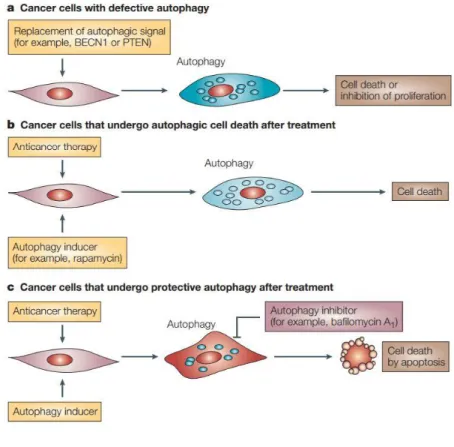
Autophagy is the catabolic degradation of cellular components in response to starvation or stress[70]. Autophagy is believed to influence tumor development. In one study, basal levels of the autophagy was found to be much less in cancer cells than their normal counterparts [71]. However, autophagy has a dichotomous role in cancer. In early stage of cancer, it was shown to have tumor suppressor effect. In a study conducted with mice bearing deletion in a major autophagy protein, Beclin1, manifested lung cancer in early ages. On the other hand, at a later stage of cancer, autophagy is believed to have tumor-promoting effect, and there are a number of clinical studies combining autophagy inhibitors with cancer drugs (Figure 5) [72] [73]. Currently, both activation and complete inhibition of autophagy approaches are being tested in cancer treatment [74].
20
Figure 5: Schematic representation of modulation of autophagy in cancer. (A) When cancer cells
are re-introduced with absent or defective autophagic proteins, autophagy is induced and results in cell death. (B) Anticancer therapy along with other autophagy inducers can enhance autophagy and cell death. (C) Cancer cells may use protective autophagy as tumor-promoting process. Those types of cells could then be treated with autophagy inhibitors to switch autophagy to apoptosis. (Taken from: Kondo et al., 2005, Nat Rev Cancer [72]) See Appendix for the copyright permission.
1.5.1 Autophagy in Breast Cancer
One of the well-studied autophagy regulator proteins, Beclin1, was reported to be decreased in breast cancer tissues compared to their normal counterparts. A key study which was conducted with MCF7 xenografts to validate the tumor suppressor role of Beclin1 in breast cancer showed that when Beclin1 expression was restored to normal levels, tumor growth was suppressed [75]. Besides Beclin1, mTOR (mechanistic target of rapamycin) has an important role in controlling autophagy. Active mTOR blocks autophagy induction while in the case of starvation, mTOR is inhibited allowing autophagy to be activated. Therefore, mTOR inhibitors are proposed to induce autophagy in breast cancer[76] [77].Importantly, tamoxifen was shown to be an autophagy inducer drug [78].
21 1.6 Hypothesis and Aim
ERα-positive breast cancer patients treated with tamoxifen initially have good response, but after a long latent period, tumors relapse. Even though there have been numerous studies on tamoxifen resistance, not all the players, especially lncRNAs, in this process are identified. A large, heterogeneous patient population with different molecular backgrounds, adverse side effects of the second-line and third-line therapeutic agents, advances in the field of personalized medicine and accumulation of the cancer biology knowledge give motive to investigate and understand more on drug resistance and enhance therapy options for the tamoxifen resistant patients.
Thus, non-coding portion of the genome has great potential to bring up new targets and/or biomarkers overcoming tamoxifen resistance. In this thesis, I aimed to take an unbiased transcriptome approach to identify novel lncRNA regulators of tamoxifen resistance. In this line, whole transcriptome analysis was performed in acquired tamoxifen resistant cell line models (MCF7-TamR) and their sensitive counterparts (MCF7-WT). After refining whole RNA-Seq results, I selected validated lncRNAs and narrowed down the list by choosing previously cancer-associated lncRNAs. LINC00152 was the most prominent candidate among 330 lncRNA transcripts in the list. It was previously shown to be prognostic marker for gastric, hepatocellular and lung carcinoma. Furthermore, a recent study by Grembergen et. al., showed that LINC00152 expression was elevated in breast cancer [79]. In line with its oncogenic potential, LINC00152 was also upregulated in TamR-resistant cell line models (both MCF7-TamR and T47D-TamR). Knockdown of LINC00152 sensitized both resistant cell lines to tamoxifen. Importantly, this sensitization was potentiated by autophagy induction and apoptosis. Finally, higher expression of LINC00152 was associated with worse survival in tamoxifen-treated ER alpha positive breast cancer patients showing potential role of LINC00152 in tamoxifen resistance.
22 CHAPTER 2
2 MATERIALS AND METHODS 2.2 Materials
2.2.1 Buffers
1x Anode Buffer I 300mM Tris, 20% (v/v) methanol
1x Anode Buffer II 25mM Tris, 20% (v/v) methanol
1x Cathode Buffer 40mM 6-aminocaparoic acid, 20% (v/v) methanol
1x SDS-PAGE Running Buffer 25mM Tris, 14.41g/l glycine, 1% (v/v) SDS
1x TBST 20nM Tris, 8g/l NaCl 0.2% (v/v) Tween20
2.2.2 Chemicals and Reagents
4x Protein Loading Dye 250mM Tris HCl (pH:6.8), 10% (w/v) SDS, 0.1% w/v), Bromophenol Blue, 50% (v/v) Glycerol, 25% (v/v) Betamercaptoethanol
6-aminocaparoic acid Sigma Aldrich, St Louis, MO, USA
Acrylamide/bisacrylamide Applichem, Darmstadt, Germany
Ammonium persulfate (APS) Carlo Erba, Cornaredu, Italy
Bovine Serum Albumin (BSA) Santa Cruz, Dallas, TX, USA
ElectroChemoLuminescence (ECL) Detection Reagent
Amersham Pharmacia Biotech, Amersham, UK
Ethanol Sigma Aldrich, St Louis, MO, USA
Isopropanol Sigma Aldrich, St Louis, MO, USA
LightCycler 480 SYBR Green I Master Mix
23
Lipofectamine 2000 Invitrogen, Carlsbad, CA, USA
Methanol Sigma Aldrich, St Louis, MO, USA
Milk Powder Sigma Aldrich, St Louis, MO, USA
Nuclease free water Applied Biosystems/Ambion, Austin, TX, USA
Page Ruler Protein Ladder Thermo Ficher Scientific, Waltham, MA, USA
Phosstop Roche Applied Science, Mannheim, Germany
Ponceu S Sigma Aldrich, St Louis, MO, USA
Protease inhibitor cocktail Roche Applied Science, Mannheim, Germany
Sodium Chloride (NaCl) Merck, Darmstandt, Germany
Sodium Dodecyl Sulfate (SDS) Merck, Darmstandt, Germany
Tetramethyl ethylenediamine
(TEMED)
Serva, Heidelberg, Germany
TRIsure Bioline, Luckenwalde, Germany
Triton X-100 Sigma Aldrich, St Louis, MO, USA
Trizma Base (Tris) Sigma Aldrich, St Louis, MO, USA
Tween-20 VWR, Radnor, PA, USA
2.2.3 Media and Supplements
DMEM Lonza, Basel, Switzerland
Fetal bovine serum (FBS) Biowest, Nuaille, France
Insulin Sigma Aldrich, St Louis, MO, USA
24
optiMEM Invitrogen, Carlsbad, CA, USA
Penicillin/Streptomycin (P/S) Lonza, Basel, Switzerland
2.2.4 Kits
BCA Protein Assay Kit Pierce, Rockford, IL, USA
Cell Titer-Glo cell viability assay kit Promega, Madison, WI, USA
MycoAlert detection kit Lonza, Basel, Switzerland
Revert Aid first strand cDNA synthesis kit Fermantas, St Leon-Roth, Germany
2.2.5 Equipment
Cell Culture Hood Nüve, Ankara, Turkey Cell Culture Incubator Nüve, Ankara, Turkey
Centrifuges Thermo Fisher Scientific, Waltham, MA, USA Beckman, Pasadena, CA, USA
Counting Chamber Marienfeld, Könighofen, Germany Freezer (-20C) Bosch, Stuttgart, Germany
Freezer (-80C) Hettich, Geldermansen, Germany
Fridge Bosch, Stuttgart, Germany
Horizontal shakers FinePCR, Seoul, South Korea Bellco, Vineland, NJ, USA
LightCycler 96 Roche Applied Science, Mannheim, Germany
25
Mini-PROTEAN Tetra Cell Biorad, Hercules, CA, USA
Multichannel Pipette Thermo Fisher Scientific, Waltham, MA, USA
NanoDrop 1000 Thermo Fisher Scientific, Waltham, MA, USA
Nikon TS300 Inverted Microscope Nikon, Tokyo, Japan
Power supplies for electrophoresis Biorad, Hercules, CA, USA
Semidry western blot transfer unit Biorad, Hercules, CA, USA
Synergy HT Multireader Biotek, Winooski, VT, USA
Vortex Isolab, Wertheim, Germany
Water Bath Nüve, Ankara, Turkey
X-ray casette Amersham Pharmacia Biotech, Amersham, UK
X-ray hyper processor Amersham Pharmacia Biotech, Amersham, UK
2.2.6 Consumables
100mm dishes Greiner bio-one, Frickenhausen, Germany
145mm dishes Greiner bio-one, Frickenhausen, Germany
6-well plates Greiner bio-one, Frickenhausen, Germany
96-well plates Greiner bio-one, Frickenhausen, Germany
Cell scrapers Greiner bio-one, Frickenhausen, Germany
Coverslips Marienfeld, Königshofen, Germany Cryovials Greiner bio-one, Frickenhausen, Germany
Cuvettes VWR, Radnor,PA, USA
26
200ul, 1000ul)
Microscope slides Marienfeld, Königshofen, Germany
Parafilm VWR, Radnor,PA, USA
PCR tubes Axygen, Corning, NY, USA
Plastic pipettes (5 ml, 10ml, 25ml) Corning Incorporated, Corning, NY, USA
PVDF Membrane Biorad, Hercules, CA, USA10
qPCR Plates Roche Applied Science, Mannheim, Germany
Reaction tubes (500ul, 1.5ml, 2ml) Axygen, Corning, NY, USA
Storage bottles (250ml, 500ml, 1L) Corning Incorporated, Corning, NY, USA
Whatmann paper GE Healthcare, Little Chalfont, UK
White plates Costar, Corning, NY, USA
-X-ray films Kodak, Rochester, NY, USA
2.3 Methods
2.3.1 Cell Culturing
2.3.1.1 Culturing Human Breast Cancer Cell Lines MCF7 and T47D
MCF7-WT and MCF7-TamR cells are developed previously [48]. T47D cells were generous gift from Işık Yuluğ’s group. Cells were grown in 100mm petri dishes at 37°C 5% CO2
condition. Cells were passaged every 4-5 days in 1-to-3 ratio. Medium of the cells was composed of phenol-red-free DMEM or RPMI supplemented with 10% heat inactivated and filtered FBS, 1% NEAA, 1% P/S and 0.01 mg/ml of insulin. For cell splitting, growth medium was removed, and cells were briefly rinsed with 2 ml PBS, then 1.5ml trypsin was added onto cells and incubated in 37° C for 5 minutes. Trypsin was inhibited adding fresh culture medium, and cells were resuspended. Petri dish was filled up to 10 ml after splitting.
27
2.3.1.1.1 Development of Tamoxifen Resistant Cell lines
T47D cells are first tested for mycoplasma contamination and tamoxifen response. TamR resistant T47D cells were developed under continuous tamoxifen exposure for more than 8 months. Briefly, 4-OH-tamoxifen was added into culture medium one day after splitting. Medium was kept with tamoxifen for 3-4 days, and then growth medium was replenished in order to allow cells to proliferate. After 2-3 days of recovery, cells were split and incubated with tamoxifen until cells develop resistance. All cell lines were tested for mycoplasma contamination periodically.
2.3.1.2 Antisense oligonucleotide (ASO) Transfections
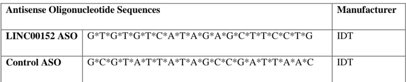
ASO was designed according to manufacturer’s recommendations. All nucleotides bear Phosphorothioate bonds (PS bonds). ASOs were aliquoted under the hood with nuclease-free, high grade water upon delivery. ASO transfections were carried out using Lipofectamine 2000TM and OptiMEM reagents. Final concentrations of ASOs were 25nM/well. Sequence of the ASOs are given in Table 1. Cells were seeded at a number of 200,000 cell/well for 6-well-plate and 6,000 cell/well for 96-well-6-well-plate. After 24 hours of cell seeding, cells received 25nM final concentration of ASO. All transfections were done with P/S-free medium.
Table 1. List of ASO sequences used in the experiments (* stands for PS bonds)
Antisense Oligonucleotide Sequences Manufacturer
LINC00152 ASO G*T*G*T*G*T*C*A*T*A*G*A*G*C*T*T*C*C*T*G IDT
Control ASO G*C*G*T*A*T*T*A*T*A*G*C*C*G*A*T*T*A*A*C IDT
2.3.2 Cell-based Assays
2.3.2.1 In Vitro Sensitization Assay
Viability of the cells was measured by Cell Titer-Glo cell viability assay kit according to manufacturer’s instructions. Briefly, cells were seeded as 4 replicates in 96-well format where each of the wells harbors 6,000 cells. Seventy-two hours after treatment, cells were incubated
28
with Cell-Titer-Glo reagent about 15 minutes on orbital shaker. After incubation, equal volume of the media was transferred onto opaque white bottom 96-well plates, and luminescence was measured on Biotek Multireader machine. Readings were converted to percent inhibition, and Student's two-tailed t-test was used for statistical analysis.
2.3.3 Molecular Biology
2.3.3.1 Quantitative Real Time Polymerase Chain Reaction (qRT-PCR) 2.3.3.1.1 RNA isolation
Cell seeding and treatments were done as described previously (2.2.1.2.). After 24, 48 and 72 hours of the incubation in cells were collected by trypsinization and rinsed once with PBS. RNA was isolated by using TRIsure, chloroform and isopropanol. After isolation, RNAs were solubilized with adequate volume of nuclease-free, high grade water and stored at -80° C freezer.
2.3.3.1.2 cDNA synthesis
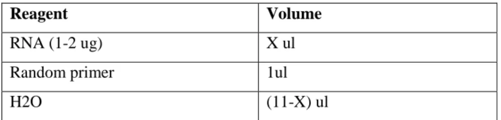
RevertAid Reverse Transcription (RT) first strand cDNA synthesis kit was used for reverse-transcription of total RNA. Total of 1-2ug RNA from each sample was converted to cDNA by using random oligomer. Protocol for RT is given at Table 2
Table 2. Components of first step of RT reaction.
Reagent Volume
RNA (1-2 ug) X ul
Random primer 1ul
H2O (11-X) ul
Samples were placed in ThermoCycler for 5 mins at 65° C. After first step was accomplished, proceeded with the last step.
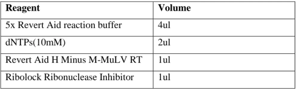
29 Table 3. Components for last step of RT reaction.
Reagent Volume
5x Revert Aid reaction buffer 4ul
dNTPs(10mM) 2ul
Revert Aid H Minus M-MuLV RT 1ul Ribolock Ribonuclease Inhibitor 1ul
Samples were placed back in ThermoCycler and incubated as stated in Table 4.
Table 4. Thermocycler program for cDNA synthesis.
Temperature Time
37o C 5 minutes
42o C 60 minutes
70o C 10 minutes
4o C ∞
All cDNA samples were diluted to 10 ng/uL of final concentration before proceeding with qRT-PCR.
2.3.3.1.3 qRT-PCR for RNA expression
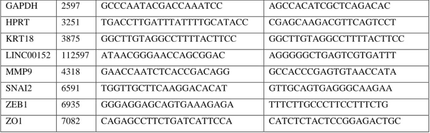
For quantitative Real-Time-PCR, SYBR Green Master mix was used along with the primers listed on Table 5.
Table 5. List of qRT-PCR primers.
Name Gene
ID Forward Primer (5' to 3') Reverse Primer (5' to 3')
ACTB 60 CCAACCGCGAGAAGATGA CCAGAGGCGTACAGGGATAG
CDH1 999 CCCGGGACAACGTTTATTAC GCTGGCTCAAGTCAAAGTCC
CDH2 1000 ACAGTGGCCACCTACAAAGG CCGAGATGGGGTTGATAATG
ESR1 2099 TTACTGACCAACCTGGCAGA ATCATGGAGGGTCAAATCCA
30
GAPDH 2597 GCCCAATACGACCAAATCC AGCCACATCGCTCAGACAC
HPRT 3251 TGACCTTGATTTATTTTGCATACC CGAGCAAGACGTTCAGTCCT
KRT18 3875 GGCTTGTAGGCCTTTTACTTCC GGCTTGTAGGCCTTTTACTTCC
LINC00152 112597 ATAACGGGAACCAGCGGAC AGGGGGCTGAGTCGTGATTT
MMP9 4318 GAACCAATCTCACCGACAGG GCCACCCGAGTGTAACCATA
SNAI2 6591 TGGTTGCTTCAAGGACACAT GTTGCAGTGAGGGCAAGAA
ZEB1 6935 GGGAGGAGCAGTGAAAGAGA TTTCTTGCCCTTCCTTTCTG
ZO1 7082 CAGAGCCTTCTGATCATTCCA CATCTCTACTCCGGAGACTGC
Reaction was performed in 96-well LightCycler plates. Each well contained 20ng cDNA and 8 uL of MasterMix (total volume of 10 uL). Reaction components were given in Table 6.
Table 6. Mastermix components for qRT-PCR reaction
Reagent Volume
dNTP 2,5 uL
Forward Primer (20uM) 0,25 uL Reverse Primer (20uM) 0,25 uL
SYBR Green 5 uL
Tightly-sealed plate was briefly centrifuged and then placed into LightCycler® 96 qRT-PCR thermocycler (Roche Applied Science, Mannheim, Germany) machine. qRT-PCR incubation program was shown on Table 7.
Table 7. qRT-PCR incubation program.
Pre-incubation Target (°C) Acquisition Mode Hold (hh:mm:ss) Ramp Rate (°C/s) Acquisitions (per °C) Sec Target (°C) Step Size (°C) Step Delay (cycles) 95 None 00:05:00 4.4 5 0 0 0 Amplification Target (°C) Acquisition Mode Hold (hh:mm:ss) Ramp Rate Acquisitions (per °C) Sec Target Step Size Step Delay
31 (°C/s) (°C) (°C) (cycles) 95 None 00:00:10 4.4 5 0 0 0 58 Single 00:00:20 2.2 5 0 0 0 72 None 00:00:20 4.4 5 0 0 0 Melting Curve Target (°C) Acquisition Mode Hold (hh:mm:ss) Ramp Rate (°C/s) Acquisitions (per °C) Sec Target (°C) Step Size (°C) Step Delay (cycles) 95 None 00:00:05 4.4 5 0 0 0 55 None 00:01:00 2.2 5 0 0 0 95 Continuous 00:00:00 0.11 5 0 0 0 Cooling Target (°C) Acquisition Mode Hold (hh:mm:ss) Ramp Rate (°C/s) Acquisitions (per °C) Sec Target (°C) Step Size (°C) Step Delay (cycles) 4 None 00:00:30 2.2 5 0 0 0 2.3.3.1.4 qRT-PCR data analysis
All qRT-PCR plates had at least two housekeeping genes. Every sample had triplicates for one gene. Data analyses were done using ΔΔCt method and normalized to geometric average of the housekeeping genes. Statistical analysis was performed using Student's two-tailed t-test.
2.3.3.2 RNA Localization
The whole protocol was modified from study of Zhang et. al. (2014) [80]. In summary, the cells were harvested fresh using a scraper when they reached around 3 million cells, and were briefly centrifuged at 4° C. Pellet was re-suspended by Cell Lysis Buffer. After the suspension was incubated on ice up to 15 mins, it was transferred into a sterile Dounce homogenizer. Ensuring not to make any bubbles, the cells were homogenized. Later, the homogenate was transferred to a fresh tube while adding Triton X-100 with a final concentration of 0.1%. After mixing the new suspension by inversion, samples were centrifuged at 1500g for 5 minutes at 4° C. While the supernatant contains cytoplasmic extract, the pellet contains nuclei and cell debris. All the supernatant was placed into a new tube without disturbing the pellet.
32
The nuclear and cytoplasmic RNAs are isolated using Tri-Reagent as described above (2.3.3.1.), and qRT-PCR was performed.
2.3.3.3 Protein Biochemistry 2.3.3.3.1 Protein isolation
Cell seeding and treatments were done as described previously (2.2.1.2.). After 24, 48 and 72 hours of the incubation, first medium was collected and centrifuged at 5,000 rpm for 5 minutes for apoptotic body collection. Cells were then trypsinized and collected. Depending on cell pellet size, 50-100μL RIPA lysis buffer was added to the pelllet and mixed thoroughly by pipetting up and down. Suspension was then transferred to 1.5 ml Eppendorf tubes and vortexed for 5-10 seconds every 5 minutes for a total of 30 minutes. Later, this suspension was centrifuged at 13,000 rpm 4oC for 30 minutes. Supernatant was collected as protein and stored at -20oC for further experiments.
2.3.3.3.2 Protein quantification
Quantity of the proteins was determined by BCA Assay Kit according to manufacturer’s instructions. Nine different standard protein solutions (BSA) with a range of 0-2 ug/uL were placed in 96-well clear plates in duplicates. Twenty uL of the standard solutions were used while only 5 uL of the unknown sample was used. Thus, standards and samples share a dilution factor of 5. A and B reagents were mixed 50:1 ratio, and total of 200 uL mixed working solution were added into each well. Proteins incubated in working solution for 30 minutes at 37° C. Colorimetric reading was obtained by BioTek Multiplate reader at 562 nm. A standard calibration curve was drawn based on absorbance readings from BSA standards, and sample concentrations were quantified from line graph of the curve.
2.3.3.3.3 Sodium dodecyl sulfate polyacrylamide gel electrophoresis (SDS-PAGE)
Concentrations and loading volumes of protein samples were equalized using 4X protein loading dye and RIPA buffer. Subsequently, samples were heated to 95°C for 3-5 minutes. Polyacrylamide-based stacking and resolving gels were prepared according to Table 8.
33 Table 8. Components of polyacrylamide gels.
Reagent 8% Resolving gel 10% Resolving gel 12% Resolving gel 5% Stacking gel
1 M Tris solution (pH 8.8) 1.3ml (pH 8.8) 1.3ml (pH 8.8) 1.3ml (pH 6.8) 260ul
10% APS 50ul 50ul 50ul 20ul
10% SDS 50ul 50ul 50ul 20ul
30% acrylamide mix 1.3ml 1.7ml 2ml 340ul
H2O 2.3ml 1.9ml 1.6ml 1.36ml
TEMED 5ul 5ul 5ul 2ul
Total volume 5 mL 5 mL 5 mL 5 mL
Using Mini-PROTEAN Gel casting module (Biorad, Hercules, CA, USA), resolving gel solution was poured initially and overlaid with isopropanol until gel polymerized. After removing isopropanol from gel casting system, stacking gel solution was poured on top of resolving gel, and 10 or 15 well comb was placed in it. 10-20 μg protein samples were loaded per well, and empty wells were filled with diluted protein loading dye. Electrophoresis was performed at 130V for 90 minutes.
2.3.3.3.4 Western Blotting
Whatmann papers with 3 mm thickness were cut in a dimension of 7cm X 9 cm. Four cut papers were soaked in anode buffer I, 2 of them in anode buffer II and 6 of them in cathode buffer. PVDF membranes were activated in 100% methanol for 3 minutes. Layers for a proper transfer from bottom to top were 4 papers for anode I, 2 for anode II, one activated PVDF membrane, polyacrylamide gel and lastly 6 papers for cathode. Semi-dry transfer was performed at 25V for 30-60 minutes. After transfer was completed, PVDF membrane was briefly stained with Ponceu S solution. Membranes were then washed with ddH2O until
Ponceu stain was completely removed and cut at specific molecular weight (kDa) of interest. Cut membranes were then blocked either in 5% (w/v) milk:TBST or in 5% (w/v) BSA:TBST for 1 hour room temperature Blocking solution was removed, and membranes were incubated with primary antibody either for 1-hour in room temperature or overnight in 4° C (Table 9). After primary antibody incubation, membranes were washed with 1X TBST and then
34
incubated with secondary antibody either for 1-hour at room temperature or overnight at 4° C. Later, membranes were again washed with 1X TBS-T three times for 10 minutes on shaker. After washing, enhanced chemiluminescence (ECL) reagent (Amersham Pharmacia Biotech) was applied on membranes, and X-ray films were exposed to membranes for different time points ranging from 3 seconds to 30 minutes and then membranes were developed.
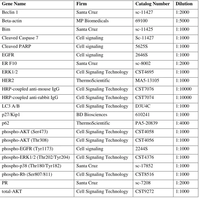
Table 9. List of Western Blot antibodies.
Gene Name Firm Catalog Number Dilution
Beclin 1 Santa Cruz sc-11427 1:2000
Beta-actin MP Biomedicals 69100 1:5000
Bim Santa Cruz sc-11425 1:1000
Cleaved Caspase 7 Cell signaling Sc-11427 1:1000
Cleaved PARP Cell signaling 5625S 1:1000
EGFR Cell signaling 2646S 1:1000
ER F10 Santa Cruz sc-8002 1:2000
ERK1/2 Cell Signaling Technology CST4695 1:1000
HER2 ThermoScientific MA5-13105 1:1000
HRP-coupled anti-mouse IgG Cell Signaling Technology CST7076 1:10000 HRP-coupled anti-rabbit IgG Cell Signaling Technology CST7074 1:10000
LC3 A/B Cell Signaling Technology D3U4C 1:1000
p27/Kip1 BD Biosciences 610241 1:1000
p62 ThermoScientific PA5-20839 1:4000
phospho-AKT (Ser473) Cell Signaling Technology CST4058 1:1000 phospho-AKT (Thr308) Cell Signaling Technology CST4056 1:1000
phospho-EGFR (Tyr1173) Cell signaling 2244S 1:1000
phospho-ERK1/2 (Thr202/Tyr204) Cell Signaling Technology CST4376 1:1000
phospho-p38 (Thr180/Tyr182) Santa Cruz sc-17852 1:1000
phospho-Rb (Ser807/811) Cell Signaling Technology CST8516 1:1000
PR Santa Cruz sc-7208 1:2000
35 2.3.4 Bioinformatics Analysis
2.3.4.1 Transcriptome Sequencing (RNA-Seq)
Whole Transcriptome sequencing was done by using Illumina HiSeq 2000 technology at McGill University, Canada. A paired-end library of 100 bp reads was produced for MCF7-WT and MCF7-TamR samples in triplicates. Total of 6 libraries were marked with different barcodes and afterwards pooled and read. Nearly 65 million paired-end 2×100bp reads/replicate were obtained.
2.3.4.1.1 RNA-Seq analysis
Data including the reads was sent to Bilkent via cloud as bmp format. Bmp was converted to FASTQ format in order to proceed with Tuxedo protocol [81]. Briefly, from tamoxifen resistant and sensitive samples were combined via TopHat v2.1.0, and mapped into human genome. As a reference genome, GH37(h19) and MiTranscriptome was used [82]. Then, CuffLinks was used to assemble transcripts, and CuffDiff identified differentially expressed transcripts between MCF7-WT and MCF7-TamR.
2.3.4.2 Patient Data Set Analysis
2.3.4.2.1 Patient Survival Analysis (Kaplan-Meier plot)
METABRIC patient data set along with NCBI Gene Expression Omnibus (GEO) database (GSE6532, GSE9195, GSE58644, GSE22220) were analyzed. By using Graphpad software (GraphPad software Inc., La Jolla, CA, USA) Kaplan-Meier patient survival graphs were generated for LINC00152 expression. Patient clinical information was given in GSE datasets. Patients with no survival information, "dead of non-cancerous cause" were censored. For each survival analysis, patients were separated from median. Log-rank (Mantel-Cox) test was applied as statistical analysis.
36 CHAPTER 3
RESULTS
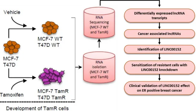
Overall workflow of the study
In order to identify lncRNA regulators of tamoxifen resistance, I utilized MCF7 and T47D cell lines, both of which are well-known ERα-positive breast cancer cell lines. MCF7-WT and MCF-TamR cells were already available and used for identification of miRNA regulators of tamoxifen resistance in Şahin Lab [48]. Firstly, RNA-Seq analysis was performed using MCF7-WT and TamR cells in order to find differentially expressed lncRNAs associated with tamoxifen resistance. After well-refining steps for filtering cancer-associated and validated lncRNAs, I chose LINC00152 as a candidate potential mediator of tamoxifen resistance. In meantime, I developed a second model of tamoxifen resistance using T47D cell line via exposing cells to tamoxifen over 9 months which are named T47D-TamR. Using sensitization assays and protein biochemistry in both MCF7-TamR and T47D-TamR cell lines, I showed that LINC00152 sensitizes resistance cells to tamoxifen via inducing autophagy and apoptosis. Finally, these findings were corroborated with patient data analyses and LINC00152 was identified as a novel lncRNA regulator of tamoxifen resistance (Figure 6)
Figure 6. Overall workflow of the study. Acquired tamoxifen resistant cell lines were developed (in
37
obtained and RNA-Seq was performed with MCF7-WT and MCF7-TamR cells. Differentially expressed lncRNA transcript lists were reduced according to criteria described in the text, and LINC00152 was selected. Later, effect of LINC00152 on tamoxifen resistance was examined in both MCF7 and T47D-TamR cells and also supported by clinical data.
3.1 Development and Characterization of Tamoxifen Resistant Cell Line Models
Tamoxifen has been the most commonly used endocrine therapy drug in the clinic to treat ER-positive breast cancer patients. However, intrinsic (de novo) or acquired resistance to tamoxifen is one of the important cause of the therapeutic failure, and it needs to be addressed. Potential mechanisms have been proposed so far in many studies, but the underlying molecular mechanisms of tamoxifen resistance are not fully understood yet. Our lab has previously studied different resistance mechanisms developed against targeted therapy agents for different subtypes of breast cancer. In this context, different resistant cell lines or animal models were developed and characterized to identify new targets that are involved in the acquisition of resistance[48] [49].
3.1.1 Developing Tamoxifen Resistant T47D Cell Line
In order to elucidate the molecular mechanisms for tamoxifen resistance, two different tamoxifen resistant cell line models were used in this thesis. Tamoxifen-resistant MCF-7 cells (MCF7-TamR) were previously developed and characterized [48] [49]. I generated a second tamoxifen resistant cell line model (Tamoxifen-resistant T47D cells (T47D-TamR)) to obtain a better representation of diverse patient background. Both cell lines are clinically classified as ER- positive, representing the luminal A molecular subtype. Therefore, these two cell lines are suitable models to study molecular events that are likely to be important in tamoxifen-treated ER-positive breast cancers.