Lipid-Laden Aqueous Humor in a Cat
Mustafa ARICAN
1,a
Kurtuluş PARLAK
1,bElgin Orçum UZUNLU
1,c 1 University of Selcuk, Faculty of Veterinary Medicine, Department of Surgery, TR-42100 Konya - TURKEY a ORCID: 0000-0001-8180-135X; b ORCID: 0000-0002-8656-037X; c ORCID: 0000-0001-5356-8968Article Code: KVFD-2018-20190 Received: 20.05.2018 Accepted: 23.08.2018 Published Online: 23.08.2018
How to Cite This Article
Arıcan M, Parlak K, Uzunlu EO: Lipid-laden aqueous humor in a cat. Kafkas Univ Vet Fak Derg, 24 (6): 909-911, 2018. DOI: 10.9775/kvfd.2018.20190 Abstract
This case report describes the clinical findings in a 5 years old neutered female cat presented with sudden onset of binocular blindness with cloudy appearance in both eyes. Systematic eye examination was performed after the anamnesis. Only intraocular pressure was increased in right eye. The examination of iris was not properly due to the opacity of humor aquosus. Liver function was checked in terms of diabetes and pancreatitis. Cholesterol and triglyceride levels were increased in blood analysis. The diagnosis was hyperlipidemia and hypertriglyceridemia. Dexamethasone (0.5 mg/kg) was applied to the subconjunctivaly; dexamethasone 0.1% w/v opthalmic solution drops (6 times in a day) and low-fat diet were recommended in the continuation of the treatment. The patient was immediately responded to the treatment within 36 hours. Aqueosus humor reached clear structure in both eyes. A visual activity was improve in the eyes. Post-operatively, the case was followed for 8 months. There were no complications encountered. This report is to contribute to increased awareness regarding the some ocular complications following diet programmes. In addition, the use of steroid ophthalmic solutions along with a diet program in the treatment protocol will accelerate the healing process quickly.
Keywords: Cat, Hyperlipidemia, Hypertriglyceridemia, Lipid-laden
Bir Kedide Lipid-Laden Humor Aquosus
ÖzBu olgu sunumunda, 5 yaşındaki dişi kedinin her iki gözünde ani olarak şekillenen bulutumsu görünüm ile karakterize körlüğe ilişkin bulgular anlatılmaktadır. Anamnez bilgileri alındıktan sonra, olguya gözde sistemik göz muayenesi yapıldı. Göz içi basıncı sağ gözde yükselmişti. İris’in muayenesi, humor aköz daki opasiteden dolayı sağlıklı yapılamadı. Karaciğer fonksiyonlarına bakılarak, diyabet ve pankreatitis yönünden muayene edildi. Kan analizi sonuçlarında kolesterol, trigliserit miktarlarındaki artışın olması, teşhisin hiperlipidemi ve hipertrigliseridemi olduğunu gösterdi. Olguya az yağlı diyet ve subkonjunktival deksametazon (0.5 mg/kg) ve deksametazon %0.1 w/v oftalmik solüsyon damlaları (günde 6 kez), tavsiye edildi. Olgu 36 saat içinde sağaltıma yanıt verdi. Humor aquosus her iki gözde de berrak yapısına ulaştı ve görüş vardı. Post-operatif olarak olgu 8 ay takip edildi. Herhangi bir komplikasyonla karşılaşılmadı. Bu olgu sunumu, kedilerin gözlerinde meydana gelen bazı problemlerde diyet programlarının önemini vurgulamak için paylaşılmıştır. Ayrıca hiperlipidemia olgularında tedavi protokolüne diyet programı ile beraber steroid oftalmik solüsyonların önerilmesi iyileşme sürecini hızlandıracaktır.
Anahtar sözcükler: Kedi, Hiperlipidemi, Hipertriglyceridemi, Lipid-laden
INTRODUCTION
Primary lipid disorders are not commonly observed in cats. Lipid disorders secondary to hepatic lipidosis, diabetes mellitus, pancreatitis, hyperadrenocorticism and administration of pregestagens are more likely to account for fasting lipaemia in this species [1]. Lipid and/or mineral
accumulation appears as sparkly, crystalline or shiny white areas in the cornea. Lipid metabolism is more affected in obese cats. Plasma triglyceride and cholesterol concentrations were significantly increased in obese
cats [2]. As a result of increasing the lipid concentration
in cats, lipids pass through the blood-aqueos barrier in the eye and cause the anterior chamber to become opaque or cloudy [3]. The incidence of obesity increases
with age and is more frequent in neutered than intact animals. After the neutered cats its important to start special diet for prevent obesity and obesity-related diseases [1]. The aim of this report is to contribute to
increased awareness among veterinary practitioners regarding the ocular complications following diet programmes.
İletişim (Correspondence)
+90 532 3675586 Fax: +90 332 2410063
[email protected]KafKas Universitesi veteriner faKUltesi Dergisi JoUrnal Home-Page: http://vetdergi.kafkas.edu.tr online sUbmission: http://submit.vetdergikafkas.org
Case Report
Kafkas Univ Vet Fak Derg 24 (6): 909-911, 2018
910
Lipid Laden in a Cat
CASE HISTORY
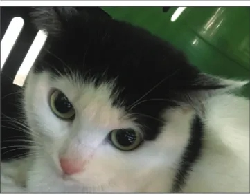
A five-years-old neutered female cat with the complaint of sudden onset of binocular blindness due to a white cloudy appearance in both eyes (Fig. 1). The owner was noted significant weight gain last 4 months after the sterilization procedure. Firstly, the examination is conducted in dim ambient light. Respectively, schirmer tear test and intra-ocular pressure were measured. Intraintra-ocular pressures was 15 mmHg in the left eye and 35 mm Hg in the right eyes, The result of schirmer tear test was shown referals. There was no obvious change in size and conformation of both eyes. The surface of cornea was clear and there was no evidence of vascularization, or ulceration during routine examination of both eyes under illumination of light. It was impossible to observe and evaluate the pole components of the anterior segment such as the anterior camera and iris because of the homogeneous blurred white color eyes. Nevertheless, iris has been evaluated for uveitis. Routine hematologic and serum biochemistry profiles were checked for the presence of inflammatory disorders. The bood was taken for complete blood count (Table 1), blood gas (Table 2) and serum biochemical profile analysis. Serum Gutamic-Pyruvic Transaminase (SGPT) 178 U/L (normal value 25-97 U/L), Cholesterol 182 mg/dL (normal value 71-156 mg/dL), Triglyceride 117 mg/dL (normal value 10-114 mg/dL), Amylase 2584 U/L (normal value 500-1800 U/L), LDH 135 U/L (normal value 58-120 U/L) [1]. The
inflammation was not observed. According to the ocular signs and laboratory findings, a diagnosis of lipemia and subsequent lipid-laden aqueous humor was considered. Treatmet started with subconjunctival dexamethasone sodium phospate injection and Dexamethasone 0.1% w/v Opthalmic Solution Drops (6 times in a day) were prescribed for white colored eye. Eye drops solution that each ml contains 22.26 mg of dorzolamide hydrochloride
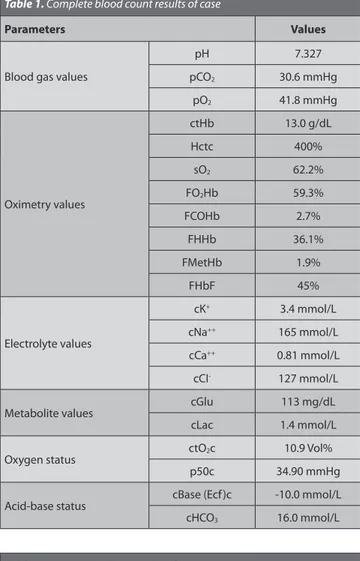
Table 1. Complete blood count results of case
Parameters Values
Blood gas values
pH 7.327 pCO2 30.6 mmHg pO2 41.8 mmHg Oximetry values ctHb 13.0 g/dL Hctc 400% sO2 62.2% FO2Hb 59.3% FCOHb 2.7% FHHb 36.1% FMetHb 1.9% FHbF 45% Electrolyte values cK+ 3.4 mmol/L cNa++ 165 mmol/L cCa++ 0.81 mmol/L cCI- 127 mmol/L
Metabolite values cGlu 113 mg/dL cLac 1.4 mmol/L
Oxygen status ctO2c 10.9 Vol%
p50c 34.90 mmHg Acid-base status cBase (Ecf)c -10.0 mmol/L cHCO3 16.0 mmol/L
Table 2. Venous blood gas results of case
Parameters Values WBC 19.41 m/mm3 Lym. 57.0% Mon. 6.9% Gra. 36.1% Lym# 11.06 m/mm3 Mon# 1.33 m/mm3 Gra# 7.02 m/mm3 RBC 10.83 m/mm3 MCV 43.6 fl Hct 47.2% MCH 13.5 pg MCHC 31.1 g/dL RDW 10.8 Hb 14.7 g/dL THR 213 m/mm3 MPV 10.2 fl Pct 0.22% PDW 9.0
Fig 1. A five-years-old neutered female cat with the complaint of
sudden onset of binocular blindness due to a white cloudy appearance in both eyes
911 ARICAN, PARLAK UZUNLU
corresponding to 20 mg dorzolamide and 6.83 mg of timolol maleate corresponding to 5 mg timolol drops were prescribed for high intraocular pressures. Urgently low-fat diet was recommended to patient. On the other hand, it was also assessed in terms of pancreatitis due to the high amylase level.
On the next day ophthalmologic examination, it was observed that the cloudy white color completely dis-appeared on both eyes and the intraocular components were clearly visible and there was no abnormality in iris shape and conformation (Fig. 2). For this reason, it was emphasized that lipidemia was the main reason. Intraocular pressures were 15 mmHg in the left eye and 37 mmHg in the right eyes for next day. The pupil size and pupillary light response were determined normal in both eyes. On direct ophthalmoscopy, optic disc and tapetal area appeared normal and both eyes seemed to be visual. Retinal examination was not shown any a pale appearance. Thus, no problems were encountered in the follow-up period of 8 months after the treatment and suggestion.
DISCUSSION
Primary lipid disorders are not commonly observed in cats. Lipid disorders secondary to hepatic lipidosis, diabetes mellitus, pancreatitis, hyperadrenocorticism and administraiton or pregestagens are more likely to account for fasting lipaemia in this species [2]. Lipid and/or mineral
accumulation appears as sparkly, crystalline or shiny white areas in the cornea. Plasma triglyceride and cholesterol concentrations were significantly increased in obese cats, compared with lean cats [2]. As a result of increasing the
lipid concentration in cats, lipids pass through the blood-aqueos barrier in the eye and cause the anterior chamber
to become opaque or cloudy [2]. The incidence of obesity
increases with age and is more frequent in neutered than intact animals [4]. Special diet programme should be
started to prevent obesity and obesity-related diseases in neutered cats [5,6]. The more common ocular consequences
of hyperlipidemia in small animals include lipemia retinalis and lipid-laden aqueous humor. In patients with lipemia retinalis, retinal vessels appear white or creamy pink, which is reminiscent of blood taken from an animal that has recently eaten a fatty meal [1]. The blood-aqueous barrier
generally prevents leakage of large molecules, like lipo-proteins, into the aqueous humor but when barrier has a disorder lipids then enter the eye and cause the aqueous humor to appear turbid, variably cloudy, and, in some cases, completely opaque [1]. Increased triglycerides may
result in brain dysfunction, acute pancreatitis, lipid-laden aqueous humor with anterior uveitis, and lipemia retinalis. Increased serum cholesterol can cause lipid keratopathy [6].
The main therapy of primary hyperlipidemia involves feeding a low-fat diet with moderate protein content. Diets low in protein may cause an increase in serum cholesterol concentration and are therefore not recommended unless the presence of other conditions warrant their use.
Although, lipid-Laden or hyperlipidemia is not seen very common in cats, but it should be considered in cats for the wrong diet programme. Cholesterol and triglycerides should be measured for diagnosis and prognosis, after routine eye examination. Uveitis must be assessed before diagnosis of lipid-Laden. Treatment options for hyperlipidemia include treatment of inciting diseases, diet modification, and pharmacologic intervention. Hyper- lipidemia secondary to an underlying disorder will probably resolve or improve after the metabolic disturbance is corrected.
REFERENCES
1. Kluger EK, Caslake M, Baral RM, Malik R, Govendir M: Preliminary
post-prandial studies of Burmese cats with elevated triglyceride concentrations and/or presumed lipid aqueous. J Feline Med Surg, 12, 621-630, 2010. DOI: 10.1016/j.jfms.2010.04.002
2. Turgut K: Erişkin hayvanlarda normal serum biyokimyası referans
değerleri. In, Veteriner Klinik Laboratuvar Teşhis. İkinci Baskı, 886, Bahçıvanlar, Konya, 2001.
3. Kluger EK, Hardman C, Govendir M, Baral RM, Sullivan DR, Snow D, Malik R: Triglyceride response following an oral fat tolerance test in
Burmese cats, other pedigree cats and domestic crossbred cats. J Feline
Med Surg, 11, 82-90, 2009. DOI: 10.1016/j.jfms.2008.05.005
4. Hoenig M, Wilkins C, Holson JC, Ferguson DC: Effects of obesity on
lipid profiles in neutered male and female cats. Am J Vet Res, 64, 299-303, 2003. DOI: 10.2460/ajvr.2003.64.299
5. Plummer CE, Specht A, Gelatt KN: Ocular manifestations of endocrine
disease. Compend Contin Educ Vet, 29 (12): 733-743, 2007.
6. Brooks DE, Sullivan M: Ocular manifestations of systemic diseases. In,
Schaer M, Gaschen F (Eds): Clinical Medicine of the Dog and Cat. 3rd ed.,
846-847, Taylor & Francis Group, New York, 2016.
Fig 2. After the drug administration intraocular components are clearly