26
Geliş(Recevied) :02/08/2018Kabul(Accepted) :20/02/2019 Araştırma Makalesi/Research Article Doi:10.30708mantar.450370
Biodiversity of Penicillium species isolated from Edirne
S
öğütlük Forest soil (Turkey)
Tugba Korur KOLANLARLI
1, Ahmet ASAN
2,
Burhan SEN
3, Suzan OKTEN
4*Corresponding author: [email protected]
1,2,3Trakya University Faculty of Science, Department of Biology. 22030 Edirne-TURKEY.
1Orcid ID: 0000-0001-6561-7384/ [email protected] 2Orcid ID: 0000-0002-4132-3848/ [email protected]
3Orcid ID: 0000-0002-8477-9570/ [email protected]
4Trakya University Faculty of Pharmacy, Department of Pharmaceutic Microbiology. 22030 Edirne-TURKEY. Orcid ID: 0000-0003-3372-5617/ [email protected]
Abstract: We studied Penicillium species isolated from forest soil and aimed to be detected with morphologically, colonial-molecular methods and species diversity. Soil sample was taken Edirne Söğütlük Forest in October, 2015 and 10 cm deep under aseptic conditions and Waksman’s ‘Soil Dilution Method’ was used for the isolation of Penicillium species; some books and articles have been used for morphological and colonial identification. The service was purchased for the molecular diagnosis of fungi and the company used the Speedy ™ Fungal DNA Isolation Kit for isolation. As PCR Kit, Bio-Speedy™, yeast and mould real-time PCR rapid detection kit was used. The obtained sequences were subjected to BLAST analysis similar sequences using the gene bank and to draw phylogenetic tree, used Mega 7 software. Twelve species belonging to the genus Penicillium were identified; these are: P. chrysogenum, P. citrinum, P. commune, P. manginii (current name: Penicillium atrosanguineum), P. ubiquetum (New record for Turkey), P. camemberti, P. sanguifluum, P. janczewskii, P. canescens, P. roseopurpureum, P. steckii (current name: Penicillium citrinum), P. hordei.
Key words: Soil, Fungi, Penicillium, Sanger Sequencing Method, ITS, Edirne.
Edirne İli (Türkiye) Söğütlük Ormanı toprağından izole edilen
Penicillium
türlerinin biyoçeşitliliği
Öz: Çalışmamızda orman toprağından izole edilen Penicillium türlerinin, morfolojik, koloniyal ve moleküler yöntemlerle tespiti amaçlanmış ve Penicillium tür çeşitliliği incelenmiştir. Toprak örnekleri, Edirne Söğütlük Ormanı’ndan 2015 yılı Ekim ayında, 10 cm derinden aseptik şartlarda alınmış olup, Penicillium türlerinin izolasyonu için Waksman’ın ‘Toprağı Sulandırma Yöntemi’ kullanılmış; morfolojik ve koloniyal tanımlaması için çeşitli eserler kullanılmıştır. Moleküler tanı amacıyla izolasyon işlemi için Bio-Speedy™ fungal DNA izolasyonu kiti kullanılmıştır. PCR kiti olarak ise Bio-SpeedyTM Maya ve Küf Real- Time PCR Hızlı Tespit Kiti kullanılmıştır. Elde edilen sekanslar gen bankası kullanılarak benzer sekanslarla BLAST analizi yapılmış ve Mega 7 yazılımı kullanılarak filogenetik ağaç çizilmiştir. Penicillium cinsine ait 12 tür tanımlanmıştır, bunlar: P. chrysogenum, P. citrinum, P. commune, P. manginii (geçerli isim: Penicillium atrosanguineum), P. ubiquetum (Türkiye için yeni kayıt), P. camemberti, P. sanguifluum, P. janczewskii, P. canescens, P. roseopurpureum, P. steckii (geçerli isim: Penicillium citrinum), P. hordei’ dir.
MANTAR DERGİSİ/The Journal of Fungus Nisan(2019)10(1)26-39
27
IntroductionPenicillium genus is common in different environments such as soil, plant, air and food products. Some species can use in cheese production, also some of them are pathogens for human; in addition, some species can use for antibiotics production. Besides, Penicillium is important genus because of commercial and industrial such as using production anti-tumoral, anti-fungal and anti-viral compounds, and using production of extra cellular enzymes. There are over 250 species of Penicillium genus (Visagie et al., 2014; Abastabar, 2016).
One of the first study on soil fungi of Turkey was performed by Oner (1970). Many studies performed after mentioned study indicated that the soils of Turkey has rich fungal biodiversity. Hasenekoglu et al. (Hasenekoğlu, 1982; Hasenekoğlu and Azaz, 1991; Hasenekoğlu and Sülün, 1991) on soil fungi especially in North-East of Anatolia. In Izmir and environs (Ekmekçi, 1975; Öner, 1974) and the other cities (İlhan and Asan, 2001; Asan and Ekmekçi, 2002; Azaz, 2003) for example.
The purpose of our study is isolation of Penicillium species from Edirne Söğütlük Forest soil and description of species by morphological, colonial and molecular methods.
Materials and Methods
Soil samples were obtained 10 cm depth in aseptic conditions from Edirne Söğütlük forest in October 03, 2015, to put sterile plastic bags and returned to laboratory. Soil samples are weighed 10 gr and put petri plates that tared. Petri plates were put to oven in 1050C during 24 h and weighed again so, we obtained air-dry-soil. After considering the moisture content, we calculated amount of moistured soil that equaled to 25 gr oven dried soil. The soil corresponding to 25 g of soil was placed into erlenmeyer flask (500 ml capacity) and a 9 % NaCl solution was added (10-1 dilution). This suspension was agitated in the shaking machine for 25 minutes for homogenization. After shaking, 10 ml of the soil particles from the 10-2 suspension were taken to the bottom, and 90 ml of NaCl solution was added there for to obtain a 10-3 dilution. Then, 10-5 and 10-6 dilution series were prepared. 1 ml samples from all dilutions, starting from 10-1 dilution series to 10-6 dilution series, were transferred to Petri dishes containing PDA (without any antibiotic) medium was poured on them and the samples incubated at 25°C for 5-7 days. PDA culture includes carbohydrate and potato. These components support the development of yeast and mould, while low pH
suppresses bacterial growth. Molds develop in this medium by showing typical morphologies. Pure culture was isolated from the growing fungal samples and the isolates were transferred to the PDA medium (Marshall, 1992; Vanderzant and Splittstoesser, 1992; Waksman, 1922).
Morphological Identification of Fungi
Species obtained from PDA medium were produced by three-point seeding on DG18 medium, from which glycerol stock solutions of molds were prepared. Czapek Yeast Extract Agar, Malt Extract Agar, 25 % Glycerol Nitrate Agar, Neutral Creatine Sucrose Agar and Yeast Extract Sucrose Agar media were prepared for the morphological and colonial identification of Penicillium species. Pitt (1979 and 2000), Samson et al. (2010) and Houbraken et al. (2010 and 2011) are used for identification of Penicillium species.
Molecular Identification of Fungi
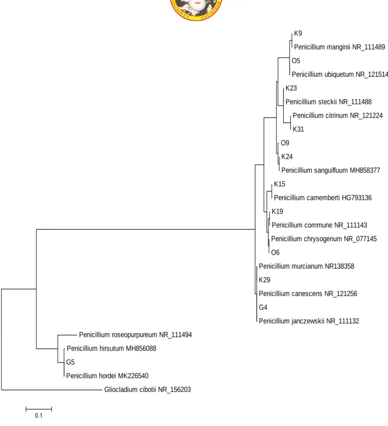
Bio-Speedy™, Fungal DNA Isolation Kit is used for isolation of fungi. As the PCR kit ( https://www.bioeksen.com.tr/fungal-dna-isolation-kit/), Bio-Speedy™, Fungal DNA İsolation Kit and Universal ITS primers (White et. al., 1990) were used for this purpose. In adition, to draw the phylogenetic tree, the evolutionary history was inferred by using the Maximum Likelihood method based on the Tamura-Nei model. The tree with the highest log likelihood (-2044.9430) is shown. Initial tree(s) for the heuristic search were obtained automatically by applying Neighbor-Join and BioNJ algorithms to a matrix of pairwise distances estimated using the Maximum Composite Likelihood (MCL) approach, and then selecting the topology with superior log likelihood value. The tree is drawn to scale, with branch lengths measured in the number of substitutions per site. Evolutionary analyses were conducted in MEGA7.
Results
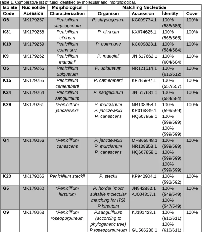
Eleven species of Penicillium genus have been identified in soil samples taken from Söğütlük Forest of Edirne City. These species are; P. chrysogenum, P. citrinum, P. commune, P. manginii (current name: Penicillium atrosanguineum), P. ubiquetum (New record for Turkey), P. camemberti, P. sanguifluum, P. janczewskii, P. canescens, P. steckii (current name: Penicillium citrinum), P. hordei’ (Table 1 and Figure 2-13). The obtained sequences were not submitted to the genbank since the most similar ITS hit of the blast search
28
was 100 % similar to the query nucleotide for all of the isolates.Table 1. Comparative list of fungi identified by molecular and morphological.
Isolate Nucleotide Acession
Morphological Matching Nucleotide
Code Characterization Organism Accession İdentity Cover
O6 MK179257 Penicillium chrysogenum P. chrysogenum KC009774.1 100% (585/585) 100% K31 MK179258 Penicillium citrinum P. citrinum KX674625.1 100% (565/565) 100% K19 MK179259 Penicillium commune P. commune KC009828.1 100% (584/584) 100% K9 MK179262 Penicillium manginii P. manginii JN 617662.1 100% (604/604) 100% O5 MK179266 Penicillium ubiquetum P. ubiquetum NR121514.1 100% (612/612) 100% K15 MK179255 Penicillium camemberti P. camemberti KF285997.1 100% (557/557) 100% K24 MK179264 Penicillium sanguifluum P. sanguifluum JN 617681.1 100% (584/584) 100% K29 MK179261 *Penicillium janczewskii P. murcianum P. janczewskii P. canescens NR138358.1 KP016839.1 HQ607858.1 100% (599/599) 100% (599/599) 100% (599/599) 100% G4 MK179256 *Penicillium canescens P. janczewskii P. murcianum P. canescens MH865548.1 NR138358.1 HQ607858.1 100% (599/599) 100% (599/599) 100% (599/599) 100%
K23 MK179265 Penicillium steckii P. steckii KP942904.1 100%
(592/592) 100% G5 MK179260 *Penicillium hirsutum P. hordei (most suitable molecular
matching for ITS) P.hirsutum JN942853.1 AJ004817.1 100% (549/549) 100% (547/549) 100% O9 MK179263 *Penicillium roseopurpureum P.sanguifluum (according to phylogenetic tree) P.roseopurpureum KJ191428.1 GU566236.1 100% (610/611) 100% (610/611) 100%
MANTAR DERGİSİ/The Journal of Fungus Nisan(2019)10(1)26-39
29
Figure 1. Molecular Phylogenetic analysis by Maximum Likelihood Method.
K9 Penicillium manginii NR_111489 O5 Penicillium ubiquetum NR_121514 K23 Penicillium steckii NR_111488 Penicillium citrinum NR_121224 K31 O9 K24 Penicillium sanguifluum MH858377 K15 Penicillium camemberti HG793136 K19 Penicillium commune NR_111143 Penicillium chrysogenum NR_077145 O6 Penicillium murcianum NR138358 K29 Penicillium canescens NR_121256 G4 Penicillium janczewskii NR_111132 Penicillium roseopurpureum NR_111494 Penicillium hirsutum MH856088 G5 Penicillium hordei MK226540 Gliocladium cibotii NR_156203 0.1
30
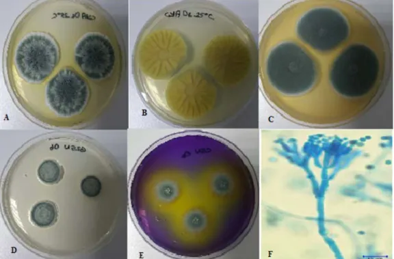
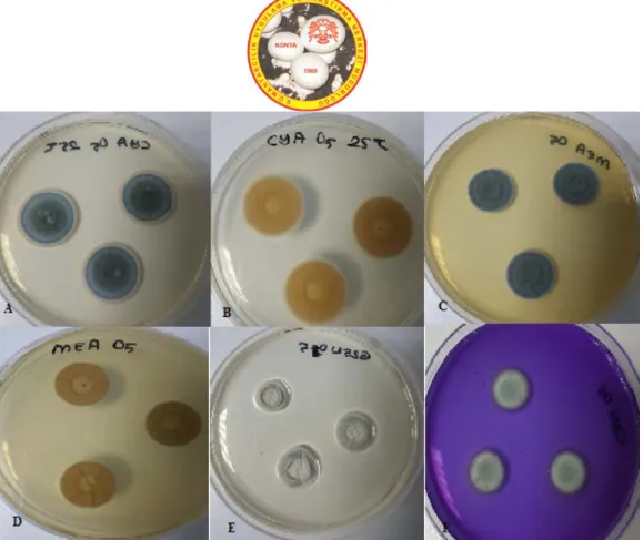
Identification of Penicillium chrysogenum (Thom 1910) was performed according to Pitt (1979 and 2000).
Figure 2. P. chrysogenum A. CYA medium, at 25°C, colonial morphology for seven days. B. CYA medium, 25°C, revers colonial morphology, C. MEA medium, 25°C, colonial morphology for seven days. D. G25N medium, 25ºC, colonial morphology for seven days, E. CSN medium, 25ºC, colonial morphology for seven days, F. Microscopic image of P. chrysogenum.
Identification of P. citrinum (Thom 1910) was performed according to the Pitt (2000), Houbraken et al. (2010, 2011).
Figure 3. P. citrinum A. CYA medium, 25°C, colonial morphology for seven days. B. CYA medium, 25°C, revers colonial morphology. C. MEA medium, 25°C, colonial morphology for seven days. D. G25N medium, 25ºC, colonial morphology for seven days, E. CSN medium, 25ºC, colonial morphology for seven days. F. Microscopic image of P. citrinum.
MANTAR DERGİSİ/The Journal of Fungus Nisan(2019)10(1)26-39
31
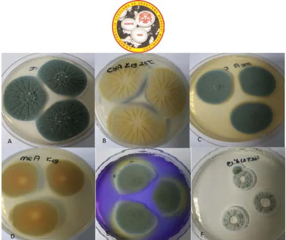
Figure 4. P. commune A. CYA medium, 25°C, colonial morphology for seven days. B. CYA medium, 25°C, revers colonial morphology for seven days. C. MEA medium, 25°C, colonial morphology for seven days. D. MEA medium, 25°C, revers colonial morphology for seven days. E. CSN medium, 25ºC, colonial morphology for seven days. F. G25N medium, 25°C, colonial morphology for seven days.
Identification of P. manginii was made by Houbraken et al. (2011).
Figure 5. P.manginii A. CYA medium, 25°C, colonial morphology for seven days. B. MEA medium, 25°C, revers colonial morphology for seven days. C. G25N medium, 25°C, colonial morphology for seven days. D. CSN medium, 25ºC, colonial morphology for seven days. E. YES medium, 25ºC, colonial morphology for seven days. F. Microscopic image of P. manginii.
32
Figure 6. P. ubiquetum A. CYA medium, 25°C, colonial morphology for seven days. B. CYA medium, 25°C, revers colonial morphology for seven days. C. MEA medium, 25°C, colonial morphology for seven days. D. MEA medium, 25°C, revers colonial morphology for seven days. E. G25N medium, 25ºC, colonial morphology for seven days. F. CSN medium, 25ºC, colonial morphology for seven days.
Identification of P.camemberti was made by Houbraken et al. (2011) and Samson et al. (2010).
Figure 7. P. camembertii A. CYA medium, 25°C, colonial morphology for seven days. B. MEA medium, 25°C, revers colonial morphology for seven days. C. G25N medium, 25°C, colonial morphology for seven days. D. CSN medium, 25ºC, colonial morphology for seven days. E. YES medium, 25ºC, revers colonial morphology for seven days. F. Microscopic image of P. camembertii.
MANTAR DERGİSİ/The Journal of Fungus Nisan(2019)10(1)26-39
33
Identification of P. sanguifluum was made by Houbraken (2011).Figure 8. P. sanguifluum A. CYA medium, 25°C, colonial morphology for seven days. B. MEA medium, 25°C, revers colonial morphology for seven days. C. G25N medium, 25°C, colonial morphology for seven days. D. CSN medium, 25ºC, colonial morphology for seven days. E. YES medium, 25ºC, revers colonial morphology for seven days. F. Microscobic image of P. sanguifluum.
Identification of Penicillium janczewskii was made by Pitt (2000).
Figure 9. P. janczewskii. CYA medium, 25°C, colonial morphology for seven days. B. MEA medium, 25°C, revers colonial morphology for seven days. C. G25N medium, 25°C, colonial morphology for seven days. D. CSN medium, 25ºC, colonial morphology for seven days. E. YES medium, 25ºC, revers colonial morphology for seven days. F. Microscopic image of P. janczewski.
34
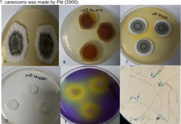
Identification of P. canescens was made by Pitt (2000).Figure 10. Penicillium canescens A. CYA medium, 25°C, colonial morphology for seven days. B. CYA medium, 25°C, revers colonial morphology for seven days. C. MEA medium, 25°C, colonial morphology for seven days. D. G25N medium, 25ºC, colonial morphology for seven days. E. CSN medium, 25ºC, revers colonial morphology for seven days. F. Microscobic image of P. canescens.
Identification of P. steckii was made by Houbraken et al. (2010 and 2011).
Figure 11. Penicillium steckii A. CYA medium, 25°C, colonial morphology for seven days. B. CYA medium, 25°C, revers colonial morphology for seven days. C. MEA medium, 25°C, colonial morphology for seven days. D. G25N medium, 25ºC, colonial morphology for seven days. E. CSN medium, 25ºC, revers colonial morphology for seven days. F. Microscobic image of P. steckii.
MANTAR DERGİSİ/The Journal of Fungus Nisan(2019)10(1)26-39
35
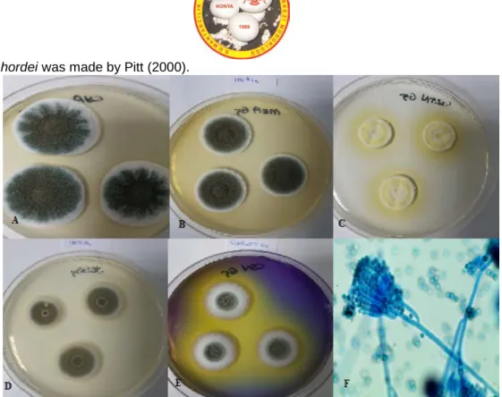
Identification of P. hordei was made by Pitt (2000).Figure 12. Penicillium hordei A. CYA medium, 25°C, colonial morphology for seven days. B. CYA medium, 25°C, revers colonial morphology for seven days. C. MEA medium, 25°C, colonial morphology for seven days. D. G25N medium, 25ºC, colonial morphology for seven days. E. CSN medium, 25ºC, ters colonial morphology for seven days. F. Microscobic image of P. hirsutum.
Identification of P. roseopurpureum was made by Houbraken et al. (2011).
Figure 13. Penicillium roseopurpureum A. CYA medium, 25°C, colonial morphology for seven days. B. CYA medium, 25°C, revers colonial morphology for seven days. C. MEA medium, 25°C, colonial morphology for seven days. D. G25N medium, 25ºC, colonial morphology for seven days. E. CSN medium, 25ºC, revers colonial morphology for seven days. F. Microcobic image of P. roseopurpureum.
36
DiscussionSoil is a complex environment containing various microorganisms; the number and diversity of fungi in this habitat varies depending to the environmental conditions. Many research has been performed about soil fungi in the world and in Turkey. For example, Kara and Asan (2007) reported that the number of fungi in the soil profile is continuously decreasing from top to bottom in all research stations.
There are some the other fungal studies in the soil of Thrace Region of Turkey such as Asan and Ekmekçi (1994), Asan (1997a, 1997b) and Asan et al. (2010). Asan
and Ekmekçi (1994) obtained soil samples for study of fungi from seven different habitats in Edirne City and one of these habitats is the Edirne Söğütlük Forest. Thirteen Penicillium species were found in mentioned habitat, but only two of them (P. camembertii and P. canescens) were also isolated in our study. There are 21 years between the two studies and there are various reasons for the different outcomes. First of all, the Meriç river is overflowing every 2-3 years in the region where it is obtaining intense alluvium from river (Türkmenoğlu, 2012; and Zal, 2006) (Figure 14 and 15).
Figure 14. Overflowing of Meriç and Tunca Rivers in March 14, 2006 (Photo by Ahmet Asan). Another important case is the methodology used in
the two studies. Only morphological and colonial methods were used in 1994, whereas molecular methods were used in this study additively. Also there are intensive human activities (backpacking, walking, etc.) in the area studied. Therefore, it was thought that it would be useful to study only one fungal genus (Penicillium) in the same region and different results were seen.
ITS region is widely used for fungi and its universal primers. However, for Penicillium and many other Ascomycetes, ITS is not sufficiently to distinguish all species for identication at species level [Visagie et al., 2014]. Skouboe et al. (1999) indicated that the ITS1 and ITS2 regions contains information can be used to support
taxonomical, ecological and physiological data of Penicillium species of common food sources, but the level of ITS region variability is low to the separation of closely related species. Additional sequences data from other fungal genes may provide sufficient character to establish phylogenetic relationships between Tervertisillate Penicillium. Because of the ITS-related limitations in Penicillium, a secondary gene region is usually required to identifying isolates for species level. This gene region should be useful for phylogenetic investigations. The second identification marker for the genus Penicillium is β-tubulin; however, there are also problems with this region. Therefore, in addition to ITS, β tubulin is useful; also ITS, β tubulin, Cam and RPB2 regions are
MANTAR DERGİSİ/The Journal of Fungus Nisan(2019)10(1)26-39
37
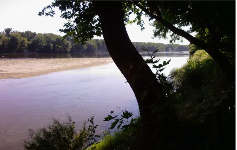
recommended for the identification of new species [Visagie et al. 2014].Figure 15. Normal state of Meriç River in August 15, 2008 (Photo by Ahmet Asan). Özdil et al. (2017) indicated that the phylogenetic
studies used protein-encoding genes and these genes contained highly variable intron regions. The most commonly used genes are elangation factor 1, calmodulin, β tubulin, actin and histone genes. These genes are more variable among species than the ITS region and are therefore more useful in identification of Penicillium species.
In our study, Penicillium chrysogenum, P. citrinum, P. commune, P. manginii, P. camemberti, P. ubiquetum and P. sanguifluum species were identified by ITS region and the identification was also supported by morphological colonial studies. The ITS gene sequences of the G4, G5, K29 and O9 isolates were matching with more than one species. However, the ITS sequence of the G5 isolate showed the optimal matching with P. hordei (Table 1) which the position in the phylogenetic tree (drawn using their related type strains) also supports this (Figure 1). Although the ITS sequence of O9 isolate shows 100% matching with two different species (Table
1), the phylogenetic tree (Figure 1) indicates P. sanguifluum.
The ITS data set and phylogenetic tree results of K29 and G4 isolates indicate Penicillium canescens, P. murcianum and P. janczewski species within the same query cover and identity. Morphological data have also been used for identification of these isolates. In conclusion, our study showed that, in addition to the ITS region for the identification of Penicillium species, the examinations of other alternative gene regions may be useful for identification of these species.
38
ReferencesAbastabar, M., Mirhendi, H., Hedayati, M.T., Shokohi, T., Matehkolaei, A.R., Mohammadi, R., Badali, H., Moazeni, M., Haghani, I., Ghojoghi, A. and Akhtari, J. (2016). Genetic and morphological diversity of the genus Penicillium from Mazandaran and Tehran Provinces. Jundishapur Microbial. 9 (1), 1-7.
Asan, A. and Ekmekçi, S. (2002). Contribution to the colonial and morphological characteristics of some Aspergillus species isolated from soil. J. Fac. Sci. Ege Univ. 25 (1), 121-139.
Asan, A. and Ekmekçi, S. (1994). The determination of Penicillium and Aspergillus species in Edirne soils and their seasonal distribution. Turk. J. Biol. 18 (4), 291-303.
Asan, A., Okten, S. and Sen, B. (2010). Airborne and soilborne microfungi in the vicinity Hamitabat Thermic Power Plant in Kırklareli City (Turkey), their seasonal distributions and relations with climatological factors. Environmental Monitoring and Assessment. 164 (1-4), 221-231.
Asan, A. (1997a). Trakya Bölgesi mısır tarlaları mikrofungus florası –I (Microfungi flora occurrence in the corn fields of European Part of Turkey-1). Turk. J. Biol. 21 (1), 89-101.
Asan, A. (1997b). Trakya Bolgesi misir tarlalari mikrofungus florasi uzerinde arastirmalar-II. (Studies on the microfungi flora occurrence in the corn fields of European Turkey-II). Kukem Derg. 20 (1), 9-18.
Azaz, A.D. (2003). Isolation of soil borne fungi in fields irrigated by GAP in Harran plain using two isolation methods, Turk. J. Bot. 27 (2), 83-92.
Ekmekçi, S. (1975). Güney yarı Ege Bölgesi topraklarından izole edilen Penicillium ve Aspergillus türleri. Bitki. 2, 19-29.
İlhan, S. and Asan, A. (2001). Soilborne fungi in wheat fields of Kırka Vicinity (Eskişehir - Turkey), Biologia. 56 (4), 363-371.
Hasenekoğlu, İ. and Sülün, Y. (1991). Erzurum Aşkale çimento fabrikasının kirlettiği toprakların mikrofunfus florası üzerine bir araştırma. Turk. J. Bot. 15 (1), 20-27.
Hasenekoğlu, İ. and Azaz, A.D. (1991). Sarıkamış civarındaki traşlanmış orman alanları topraklarının mikrofungus florası ve bunun normal orman toprakları florası ile karşılaştırılması üzerine bir araştırma. Turk. J. Bot. 15, 214-226.
Houbraken, J., Frisvad, J.C. and Samson, R.A. (2011). Taxonomy of Penicillium Section Citrina. Stud. In Mycol. 70, 53-138.
Houbraken, J., Frisvad, J.C. and Samson, R.A. (2010). Taxonomy of Penicillium citrinum and related species. Fungal Diversity. 44, 117-133.
Kara, Ö. and Asan, A. (2007). Microbial community structure from forest soils in Northern Thrace Region, Turkey. Annals of Microbiol. 57 (2), 149-155.
Marshall, R.T. (1992). Standard methods for the examination of dairy products, 16th ed. APHA, Washington, D.C.
Öner, M. (1970). Soil microfungi of Turkey. Mycopathol. Mycol. Appl. 42, 81-87.
Öner, M. (1974). Seasonal distribution of some Fungi Imperfecti in the soils of Western part of Anatolia. Mycopathol. Mycol. Appl. 52, 267-268.
Ozdil, S., Asan, A., Sen, B. and Okten, S. (2017). Evlerde kullanılan buzdolaplarının iç ortamında havayla taşınan fungusların biyoçeşitliliği (Biodiversity of airborne fungi in the indoor environment of refrigerators using in houses). Mantar Derg. 8 (2), 109-124.
Pitt, J.I. (2000). A laboratory guide to common Penicillium species. Food Science, Australia.
Pitt, J.I. (1979). The genus Penicillium and its teleomorphic states Eupenicillium and Talaromyces. Academic Press, London.
Samson, R.A., Houbraken, J., Thrane, U., Frisvad, J.C. and Andersen, B. (2010). Food and Indoor Fungi. CBS KNAW Fungal Biodiversity Centre. Utrecht, The Netherlands.
Skouboe, P., Frisvad, J.C., Taylor, J.W., Lauritsen, D., Boysen, M. and Rossen, L. (1999). Phylogenetic analysis of nucleotide sequences from the ITS region of Terveticillate Penicillium species. Mycol. Res. 103 (7), 873-881.
Thom, C. (1910). Cultural studies of species of Penicillium. U.S. Dept. Agriculture. Bur. Anim. Ind. Bul. Washington. 118, 1-109.
Türkmenoğlu, Y. (2012). Meriç Nehri’nin Kapıkule-Enez arasındaki yatak değişimlerinin taşkınlar ile ilişkisi. MSc Thesis. Istanbul Univ. Inst. Soc. Sci. Istanbul. Vanderzant, C. and Splittstoesser, D.F. (eds.). (1992).
Compendium of methods for the microbiological examination of foods. 3rd ed. APHA, Washington, D.C.
MANTAR DERGİSİ/The Journal of Fungus Nisan(2019)10(1)26-39
39
Visagie, C.M., Houbraken, J., Frisvard, J.C., Hong, S.B.,Klaassen, C.H.W., Perrone, G., Seifert, K.A., Varga, J., Yaguchi, T. and Samson, R.A. (2014). Identification and nomenclature of the genus Penicillium. Stud. In Mycol. 78, 343-371.
Waksman, S.A. (1922). A method for counting the number of fungi in the soil. J. Bact. 7 (3), 339-341. White, T.J., Bruns, T.D., Lee, S.B. and Taylor, J.W.
(1990). Amplification and direct sequencing of fungal ribosomal RNA genes for phylogenetics. 15, 315-322. In: Innis, M.A., Gelfand, D.H., Sninsky,
J.J., White, T.J. (Eds). PCR protocols: a guide to methods and applicationsPCR protocols: A guide to methods and applications. Academic Press, Inc., New York.
Zal, N. (2006). Aşağı Meriç Vadisi taşkın ovası’nın biyosfer rezervi olarak belirlenmesi üzerine bir araştırma (A study on the implementation of lower Meric Valley flood plain as a biosphere reserve). PhD Thesis. Ankara Univ. The Graduate School of Natural and Applied Sciences. Ankara.