Protective Effects of Ascorbic Acid,
DL
-
α-Tocopherol Acetate, and Sodium
Selenate on Ethanol-Induced Gastric
Mucosal Injury of Rats
S
ADAKATO
ZDIL,
1R
EF·
IYEY
ANARDAG,
2M
ERALK
OYUTURK,*
,3S
EHNAZB
OLKENT,
4 ANDS
ERAPA
RBAK51
Department of Internal Medicine, Istanbul Medical Faculty,
Istanbul University, 34390 Capa, Turkey;
2Department of
Chemistry, Faculty of Engineering, Istanbul University, 34850
Avcılar, Turkey;
3Department of Histology and Embryology,
Faculty of Medicine, Kadir Has University, 80810 Gayrettepe,
Turkey;
4Department of Biology, Faculty of Science, Istanbul
University, 34459 Vezneciler, Turkey; and
5Department of
Histology and Embryology, Marmara Medical Faculty,
Marmara University, 81326 Haydarpasa, Istanbul, Turkey
Received August 7, 2003; Revised September 25, 2003; Accepted October 14, 2003
ABSTRACT
In this study, the effect of ascorbic acid (vitamin C), DL-α-tocopherol
acetate (vitamin E), and sodium selenate (selenium) on ethanol-induced gastric mucosal injury in rats was investigated morphologically and bio-chemically. The gastric mucosal injury was produced by administration of 1 mL of absolute ethanol to each rat. Animals received vitamin C (250 mg/kg), vitamin E (250 mg/kg), and selenium (0.5 mg/kg) for 3 d 1 h prior to the administration of absolute ethanol. In gastric mucosa of rats given ethanol according to control groups, neuronal nitric oxide expression decreased. This immunoreactivity was much lower in the group given ethanol+vitamin C+vitamin E+selenium than the control group and the ethanol-induced group. Scanning electron microscopic evaluation of the ethanol-induced group, when compared to control groups, revealed degenerative changes in gastric mucosa, whereas a good arrangement in surface topography of gastric mucosa in the group given ethanol + vitamin C+vitamin E + selenium was observed. In the group administered ethanol,
© Copyright 2004 by Humana Press Inc. All rights of any nature, whatsoever, reserved. 0163-4984/04/99(1–3)–0173 $25.00
a reduction of the stomach glutathione (GSH) and serum total protein lev-els and increases in serum sialic acid, triglycerides, and stomach lipid per-oxidation (LPO) levels were observed. Vitamin C+vitamin E+Se administration to alcohol-treated rats significantly increased the serum total protein, triglyceride levels, and stomach GSH levels and significantly lowered the levels of serum sialic acid and stomach LPO compared to untreated alcohol-supplemented rats. As a result of these findings, we can say that the combination of vitamin C, vitamin E, and selenium has a pro-tective effect on ethanol-induced gastric mucosal injury of rats.
Index Entries:Ethanol; gastric mucosa; ascorbic acid; α-tocopherol acetate; sodium selenate; rat.
INTRODUCTION
Acute gastric mucosal injury is caused by various agents such as ethanol (1,2), stress (3), ischemic reperfusion (4), nonsteroidal anti-inflam-matory drug (5), acetic acid (6), indomethacin, and reserpine (7). Many stud-ies have demonstrated that inhibition of the enzyme involved in oxygen radical generation markedly reduced gastric mucosal injury induced by various agents. Oxygen radicals have been proposed to be pathogenic fac-tors in acute gastric mucosal injury in animals and human, and extracellu-larly generated reactive oxygen metabolites can be directly toxic to gastric mucosal cells in vitro and in vivo (8). The mechanism of ethanol-induced gastric mucosal injury is unclear and several factors have been implicated. It has been reported that oxygen-derived radicals could play an important role in the pathogenesis of ethanol-induced gastric mucosal damage and that free-radical production increased during exposure to ethanol (9–11).
Selenium (Se) is an essential component of the antioxidant defense system that is known to protect DNA and other cellular components from damage by oxygen radicals (12,13). It is located at the catalytic site of the enzyme glutathione peroxidase (GPx), and gastrointestinal glutathione peroxidase (GI-GPx) is a member of the glutathione peroxidase family
(14). One of the other antioxidant agents in our study is vitamin E, known
as dietary antioxidant and it inhibits peroxidation of membrane lipids (15) in gastric mucosal injury (16). Vitamin C scavenges reactive oxygen and nitrogen species and can thereby prevent oxidative damage to important biological macromolecules such as DNA, lipids, and proteins (17). High dietary ascorbic acid intake appears to protect against gastric cancer. This could be because of its action as a scavenger of reactive radical species formed in the gastric mucosa (18). Neuronal nitric oxide expression has been reported in rat gastric mucosa and NO could influence muscle tone as well as exocrine and endocrine functions (19). Nitric oxide plays a cyto-protective or cytotoxic role depending on its concentration (20).
The aim of the present study was to determine whether a combination of vitamin C, vitamin E, and selenium had a protective effect on gastric
mucosal injury induced by ethanol and the effects of antioxidant combi-nation to regulation of neuronal nitric oxide synthase activity.
MATERIALS AND METHODS
Animals
In this study, 40, 4- to 5-mo-old female Sprague–Dawley rats (Istanbul University Centre for Experimental Medicine Research and Application [DETAM]) were used in metal cages maintained at normal room tempera-ture. The animals were given a standard chow diet and tap water ad
libi-tum before the experiments and fasted for 24 h prior to the experiments.
All rats were clinically healthy.
Experimental Design and Treatment of Animals
A total of 40 rats were divided into 4 groups: group I: intact animals (control) (n=10); group II: control animals receiving vitamin C (250 mg/kg, orally, daily, for 3 d), vitamin E (250 mg/kg, orally, daily, for 3 d), and Se (0.5 mg/kg, orally, daily, for 3 d) (n=10); group III: animals receiving 1 mL absolute ethanol (n=10); group IV: animals receiving vitamin C, vitamin E, and Se for 3 d 1 h prior to the administration of absolute ethanol (in the same dose and time) (n=10). Animals were fasted overnight (18 h) prior to the experiment, but they were allowed free access to water. The rats were sacrificed at 1 h after ethanol exposure to ether.
Animal Model for Gastric Mucosal Lesions
The gastric mucosal lesions were produced intragastrically at a con-stant volume by 1 mL absolute ethanol per rat. The animals were killed 1 h after treatment with absolute ethanol and the stomachs were removed and then opened along the lesser curvature.
Immunohistochemical Study
The same paraffin block of morphological study of 3 µm was cut and then placed on poly-L-lysine-coated glass slides. Slides were deparaf-finized in toluol and hydrated in an ethanol series. The tissue was perme-abilized with 0.3% Triton X-100 for 10 min and then rinsed in phosphate-buffered saline (PBS) (10 mM, pH 7.5). For antigen retrieval, the slides were pressure cooked in 0.01 M citrate buffer using a standard household pressure cooker. When the pressure indicator valve has risen after about 5 min, sections were incubated for 5 min. Endogenous peroxi-dase was blocked with 3% hydrogen peroxide. A Histostatin Plus (Zymed Laboratories, USA) broad-spectrum kit of the streptavidin–biotin system was then employed. Sections were covered with blocking serum for 20 min to block nonspecific binding sites. They were then incubated with
ronal nitric oxide synthase (nNOS) antibody overnight (Transduction Lab-oratories, Lexington, KY) at 4°C at 1 : 100 dilution. Slides were incubated for 20 min with biotinylated secondary antibody and then incubated with the streptavidin–peroxidase conjugate for 20 min. The enzyme activity was developed using aminethylcarbazole (AEC) and then the sections were counterstained with hematoxylin. Negative control sections were prepared by substituting the nNOS antibody with PBS.
Electron Microscopical Study
For scanning electron microscopy, tissue samples were prefixed for 2 h in a 2% phosphate-buffered glutaraldehyde solution (0.1 M, pH 7.2) and postfixed for 1 h in a 1% phosphate-buffered osmium tetroxide solution and passed from increasing alcohol and amyl acetate series. After drying the tissue samples with a Bio-Rad “Critical Point Dryer” and gold coating with a Bio-Rad Sputter Coater (SC 502), tissue samples were examined under a JEOL 5200 JSM scanning electron microscope.
Biochemical Assays
Biochemical investigations were made in serum and tissue. Biochem-ical investigations of triglyceride and total protein in serum were meas-ured by means of an autoanalyzer (Targa 3000; Biotechnica). Serum sialic acid level was determined by the Lorentz methods (21).
For biochemical analyses, tissue samples of the stomach were washed with physiological saline and kept frozen until the day of the experiments. Stomach was then homogenized in cold 0.9% NaCl with a glass homoge-nizer to make up 10% homogenate (w/v). These homogenates were cen-trifuged. The clear supernatants were used for protein, lipid peroxidation (LPO), and glutathione (GSH) analyses.
The LPO levels were determined according to the method of Led-wozwy (22). In brief, the adducts formed following boiling tissue homogenate with thiobarbituric acid and extracted with n-butanol. The difference in optical density at 532 nm is a measure of the stomach malon-dialdehyde (MDA) content as a measure of thiobarbituric acid reactive species (TBARS), which is undertaken as an index of lipid peroxidation.
Reduced glutathione (GSH) was determined according to the method by Beutler using Ellman’s reagent. The procedure is based on the reduc-tion of Ellman’s reagent by SH groups to form 5,5′-dithio-bis(2-nitroben-zoic acid), which has an intense yellow color that is measured spectrophotometrically at 412 nm using a Shimadzu spectrophotometer
(23). The protein content in the supernatants was determined by the
Lowry method (24).
Statistical Study
The results were evaluated using an unpaired t-test and analysis of variance (ANOVA) using the NCSS statistical computer package (25).
RESULTS
Immunohistochemical Results
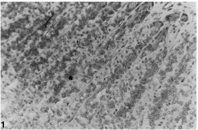
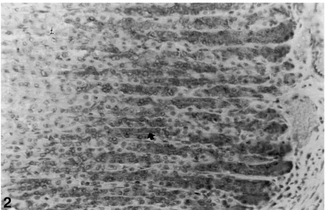
The nNOS immunoreactivity was determined in the lower half of the oxyntic gland in the gastric mucosa. Strong immunoreactions were located in parietal and chief cells of control gastric mucosa and also the intensity of enteroendocrine cells immunoreactions was quite lower than the other cells (see Fig. 1). Similar reactions were found in the control group given an antioxidant (see Fig. 2). The intensity and number of immunoreactive cells decreased in the ethanol-induced gastric mucosa (see Fig. 3). The reaction of the group given ethanol+antioxidant was lower than control group and much lower than the ethanol-induced group (see Fig. 4).
Electron Microscopic Results
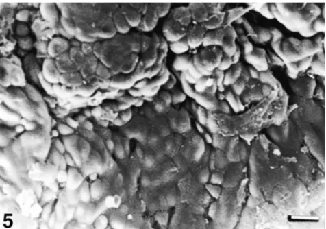
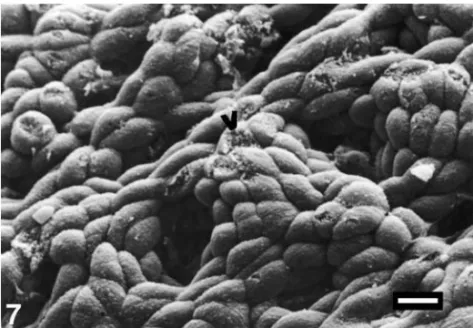
Scanning electron microscopic evaluation of the control groups revealed a good surface topography of gastric mucosa (see Fig. 5). In the group given absolute ethanol, extreme mucosal degeneration, exfoliation in epithelial cells, epithelial cell separation, deep erosions and gastric pits, exposure of basal lamina, fibrin deposits, and deformation in erythrocytes were revealed (see Fig. 6). In the group given ethanol +vitamin C+vitamin E+ Se, a good epithelial arrangement in surface topography of gastric mucosa except for minimal epitelial cell separation was observed (see Fig. 7).
Vitamins and Se Protection Against Gastric Injury 177
Fig. 1. nNOS immunreactivity (➧) in the oxyntic gland in gastric mucosa of control rats (magnification: ×520).
Fig. 2. nNOS immunre activity (➧) in the oxyntic gland in gastric mucosa of control rats given vitamin C, vitamin E, and selenium (magnification: ×520).
Fig. 3. A decrease in nNOS immunreactivity (➧) in the oxyntic gland in gas-tric mucosa of the rats given ethanol (magnification: ×520).
Biochemical Results
The results of the present experiment are shown in Tables 1 and 2. The serum total protein levels in the ethanol group (7.69 ± 0.22 g%) were found to be significantly lower than in the control group (8.12 +0.47 g%)
Vitamins and Se Protection Against Gastric Injury 179
Fig. 4. The much lower immunreactivity (➧) in the oxyntic gland in gastric mucosa of the rats given ethanol+vitamin C+vitamin E+ Se according to control groups and ethanol-induced group (magnification: ×520).
(p=0.005). In the ethanol + vitamin C+vitamin E+Se group (9.02 ± 0.30 g%), they were high compared to the ethanol group (p= 0.0001) (see Table 1).
Serum triglyceride levels in ethanol group (76.34 ±2.83 mg%) were significantly increased compared to control group (64.45 ±1.35 mg%). In ethanol +vitamin C+vitamin E+Se group, serum triglyceride levels were high compared to the ethanol group (p=0.027) (see Table 1). Serum sialic acid levels in the ethanol group (2.62 ± 0.22 mmol/L) were significantly higher than in the control group (1.95 ± 0.30 mmol/L). Serum sialic acid
Fig. 6. Separation in epitheliall cells (➧), exposure of basal lamina (V), fibrin deposits (䉴), and deformation in erythocytes (夹) (bar= 10 µm).
Fig. 7. Same topography as those of the controls in the group given ethanol +vitamin C+vitamin E+ Se (bar= 10 µm).
Vitamins and Se Protection Against Gastric Injury 181
T
able 1
Mean Levels of Ser
um Parameters* * Mean ±SD; n =number of animals. ap=0.0001 versus contr ol gr oups. bp=0.0001 versus contr ol gr oups. cp=0.001 versus contr ol gr oups.
T
able 2
Mean Levels of Stomach Glutathione and Lipid Per
oxidation for All Gr oups* * Mean ± SD; n =number of animals. ap=0.0001 versus contr ol gr oups. bp=0.0001 versus contr ol gr oups.
levels were decreased in the ethanol +vitamin C+vitamin E+Se group (1.92 ± 0.46 mmol/L) compared to the ethanol group (see Table 1).
The mean stomach LPO and GSH levels of four groups are given in Table 2. According to Table 2, a significant difference in the stomach GSH levels of the four groups was observed (p=0.0001). Alcohol administration significantly reduced the GSH levels in rat stomach tissues compared to control animals. Administration of vitamin C+vitamin E+Se to rats increased the stomach GSH content in ethanol groups.
Stomach LPO levels in ethanol group were significantly increased compared to the control group (p= 0.0001). Administration of vitamin C+vitamin E+Se caused a significant decrease in the LPO levels in the ethanol group (p=0.0001), but vitamin C+vitamin E+Se caused a signifi-cant increase in LPO levels in the control groups (p= 0.0001).
DISCUSSION
Ethanol is a direct systemic toxin that produces injury to all tissue, depending on dose and duration of exposure (26,27). Acute gastric mucosal damage is produced by various mucosal damaging agents such as ethanol, aspirin, strong acids and bases, and stress (28). It has been reported that free radicals could play an important role in pathogenesis of acute gastric lesions induced by ethanol and that free-radical production increased during exposure to ethanol (29,30).
The low amounts of GI-GPx protein has been reported in selenium deficiency. The function of GI-GPx has been discussed as a primary barrier against the absorbtion of ingested hydroperoxides (31). The protective effects of selenium reported via inhibition of gastric lesions by the mucosal regeneration of prostaglandins (32). Also, an increasing GPx level was reported in combined protective effect of selenium and ascorbic acid against alcohol-induced oxidative stress (33).
α-Tocopherol interacted with the biological reductants NADH and NADPH in microsomal membranes. Protection of vitamin E was reported by inhibiting lipid peroxidation and accumulation of activated neutrophils
(5). Another study on ethanol-induced gastric lesions reported reduced
basal gastric acid secretion with treatment of Se and vitamin E when given individually. The better inhibition of gastric acid secretion was reported with the combination effects of these agents (7). However, some authors reported that neither gastric acidity nor mucus protection are involved in the gastroprotective effects of vitamin E (34,35). The antioxidant synergism was reported also between α-tocopherol and ascorbic acid (36). Vitamin E shares with vitamin C the ability to inhibit nitrosamine formation in the stomach (37). Vitamin C acts as a reducing compound in the aqueous part of the cell and vitamin E acts as scavenging free radicals and stabilizes the membrane in the cell (38). Vitamin E administration was reported to reduce protein glycosylation in diabetic patients and a similar inhibiting
effect of vitamin C (39). Vitamin C also prevents oxidative lesions in DNA that include base modifications, sugar damage, and strand breaks (40).
Several findings reported that NO is important for normal gastric function. NO is necessary for the intracellular barrier of the gastrointesti-nal epithelium (41). The protective effects of NO was also reported via increasing mucus secretion and regulation of gastric acid secretion (42,43). Nitric oxide is a molecule capable of reacting via multiple pathways to modulate lipid oxidation reactions and effects on the inflamatory process
(44). Nitric oxide synthase decreased the ischemia-reperfusion-induced
gastric mucosal injury and the effects of NO was reported as an antioxi-dant defensive role by maintaining mucus, glutathione, and glutathione peroxidase (45). Neuronal nitric oxide expression was determined in pari-etal cells, chief cells, mucosecretory cells of gastric epithelium, and some enteroendocrine cells. The findings of studies reported that NO might influence parietal cell secretion directly as an intracellular signaling mole-cule and/or indirectly by acting on adjacents cells (19,46). In our study, immunoreactions of nNOS decreased ethanol-induced gastric mucosa lower than the normal group. The antioxidant and ethanol group immunreactions were decreased compared to the ethanol-induced group. According to these findings, the visible healing in the cases of antioxidant and ethanol group is independent of nNOS expressions.
Our study showed that antioxidants improved the integrity of gastric mucosal epithelium and reduced the degree of damage in the mucosal architecture. The mucosal injury is the result of the hazardous effect of ethanol, which rapidly penetrates gastric mucosa, causing membrane damage. The prominent epithelial damage of the ethanol-induced group could be the result of increased lipid peroxidation of cell membranes, lead-ing to cell death. It is reported that pharmacological antioxidants could have beneficial effects in reducing the incidence of ethanol-induced changes in cellular lipids, proteins, and nucleic acids (47). Microscopic evaluation of gastric mucosa of the antioxidant treated group revealed a significant reduction in injury formation. We could correlate these findings with the free-radical-trapping activity of antioxidants.
Antioxidants are essential in preventing the cellular damage caused by free radicals and free-radical-modified LPO. In normal metabolism, there is a balance between the generation of free radicals and the antioxi-dant defense mechanism. Chronic ingestion of alcohol upsets this balance, and there is ample evidence to demonstrate the oxidative stress induced by ethanol (3). Antioxidants prevent new radical species formation by con-verting existing free-radical species into less harmful molecules or by pre-venting the transformation of free radicals from other molecules (48). The protective effect of some antioxidants has been demonstrated light micro-scopically in the treatment of gastric mucosal injury (28). Low-molecular-weight antioxidants also play important roles in preventing free-radical damage. Vitamin E disrupts the chain reaction of lipid peroxidation by scavenging intermediate peroxyl radicals and also traps mutagenic
elec-trophilics such as NO (49). Some studies have shown that vitamin E has a protective effect against gastric mucosal injury induced by ischemia reper-fusion and other insults (50). In addition, it has been demonstrated that macroscopically vitamin E has a protective effect against gastric mucosal injury induced by NSAIDs (nonsteroidal anti-inflammatory drugs) (50). Vitamin C or ascorbic acid is a naturally occurring free-radical scavenger, and as such, its presence assists various other mechanisms in decreasing the numerous distruptive free-radical processes (3) from taking place, including LPO. Recent studies have shown that both vitamins E and C are reduced in alcoholics. Chakrabarthy et al. (51) have shown that ascorbate protects guinea pig tissues from LPO both in vivo and in vitro. Selenium is an essential part of the enzyme glutathione peroxidase, which functions as part of an antioxidant.
Lipid peroxidation mediated by free radicals is considered to be a pri-mary mechanism of cell membrane destruction (52). Ethanol has been shown to increase LPO in gastric mucosa (53). Some publications have indicated high levels of malondialdehyde (MDA) and conjugated dienes in cell membranes of animals subjected to acute or chronic ethanol intoxi-cation (54). Oxidative injury can cause inflammation in the gastric mucosa as a result of infiltration of polymorphonuclear leukocytes into the lesion
(53). MDA and hydroperoxides levels increased in the alcohol-fed animals
and were substantially lower in the alcohol + ascorbic acid-fed animals
(55). MDA levels in gastric mucosa, which is an index of LPO, increased
significantly with ethanol administration (see Table 2). The ethanol-induced LPO in gastric mucosa was inhibited by vitamins C and E and Se. The inhibitor activity of antioxidant (vitamins C and E and Se) could result from scvanging reactive oxygen species, which initate LPO. Alternatively, the inhibition could occur at the membrane level by inhibiting oxidore-ductase activity (53). Glutathione is an important constituent of an intra-cellular protective mechanism against various toxic stimuli, including oxidative stress. Reduced glutathione is known to be a major low-molecu-lar-weight scavenger of free radicals in the cytoplasm (56). The stomach is rich in GSH, where it might serve a protective role similar to that observed in the liver, namely removal of free radicals and maintaining mucosal integrity (56). Ethanol rapidly penetrates gastroduodenal mucosa, causing membrane damage, exfoliation of cells, and erosion. Ethanol-induced gas-tric mucosal damage is associated with a significant reduction in nonpro-tein sulfhydryl concentration in the rat, dog, and humans (56). In our study, ethanol significantly decreased gastric GSH concentration. This reduction could be the result of the oxidation of GSH because of the ethanol-induced generation of toxic oxygen metabolities or the binding of GSH to acetaldehyde generation through the oxidation of ethanol by the gastric alcohol dehydrogenase (57). Alternatively, a block in the synthesis of the tripeptide induced by ethanol-generated free radicals might occur. In our study, administration of vitamins C and E and Se significantly increased stomach GSH levels in the ethanol group. This effect might be
the result of the activator role of Se on GSH-Px activity and indicate that these three substances effectively protect membrane integrity.
Many reports indicated that alcohol intake significantly increases serum triglyceride levels resulting in hypertriglyceridemia and fatty liver
(58). Ethanol oxidation by the alcohol dehydrogenase pathway results in
the production of NADH, which might contribute to enhanced lipid syn-thesis. Ethanol itself is converted into acetate, which is the building block of fatty acid synthesis. Ethanol oxidation has been shown to increase α-glycerophosphate levels (59), which can lead to enhance triglyceride syn-thesis. In our study, serum triglyceride levels were increased by the application of ethanol, but vitamins C and E and Se show that antioxidants prevent the damage caused by ethanol.
Sialic acid is an acetylated derivative of neuraminic acid (60). It attaches to nonreducing residues of the carbohydrate chains of glycopro-teins and glycolipids (61). An increase in serum total sialic acid levels has been previously reported in diabetes, cardiovascular disease, inflamma-tory disorders, and malignant disease, including breast cancer and differ-ent solid tumors and leukemias. Recdiffer-ent studies have shown that sialic acid concentration in serum might be increased in alcoholics (61). In our study, the sialic acid levels were significantly higher in the ethanol groups that in control groups. Administration of vitamin C+vitamin E+Se significantly decreases sialic acid in the serum of the ethanol group.
As a result, the microscopical and biochemical evaluations reveal that the combination of vitamin C, vitamin E, and Se has a protective effect on ethanol-induced gastric mucosal injury of rats. However, this protective effect is independent of nNOS expressions.
REFERENCES
1. E. S. L. Liu and C. H. Cho, Relationship between ethanol-induced gastritis and gastric
ulcer formation in rats, Digestion 62, 232–239 (2000).
2. S. Ozdil, S. Bolkent, R. Yanardag, et al., Protective effects of ascorbic acid, DL
-α-toco-pherol acetate and sodium selenate on ethanol-induced liver damage of rats, Biol. Trace
Element Res., 97, 149–162 (2004).
3. M. V. Suresh, S. Kumar, J. J. Lal, et al., Impact of massive ascorbic acid supplementation
on alcohol induced oxidative stress in quinea pigs, Toxicol. Lett. 104, 221–229 (1999).
4. G. S. Smith, D. W. Mercer, J. M. Cross, et al., Gastric injury induced by ethanol and
ischemia-reperfusion in the rat, Dig. Dis. Sci. 41, 1157–1174 (1996).
5. N. Sugimoto, N. Yoshida, T. Yoshikawa, et al., Effect of vitamin E on aspirin-induced
gastric mucosal injury in rats, Dig. Dis. Sci. 45, 599–605 (2000).
6. M. Ito, T. Segami, T. Tsukahara, et al., Effect of cimetidine and omeprazole on gastric
ulcer healing of rats with limited food intake time, Eur. J. Pharmacol. 263, 245–251 (1994).
7. A. R. Al-Moutary and M. Tariq, Effect of vitamin E and selenium on hypothermic
restraint stress and chemically-induced ulcers, Dig. Dis. Sci. 41, 1165–1171 (1996).
8. T. Yoshikawa, Y. Minamiyama, H. Ichikawa, et al., Role of lipid peroxidation and
antioxidants in gastric mucosal injury induced by the hypoxanthine–xanthine oxidase system in rats, Free Radical Biol. Med. 23, 243–250 (1997).
1 2 3 4 5 6 8
9. H. Mutoh, H. Hiraishi, S. Ota, et al., Role of oxygen radicals in ethanol-induced
dam-age to cultured gastric mucosal cells, Am. J. Physiol. Gastrointest. Liver Physiol. 258, 603–609 (1990).
10. D. Rachmileewitz, F. Karmeli, E. Okon, et al., A novel antiulcerogenic stable radical
pre-vents gastric mucosal lesions in rats, Gut 35, 1181–1188 (1994).
11. P. R. Kvietys, M. A. Perry, T. S. Gaginella, et al., Ethanol enhances
leukocyte–endothe-lial cell interactions in mesenteric venules, Am. J. Physiol. Gastrointest. Liver Physiol. 259, 578–583 (1990).
12. G. N. Schrauzers. Anticarcinogenic effects of selenium, Cell. Mol. Life Sci. 57, 1864–1873
(2000).
13. R. D. Baker, Jr., S. S. Baker, and R. Rao, Selenium deficiency in tissue culture:
implica-tions for oxidative metabolism, J. Pediatr. Gastroenterol. Nutr. 27, 387–392 (1998).
14. K. M. Brown and J. R. Arthur, Selenium, selenoproteins and human health: a review, Public Health Nutr. 4, 593–599 (2001).
15. J. M. Patel and D. A. Edwards, Vitamin E, membrane order, and antioxidant behavior
in lung microsomes and reconstituted lipid vesicles, Toxicol. Appl. Pharmacol. 96, 101–114 (1988).
16. T. Yoshikawa, M. Yasua, S. Udea, et al., Vitamin E in gastric mucosal injury induced by
ischemia-reperfusion, Am. J. Clin. Nutrit. 53, 210–214 (1991).
17. A. Carr and B. Frei, Does vitamin C act as a pro-oxidant under physiological
condi-tions, FASEB J. 13, 1007–1024 (1999).
18. I. M. Drake, M. J. Davies, N. P. Mapstone, et al., Ascorbic acid may protect against
human gastric cancer by scavenging mucosal oxygen radicals, Carcinogenesis 17, 559–562 (1996).
19. S. Premaratne, C. Xue, J. M. McCarty, et al., Neuronal nitric oxide synthase: expression
in rat parietal cells, Am. J. Physiol. 280, G308–G313 (2001).
20. S. Zöllner, R. F. Haseloff, I. A. Kirilyuk, et al., Nitroxides increase the detectable amount
of nitric oxide released from endothelial cells, J. Biol. Chem. 272, 23,076–23,080 (1997).
21. K. Lorentz and E. Kraas, Sialic acid in human serum and cerebrospinal fluid, J. Clin. Chem. Clin. Biochem. 24, 189–198 (1986).
22. A. Ledwozyw, J. Michalak, A. Stepien, et al., The relationship between plasma
triglyc-erides, cholesterol, total lipids and lipid peroxidation products during human athero-sclerosis, Clin. Chim. Acta 155, 275–284 (1986).
23. E. Beutler, Glutathione in Red Blood Cell Metabolism: A Manual of Biochemical Methods, 2nd
ed., Grunne and Stratton, New York, pp. 112–114 (1975).
24. O. H. Lowry, W. I. Rosebrough, A. L. Farr, et al., Protein mesurement with the Folin
Phenol reagent, J. Biol. Chem. 193, 265–275 (1951).
25. J. L. Hintze, Copyright C, 865, East 400. North Kaysville, Utah. 84, 037 (801), 546-0445
(1986).
26. H. M. M. Arafa and M. M. Sayed-Ahmed, Protective role of carnitine esters against
alcohol-induced gastric lesions in rats, Pharmacol. Res. 48, 285–290 (2003).
27. G. Kanbak, M. Inal, and C. Bayçu, Ethanol-induced hepatotoxicity and protective effect
of betaine, Cell Biochem. Funct. 19, 281–285 (2001).
28. S. Ozdil, Protective effect of vitamin C, vitamin E and selenium on acute experimental
gastric lesions induced by ethanol in the rats: a morphological study, J. Morphol. 9, 50–53 (2001).
29. R. Hernandez-Munoz, C. Montiel-Ruiz, and O. Vazquez-Martinez, Gastric mucosal cell
proliferation in ethanol-induced chronic mucosal injury is related to oxidative stress and lipid peroxidation in rats, Lab. Invest. 80, 1161–1169 (2000).
30. P. Navasumrit, T. H. Ward, N. J. Dodd, et al., Ethanol-induced free radicals hepatic
DNA strand breaks are prevented in vivo by antioxidants: effects of acute and chronic ethanol exposure, Carcinogenesis 21, 93–99 (2000).
Vitamins and Se Protection Against Gastric Injury 187
13 14 15 17 18 22 24 26 27 29 30
31. K. Wingler and R. Brigelius-Floh, Gastrointestinal glutathione peroxidase, Biofactors 10,
245–249 (1999).
32. N. S. Parmar, M. Tariq, and M. Ageel, Gastric anti-ulcer and cytoprotective effect of
selenium in rats, Toxicol. Appl. Pharmacol. 92, 122–130 (1988).
33. A. G. Sivaram, M. V. Suresh, and M. Indira, Combined effect of ascorbic acid and
sele-nium supplementation on alcohol-induced oxidative stress in guinea pigs, Comp.
Biochem. Physiol. C: Toxicol. Pharmacol. 134, 397–401 (2003).
34. K. Jaarin, M. Renuvathani, M. I. Nafeeza, et al., Effect of palm vitamin E on the healing
of ethanol-induced gastric injury in rats, Int. J. Food Sci. Nutr. 51, S31–S41 (2000).
35. K. Jaarin, M. Renuvathani, M. I. Nafeeza, et al., Comparative effect of palm vitamin E
and ranitidine on the healing of ethanol-induced gastric lesions in rats, Int. J. Exp.
Pathol. 80, 259–263 (1999).
36. D. C. Liebler, The role of metabolism in the antioxidant function of vitamin E, Crit. Rev. Toxicol. 23, 147–169 (1993).
37. T. Byers and G. Perry, Dietary carotenes, vitamin C, and vitamin E as protective
antiox-idants in human cancers, Annu. Rev. Nutr. 12, 139–159 (1992).
38. L. H. Chen, G. A. Boissonneault, and H. P. Glauert, Vitamin C, vitamin E and cancer, Anticancer Res. 8, 739–748 (1988).
39. A. Ceriello, D. Giugliano, A. Quatraro, et al., Vitamin E reduction of protein
glycolisa-tion in diabetes, Diabetes Care 14, 68–72 (1991).
40. E. A. Lutsenko, J. M. Carcomo, and D. W. Golde, Vitamin C prevents DNA mutation
induced by oxidative stress, J. Biol. Chem. 277, 16,895–16,899 (2002).
41. A. Banan, J. Z. Fields, H. Decker, et al., Nitric oxide and its metabolites mediate
ethanol-induced microtubule disruption and intestinal barrier dysfunction, J. Pharmacol. Exp.
Ther. 294, 997–1008 (2000).
42. A. Berg, S. Kechagias, S. E. Sjöstrand, et al., Morphological support for paracrine
inhi-bition of gastric acid secretion by nitric oxide in humans, Scand. J. Gastroenterol. 36, 1016–1021 (2001).
43. J. F. Brown, A. C. Keates, P. J. Hanson, et al., Nitric oxide generators and cGMP
stimu-late mucus secretion by rat gastric mucosal cells, Am. J. Physiol. 265, G418–G422 (1993).
44. A. Bloodsworth, V. B. O’Donnell, and B. A. Freeman, Nitric oxide regulation of free
rad-ical- and enzyme-mediated lipid and lipoprotein oxidation, Arteriosclerosis Thromb.
Vasc. Biol. 20, 1707–1715 (2000).
45. H. Kim and K. K. Hwan, Role of nitric oxide and mucus in
ischemia/reperfusion-induced gastric mucosal injury in rats, Pharmacology 62, 200–207 (2001).
46. M. G. Vitoria, C. G. Corchon, J. A. Rodriguez, et al., Exspression of neuronal nitric oxide
synthase in several cell types of the rat gastric epithelium, J. Histochem. Cytochem. 48, 1111–1119 (2000).
47. R. Nordmann, Alcohol and antioxidant systems, Alcohol Alcohol. 29, 513–522 (1994). 48. J. Ren, Z. K. Roughead, L. E. Wold, et al., Increases in insulin-like growth factor-1 level
and peroxidative damage after gestational ethanol exposure in rats, Pharmacol. Res. 47, 341–347 (2003).
49. D. Anderson and B. J. Phillips, Comparative in vitro and in vivo effects of antioxidants, Food Chem. Toxicol. 37, 1015–1025 (1999).
50. N. Sugimoto, N. Yoshida, T. Yoshikawa, et al., Effect of vitamin E on aspirin-induced
gastric mucosal injury in rats, Dig. Dis. Sci. 45, 599–605 (2000).
51. S. Chakraborthy, A. Nandy, M. Mukhopadhyay, et al., Ascorbate peotects guanea pigs
tissue against lipid peroxidation, Free Radical Biol. Med. 16, 417–426 (1994).
52. V. Balasubramaniyan, J. K. Sailaja, and N. Nalini, Role of leptin on alcohol-induced
oxidative stress in Swiss mice, Pharmacol. Res. 47, 211–216 (2003).
53. J. S. Park, M. A. Choi, B. S. Kim, et al., Capsaicin protects against ethanol-induced
oxidative injury in the gastric mucosa of rats, Life Sci. 67, 3087–3093 (2000).
31 32 33 34 35 36 37 38 39 40 41 42 43 45 47 48 49 50 51 52 53
54. C. Coudray, M. J. Richard, H. Faure, et al., Blood and liver lipid peroxide status after
chronic ethanol administration in rats, Clin. Chim. Acta 19, 35–45 (1993).
55. K. H. McDonough, Antioxidant nutritients and alcohol, Toxicology 189, 89–97 (2003). 56. G. S. Muratoglu, K. Paskaloglu, S. Arbak, et al., Protective effect of famotidine,
omepra-zole, and melatonin against acetylsalicylic acid-induced gastric damage in rats, Dig.
Dis. Sci. 46, 318–330 (2001).
57. D. Bilici, H. Suleyman, Z. N. Banoglu, et al., Melatonin prevents ethanol-induced
gas-tric mucosal damage possibly due to its antioxidant effect, Dig. Dis. Sci. 47, 856–861 (2002).
58. K. J. Park, M. J. Lee, H. Kang, et al., Saeng-maek-san, a medicinal herb complex,
pro-tects liver cell damage induced by alcohol, Biol. Pharm. Bull. 25, 1451–1455 (2002).
59. J. Kaur, Chronic ethanol feeding affects intestinal mucus lipid composition and
glyco-sylation in rats, Ann. Nutr. Metab. 46, 38–44 (2002).
60. M. Ponnio, H. Alho, P. Heinala, et al., Serum and saliva levels of sialic acid are elevated
in alcoholics, Alcoholism: Clin. Exp. Res. 23, 1060–1064 (1999).
61. J. Romppanen, K. Punnonen, P. Anttila, et al., Serum sialic acid as a marker of alcohol
consumption: effect of liver disease and heavy drinking, Alcoholism: Clin. Exp. Res. 26, 1234–1238 (2002).
Vitamins and Se Protection Against Gastric Injury 189 54 55 56 57 58 59