INTRODUCTION
T
he oxygenation of tissues depends on several factors such as blood flow, oxygencarrying capacity of the blood and the affinity of the hemoglobin for oxygen. Patients with an abnormality of one of these factors depend on adjustment in one or more other factors in order to maintain optimal tissue oxygenation. For instance, people who reside at high altitudes have two available modes of compensation: enhanced oxygen carrying capacity and decreased oxygen affinity, mediated by increased levels of 2,3 diphosphoglycerate (DPG). In human red blood cells, 2,3 DPG appears to be an important regulator of hemoglobin functions. It
Adaptation to Nocturnal Intermittent
Hypoxia in Sleep-Disordered Breathing:
2,3 Diphosphoglycerate Levels: A
Preliminary Study
Levent Öztürk, M.D., Banu Mansour, M.D., Zerrin Pelin, M.D., Firuz Çelikoğlu, M.D., Nuran Gökhan, M.D.
An inexpensive method for determining significant sleep-related hypoxemia would be beneficial for patients being investigated and treated for sleep-disordered breathing in order to distinguish hypoxic patients from those without nocturnal hypoxemia. Since 2,3 diphosphoglycerate (DPG) which profoundly affects dissociation of O2 from hemoglobin is an instrumental subtance in determining the O2 affinity of blood, it becomes important to consider in what extent the nocturnal intermittent hypoxemia may change the levels of 2,3 DPG. In this study, we tested the hypothesis that 2,3 DPG levels of hypoxic SDB patients may be higher than that of nonhypoxic SDB patients and normal controls. Fourteen SDB patients were participated the study, seven with hypoxia (hypoxic group) who spent more than 10 minutes during sleep with SaO2<90% (mean 71 minutes), and seven SDB patients (nonhypoxic group) who spent less than 10 minutes during sleep with SaO2<90% (mean 6 minutes). Seven healthy non-smoking subjects were included as a control group. After giving informed consent, all participants underwent a venous blood sampling performed between 07:00 and 08:00 a.m. Blood 2,3 DPG levels were measured by using spectrophotometry. 2,3 DPG levels were 1.84±0.44, 1.68±0.18 and 1.71±0.11 mmol/mL in hypoxic, nonhypoxic and control groups respectively (p>0.05). In conclusion, to our knowledge this is the first study that has evaluated 2,3 DPG levels in hypoxic and non hypoxic types of SDB patients. Chronic intermittent hypoxia which is entirely a different entity from sustained hypoxia did not lead to increases in 2,3 DPG levels in both hypoxic and nonhypoxic SDB patients. (Sleep and Hypnosis 2002;4(3):143-148) Key words: 2,3 diphosphoglycerate, sleep apnea, oxyhemoglobin desaturation,
intermittent hypoxia
From the Department of Physiology (Drs. ÖZTÜRK and GÖKHAN); Biochemistry (Dr. Mansour); Pneumology (Dr. ÇELIKOGLU), Kadir Has University, Faculty of Medicine, Istanbul; and EEG Laboratory, Department of Neurology (Dr. Pelin), Pendik State Hospital, ‹stanbul, TURKEY Acknowledgements: We are grateful to Türkan Sarioglu and Melike Ersöz for their invaluable technical assistance. The 2,3 diphosphoglycerate kit was a kind gift of Zehra Sayers.
Address reprint requests to: Y›ld›ztabya Cad. No:157 D:8 34120 Gaziosmanpafla, ‹stanbul 34120, TURKEY Phone: +90 212 5642855 Fax: +90 212 2756108 e-mail: [email protected]
is formed by utilization of a shunt along the glycolytic pathway in red blood cells. 2,3 DPG is bound much more strongly by deoxygenated hemoglobin than by oxygenated hemoglobin. Thus, It leads to improved unloading of oxygen in peripheral tissues because of decreased affinity of hemoglobin for oxygen. Elevated levels of 2,3 DPG have been noted in chronic hypoxia (1). The resulting decrease in oxygen affinity permits enhanced oxygen release. Although the effects of chronic continuous hypoxia are well known, manifestations and adaptation mechanisms to intermittent hypoxia are less well characterized. Interest in the effects of intermittent hypoxia is both clinically relevant to discerning the pathophysiological mechanisms of sleep-disordered breathing syndromes and physiologically relevant to understanding the adaptive changes that occur in response to hypoxia-reoxygenation episodes. Sleep-disordered breathing syndromes are a spectrum of diseases which can be classified as simple snoring (2), obstructive sleep apnea syndrome (3), central sleep apnea syndrome (4), upper airway resistance syndrome (5) and overlap syndromes. Sleep apnea syndrome is associated with frequent episodes of apnea and hypopnea during sleep, accompanied by marked abnormalities of ventilation, gas exchange and pulmonary and systemic arterial pressures (6). The pathophysiology of apnea syndromes has spurred a recent surge of interest in the physiological and genomic effects of these persistent, intermittent episodes of hypoxemia that produce a variety of comorbid disorders such as pulmonary and systemic hypertension or cerebrovascular diseases (7). During each apnea or hypopnea, arterial blood composition changes in hypoxemia and hypercapnia which turn to baseline values within the rebreathing period. Despite the absence of patent hypoxemia, a right-shift of the oxyhemoglobin dissociation curve and increased 2,3 DPG levels were reported in those patients with obstructive sleep apnea (8).
Upper airway resistance syndrome is
characterized by excessive daytime sleepiness and frequent electroencephalographic arousals due to increased respiratory effort during sleep
with the absence of oxyhemoglobin
desaturation (5,9). In this study, we investigated the 2,3 DPG levels as an adaptation to nocturnal intermittent hypoxia in hypoxic (obstructive sleep apnea syndrome) and
nonhypoxic (upper airway resistance
syndrome) type sleep-disordered breathing patients.
METHODS Subjects
Fourteen patients (Age, yr: 52±11) with
polysomnographically diagnosed sleep
disordered breathing and seven healthy nonsmoker controls (Age, yr: 25±6) were participated. The study was approved by the local Ethical Committee. A written informed consent was signed before recruitment. All participants underwent a venous blood sampling performed between 07:00 and 08:00 am after an overnight fast following the polysomnographic recording. Blood samples were immediately taken to the laboratory to measure 2,3 DPG levels.
Sleep Study
Polygraphic sleep study was performed by using a computerized polysomnography system (Alice 3, Healthdyne, Respironics). To determine the stages of sleep, two channels electroencephalogram (C4-A1, C3-A2), chin
electromyogram, and left and right
electrooculograms were obtained.
Thoracoabdominal movements were monitored with thoracic and abdominal strain gauges. Airflow was monitored with oronasal thermistor. Arterial oxyhemoglobin saturation was recorded with the use of a pulse oxymeter. Electrocardiogram, snoring and body position were also recorded. Recordings were manually
scored according to standard criteria (10). An episode of obstructive apnea was defined as the absence of airflow for at least 10 seconds, in the presence of rib-cage and abdominal excursions. Hypopnea was defined as a 50% reduction in airflow with respect to baseline lasting 10 seconds or more and associated with at least a 4 percent decrease in arterial oxyhemoglobin saturation, an electroencephalographic arousal (11) or both. The number of episodes of apnea and hypopnea per hour is referred to as the apnea-hypopnea index.
2,3 DPG Analysis
Blood 2,3 DPG levels were measured using spectrophotometry with commercially available kit (Sigma Diagnostics-665PA). The lowest level of measurement was 0.4 mmol/ml. The procedure was established linear up to a concentration of 7.4 mmol/mL. The 2,3 DPG values obtained for whole blood also used to calculate 2,3 DPG levels on the basis of packed cells as follows:
Packed cell 2,3 DPG = {(Blood 2,3 DPG mmol/ml) / (Hematocrit %)} x 100
Statistical analysis
Kolmogorov-Smirnov test was used to determine the distribution of variates. Kruskal-Wallis test was used to compare the three groups since the distributions of variates were not normal. In two group comparisons, we performed Mann-Whitney U test. Spearman’s
test was used for correlation analysis. P value of less than 0.05 was considered to indicate statistical significance.
RESULTS
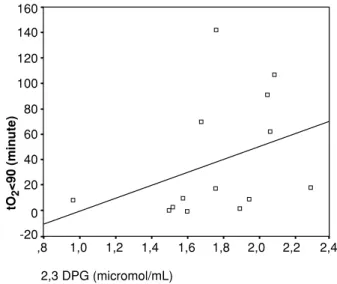
Characteristics of study groups were given in Table 1, 2, 3 Diphosphoglycerate and Packed Cell 2,3 DPG levels were not differed between hypoxic and non-hypoxic types of sleep disordered breathing and healthy control groups (Figure 1 and 2). 2,3 DPG levels were within the range of expected normal values (12) in control group. Spearman correlation analysis showed a negative relationship between mean oxyhemoglobin saturation and 2,3 DPG (r =-0.592; p=0.02) and a positive relationship between time spent below 90% and 2,3 DPG (r =0.579; p=0.03) in the whole patient group (Figure 3 and 4).
N = 7
CONTROL NONHYPOXIC7 HYPOXIC7
0,8 1,0 1,2 1,4 1,6 1,8 2,0 2,2 2,4 2,3 DPG (micromol / mL)
Figure 1. 2,3 DPG values were not differed statistically among the study groups. N: number of subjects.
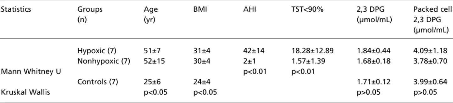
Table 1. BMI: Body-mass index; AHI: apnea-hypopnea index; TST<90%: total sleep time percent with oxygen saturation below 90%
Statistics Groups Age BMI AHI TST<90% 2,3 DPG Packed cell
(n) (yr) (µmol/mL) 2,3 DPG (µmol/mL) Hypoxic (7) 51±7 31±4 42±14 18.28±12.89 1.84±0.44 4.09±1.18 Nonhypoxic (7) 52±15 30±4 2±1 1.57±1.39 1.68±0.18 3.78±0.70 Mann Whitney U p<0.01 p<0.01 Controls (7) 25±6 24±4 1.71±0.12 3.99±0.64 Kruskal Wallis p<0.05 p<0.05 p>0.05 p>0.05
DISCUSSION
We found that the 2,3 DPG level was not a distinguishing factor between hypoxic and nonhypoxic types of sleep-disordered breathing patients. There are several reasons why we had expected to find increased 2,3 DPG levels in hypoxic group with respect to nonhypoxic group. Increase in 2,3 DPG levels is a well-known consequence of hypoxia, and there is a considerable variability in the degree of associated nocturnal hypoxemia among patients with similar degrees of obstructive
sleep apnea. The factors responsible for this variability have not been clearly defined. Bradley and colleagues demonstrated that the degree of arterial O2desaturation that develops during an obstructive apnea is dependent on the PaO2at the beginning of the apnea, the lung volume at which the apnea occurs, and the duration of the apnea (13). The decrease in functional residual capacity identified during sleep was also suggested to impair ventilation and contribute to the hypoxemia seen in patients with airflow limitation (14). Data on the impact of 2,3 DPG levels on oxyhemoglobin desaturations during sleep apnea, are more conflicting and limited. There are two studies which focused on diphosphoglycerate levels in sleep apnea patients. Maillard and colleagues demonstrated that the shift to the right of oxyhemoglobin dissociation curve occurred in patients with sleep apnea syndrome along with an increase in blood 2,3 DPG concentrations (8). An increase in 2,3 DPG within the red
blood cell shifts the oxyhemoglobin
dissociation curve to the right. The resultant enhanced unloading of oxygen in the peripheral tissues can be regarded as an intrinsic adaptive mechanism in response to hypoxemia. In the other study, it is found that there were no significant differences between
N = 7
CONTROL NONHYPOXIC7 HYPOXIC7
1 2 3 4 5 6
packed cell 2,3 DPG (micromol / mL)
Figure 2. Packed cell 2,3 DPG values of the study groups. N: number of subjects. 2,3 DPG (micromol/mL) ,8 1,0 1,2 1,4 1,6 1,8 2,0 2,2 2,4 -20 tO 2 <90 (minute) 0 20 40 60 80 100 120 140 160
Figure 4. There is a positive correlation (r = .579, p = 0.03) between 2,3 DPG and the total time spent below oxyhemoglobin saturation of 90% (t=2<90). ,8 1,0 1,2 1,4 1,6 1,8 2,0 2,2 2,4 90 92 94 96 98 100 2,3 DPG (micromol/mL) Mean O 2 saturation (%)
Figure 3. There is a negative correlation (r = -.592, p = 0.02) between mean oxyhemoglobin (MeanO2) saturation and 2,3
pretreatment and posttreatment values of blood 2,3 DPG and is concluded that although serum 2,3 DPG may be physiologically related to hypoxemia, this measure cannot be used to predict accurately the presence of moderate nocturnal hypoxemia in patients with sleep apnea or in monitoring the effect of their therapy (15).
In this study we examined two major issues on 2,3 DPG, namely, whether the adaptation mechanisms to intermittent hypoxia in obstructive sleep apnea includes increase in 2,3 DPG levels and whether the change in 2,3 DPG is in correlation with blood oxyhemoglobin saturation parameters. We failed to find any significant increase of 2,3 DPG in hypoxic group when compared with nonhypoxic and healthy control groups. In the whole patient group, there was relation between oxyhemoglobin saturation parameters such as mean oxyhemoglobin saturation (Figure 3) and total sleep time spent below oxyhemoglobin saturation of 90% (Figure 4) and 2,3 DPG. These results may be important in prediction of difference in adaptation to intermittent hypoxia and sustained hypoxia. We also suggest that 2,3 DPG level is not a distinguishing factor between hypoxic and nonhypoxic sleep disordered breathing patients, although there are correlations between oxygen parameters and 2,3 DPG.
There are several limitations of the study. We studied only fourteen patients and seven controls. Extrapolation of the results of these small number of patients may lead to
misinterpretations. Healthy control subjects were not age and body-mass index matched with the patient group. Nevertheless, There is no evidence that changing body-mass index leads to change in 2,3 DPG levels and also values of 2,3 DPG in our control group were within the range of expected normal values (12). Thus, the results of our control group served as an indicator for the accuracy of our 2,3 DPG procedure. In fact, our primary control group was nonhypoxic group and it was age and body mass index matched with hypoxic group. Another important limiting factor is the absence of blood pH and PaO2 values. Adding the blood pH and PaO2 assessment for the same blood samples would increase the validity of our results.
To our knowledge this is the first study that has evaluated 2,3 DPG levels in hypoxic and non hypoxic sleep disordered breathing patients. Intermittent hypoxia, which is more likely encountered in life to the majority of the population, may exert different adaptation mechanisms when compared with chronic or acute continuous hypoxia. Further studies are needed with increased number of patients to clarify the role of 2,3 DPG in adaptation to nocturnal intermittent hypoxia in sleep
disordered breathing patients. By
understanding these mechanisms, we may gain insights into natural compensatory mechanisms during the disease and the rationale for therapeutic intervention which can be either pharmacological or physical treatment.
REFERENCES
1. Oski FA, Gottlieb AJ, Delivora-Papadopoulos M, Miller WW. Red-cell 2,3 diphosphoglycerate levels in patients with chronic hypoxemia. N Engl J Med 1969;280:1165.
2. Hoffstein V. Snoring. Chest 1996;109:201-222.
3. Strohl K, Redline S. Recognition of obstructive sleep apnea. Am J Respir Crit Care Med 1996;154:279-289.
4. Javaheri S. A mechanism of central sleep apnea in patients with heart failure. N Engl J Med 1999;341:949-954.
5. Guilleminault C, Stoohs R, Clerk A, Cetel M, Maistros P. A cause of excessive daytime sleepiness: The upper airway resistance syndrome. Chest 1993;104(3):781-787.
6. Tilkian AG, Guilleminault C, Schroeder JS, Lehrman KL, Simmons FB, Dement WC. Hemodynamics in sleep-induced apnea. Ann Intern Med 1976;85:714-719.
7. Koskenvuo M, Kaprio J, Telakivi T, Partinen M, Heikkila K, Sarna S. Snoring as a risk factor for ischaemic heart disease and stroke in men. Br Med J 1987;294:16-19.
8. Maillard D, Fleury B, Housset B, Laffont S, Chabolle J, Derenne JP. Decreased oxyhemoglobin affinity in patients with sleep apnea syndrome. Am Rev Respir Dis 1991;143:486-489.
9. Guilleminault C, Stoohs R, Duncan S. Snoring: daytime sleepiness in regular heavy snorers. Chest 1991;99:40-48. 10. Rechtschaffen A, Kales A. A manual of standardized terminology,
techniques, and scoring system for sleep stages of human subjects. Washington DC: Government Printing Office, NIH publication. Nr. 204, 1968.
11. ASDA Report: EEG Arousals: scoring rules and examples. Sleep 1992;15:173-184.
12. Grisola S, Moore K, Luque J, Grady H. Automatic procedure for the micro estimation of 2,3 diphosphoglycerate. Anal Biochem 1969;31:235.
13. Bradley TD, Martinez D, Rutherford R, Lue F, Grossman RF, Moldofsky H, Zamel N, Phillipson EA. Physiological determinants of nocturnal arterial oxygenation in patients with obstructive sleep apnea. J Appl Physiol 1985;59:1364-1368. 14. Hudgel DW, Devadatta P. Decrease in functional residual
capacity during sleep in normal humans. J Appl Physiol 1984;57:1319-1322.
15. McKeon JL, Saunders NA, Murree-Allen K, Olson LG, Gyulay S, Dickeson J, Houghton A, Wlodarczyk J, Hensley M.J. Urinary uric acid: creatinine ratio, serum erythropoietin, and blood 2,3-diphosphoglycerate in patients with obstructive sleep apnea. Am Rev Respir Dis 1990;142:8-13.