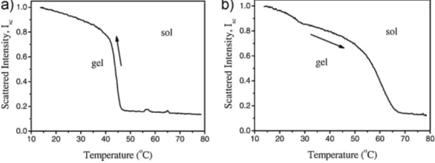
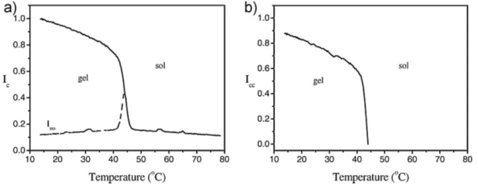
Critical Exponents of Thermal Phase Transitions of
k-Carrageenan in Various Salt Solutions
O¨ zlem Tarı,*1O¨ nder Pekcan2
Summary: The steady state fluorescence (SSF) technique was employed to study the phase transitions of k-carrageenan in NaCl and KCl solutions. Pyranine was used as a fluorescence probe for monitoring these transitions. Scattered light, Isc, and
fluor-escence intensity, I, was monitored against temperature to determine transition temperatures and exponents. It was observed that transition temperatures are strongly correlated with the NaCl and KCl contents. The weight-average degree of polymerization, DPw and gel fraction G, exponents (g and b) were measured and
found to be in accord with the classical Flory-Stockmayer model.
Keywords: carrageenan; critical exponents; fluorescence; gelation; percolation
Introduction
Carrageenans are water-soluble sulfated anionic galactans, extracted from species of marine red algae. k-carrageenan forms gels through specific interactions with metal ions.[1] Biocompatibility of these gels makes them valuable in a number of applications. For example they are used in dairy and household products such as toothpastes and lotions. In food applica-tions, carrageenans are used for gelation, thickeninig and stabilization.[2] They are also investigated for pharmaceutical and environmental applications due to their ability to immobilize micro-organisms.[3]
In k-carrageenan gels ionic interaction between OSO3 groups and Kþ ions together with intra and interchain hydrogen bonds give rise to helical structures. k-carrageenan assumes a random coil conformation in the sol state; low tempera-ture induce twisting of anhydrogalactose sequences into double helices. Gelation of k-carrageenan has been investigated by a variety of techniques such as small angle
neutron scattering,[4] rheology,[5–7] light scattering,[5] photon transmission,[8] small angle x-ray scattering.[9,10]
There are extensive studies on the effect of salts on macroscopic properties of the k-carrageenan.[11–13]In terms of the effect on gelation, monovalent alkali metal ions are classified into two groups. One group includes cations such as potassium, rubi-dium and cesium, which strongly promote gelation of k-carrageenan. The other group includes cations such as sodium and lithium, which scarcely promote the gelation. The strength of the promoting effects of diva-lent cations such as calcium, magnesium and strontium is intermediate.[14]
In this work, the thermal phase transi-tions of k-carrageenan in NaCl and KCl solutions were studied using the steady state fluorescence (SSF) technique. Pyra-nine (P) (a derivative of pyrene molecule) was introduced as fluorescence probe. It was observed that during the sol-gel transition of carrageenan, pyranine inten-sity, I, showed a continuous increase. Scattered light intensity, Isc, was also
monitored to detect the changes of turbid-ity during the sol-gel phase transition. However, on reheating during the gel-sol transition, I and Isc intensities decreased.
The necessary correction for the pyranine intensity was made to produce the real
1Department of Physics, Istanbul Technical Univer-sity, Maslak, 34469 Istanbul, Turkey
Fax: (þ90) 212 2856386; E-mail: otari@itu.edu.tr 2Kadir Has University, Cibali, 34320, Istanbul, Turkey
sol-gel and gel-sol transition curves. Sol-gel and gel-sol transition temperatures were determined for each curve of samples in various NaCl and KCl solutions. It was observed that Tsgand Tgsvalues increased
by increasing NaCl and KCl content. The measured weight-average degree of poly-merization exponents, g and gel fraction exponents, b, were found to be in accord with the classical Flory – Stockmayer model in thermal phase transitions. This theory predicts that helices and double helices should form the Cayley tree structure in the gel network.
Theoretical Considerations
The mechanism of the sol-gel transition has been described in terms of the percolation theory by a number of authors.[15,16]
According to this theory, in the sol state molecules of the solute join into small aggregates, called clusters, which grow in size during gelation. The sol-gel phase transition from sol state to the gel state occurs when small clusters link together and form a single giant cluster, which occupies most of the volume. The moment at which the giant cluster just starts to appear, indicates the gel point, p¼ pc, where the conversion factor p is the fraction of the bonds which have been formed between the molecules. Therefore, the system is called a gel for p above pc, a sol
for p below pc. In the gel state, the number
of finite clusters decreases during the gelation process, whereas the size of the giant cluster grows until all molecules are involved in its network. For the critical exponents near the sol-gel phase transition, classical theories like those of Flory–Stock-mayer predict one set of exponents, whereas scaling theories based on lattice percolation predict different exponents. The two groups of theories differ in their treatment of intramolecular loops, space dimensionality, and excluded volume effects.
Historically the exact solution of the sol-gel transition was given first by Flory and
Stockmayer on a special lattice called Bethe lattice where the closed loops were ignored. The critical exponents for the weight average degree of polymerization, DPw
and the gel fraction, G, are equal to unity independent of the dimensionality in the Flory–Stockmayer model which is also called classical theory or kinetic theory.[17,18] The dependence of the gel fraction G and the weight average degree of poly-merization, DPwon the fraction of bonds,
p is given by the following power law G/ p pð cÞb; p! pþc (1) DPw/ pð c pÞg; p! pc (2) where b and g are the critical exponents.
In this work, it can be argued that the total fluorescence intensity from the bonded pyranines follows the weight-aver-age degree of polymerization and the growing gel fraction below and above the gel point, respectively. This proportionality can be easily shown for site percolation as follows. The probability that a site belongs to a cluster of size s is given by nss, where
ns is the number of s-cluster (number of
clusters including s sites) per lattice site. The probability that an arbitrary site belongs to any cluster is p, this is simply the probability of arbitrary occupation of site. Thus, the probability ws¼Pnss
s
nssis
the cluster to which an arbitrary occupied site belongs contains exactly s site, and thus the average cluster size, S is calculated by the following relation
S¼X s ws s ¼ P ns s2 P ns s (3) Definition of the average cluster size is the same for all dimensions, although ns
cannot be calculated exactly in higher dimensions. This definition is also true for the bond percolation.
Now, to show that below pc, pyranine
intensity is proportional to S, let Npbe the
number of pyranine molecules and Nmthat
of the other molecules in the lattice. Thus, the total lattice site, N is equal to Npþ Nm. The probability, ppthat an arbitrary site is a
pyranine molecule is Np=N. The probabil-ity, Py, that an arbitrary site is both a
pyranine and belongs to the s-cluster can be calculated as a product of ws and pp as
follow Py¼ ppws¼ ppnss P nss (4) Thus the total number of pyranine molecules in the clusters including s sites will be Pys. The total fluorescence intensity,
I, which is proportional to the total number of pyranines trapped in the finite clusters, can be calculated as a summation over all s-clusters IX s Pys¼ X s ppnss P nss s¼ P ppnss2 P nss (5) where ppcan be taken out of the summation
since the concentration of the pyranine is fixed for our work,
I pp Pn ss2 P nss ¼ ppS (6)
Thus, the last expression shows that the total normalized fluorescent intensity, I is proportional to the average cluster size, S. Note that the proportionality factor, ppis
simply the concentration of the pyranine molecules in the sample cell (or the number of pyranine molecules in the lattice). The
intensity I will be linearly proportional to the average cluster size, provided that the pyranine concentration is not high enough to quench the fluorescence intensity by the reabsorption mechanism and no other parameter like viscosity influencing the fluorescence intensity in addition to the concentration of pyranine.
Experimental Part
k-carrageenan (Sigma) and pyranine were used for gel preparation by dissolving them in hot distilled water (pH 6.5) with KCl and NaCl solution. Pyranine concentration was taken as 4 104M for all samples. Mainly
two different set of experiments were carried out: The first set samples were prepared with constant carrageenan con-tent (2 wt.%) in various KCl concon-tents. These samples were named as C2K0 (no KCl), C2K02 (0.2 wt.%), C2K04 (0.4 wt.%) and C2K08 (0.8 wt.%). The second set samples were prepared with various NaCl contents from 0.4 to 1.0 (wt.%). These samples were named as C2Na04, C2Na06, C2Na08 and C2Na1, respectively. The compositions and symbols of the studied gels with various KCl and NaCl solutions are listed in Table 1 and 2.
Table 1.
The compositions, symbols, sol-gel and gel-sol transition temperatures and critical exponents for the gels in various KCl solutions.
Gel KCl content (wt. %) Sol – gel transition Gel – sol transition
Tsg(8C) b g Tgs(8C) b g C2K0 0 39.1 1.01 0.76 55.6 0.95 0.55 C2K02 0.2 43.9 0.93 0.60 60.7 0.93 0.54 C2K04 0.4 49.7 0.85 0.64 65.9 1.03 0.65 C2K08 0.8 60.9 1.01 0.31 76.2 0.93 0.20 Table 2.
The compositions, symbols, sol-gel and gel-sol transition temperatures and critical exponents for the gels in various NaCl solutions.
Gel NaCl content (wt. %) Sol – gel transition Gel – sol transition
Tsg(8C) b g Tgs(8C) b g
C2Na04 0.4 37.9 0.75 0.81 54.5 0.95 0.79
C2Na06 0.6 40.1 0.92 0.85 58.5 0.98 0.82
C2Na08 0.8 42.0 1.13 1.27 59.0 1.07 1.05
The fluorescence intensity measure-ments were carried out using a Perkin Elmer spectrometer Model LS-50, equipped with temperature control. The carrageenan sol at 80 oC was transferred into the glass cell and left to cool to room temperature. All measurements were made at the front face position; slit widths were kept at 5 nm. P was excited at 360 nm and emission was detected at 515 nm in in situ experiments. Variations in the scattered and fluorescence emission intensity of pyranine were monitored as a function of temperature.
Results and Discussion
Figure 1a and 1b show the temperature variation of scattered light intensity, Iscfor
C2K02 gel in the sol-gel and gel-sol transition, respectively. It can be seen that
Iscincreased dramatically upon cooling the
carrageenan samples in both cases indicat-ing that the gel turbidity increased con-siderably.
Fluorescence intensity, I, shown in Figure 2a and 2b was also measured. It increased continuously upon cooling pre-dicting that pyranine molecules are trapped in the gelling environment which prevents its quenching. However, Iscdecreased and
I increased upon heating the carrageenan gels.
It can be postulated that during gelation (cooling process) helices and double helices are formed through the association of carrageenan molecules. Then they aggre-gate to higher-order assemblies and create an infinite network. On reheating, the initial double helix aggregates are destroyed and then the double helices are decomposed to carrageenan molecules, which results in the destruction of the gel
Figure 1.
Temperature variation of scattered ligt intensities for C2K02 sample during (a) sol-gel transition and (b) gel-sol transition.
Figure 2.
Temperature variation of fluorescence intensities for C2K02 sample in (a) sol-gel transition and (b) gel-sol transition.
structure. A representation of the above process is shown in Figure 3.
In order to elaborate the above results, intensity I has to be corrected dividing it by transmitted light intensity. Since the shape of the excitation light has the character of Itr, the observed fluorescence intensity is
the convolution of Itr with the desired
fluorescence intensity, Ic. Figure 4a and 4b
present the corrected fluorescence intensi-ties, Ic,for C2K02 sample in the sol-gel and
gel-sol transition, respectively.
The peak positions of the first derivatives (dIc/dT) of these sigmoidal curves
can produce the sol-gel and gel-sol transi-tion temperatures (Tsgand Tgs), see Figure 5.
Figure 3.
Sol-gel and gel-sol transition and the structure of the helix network.
Figure 4.
Temperature variation of corrected fluorescence intensities for C2K02 sample in (a) sol-gel transition and (b) gel-sol transition.
Figure 5.
These temperatures are listed in Table 1 and 2 for all gel samples. It can be observed that Tsg values are much lower than Tgs
values for all carrageenan samples. In the case of cation content, both Tsg and Tgs
values are higher than the gel containing no external cation. Thus, in the presence of cations, stronger gels can be formed, which require higher temperatures due to the cation attraction.
The gelation theory often makes the assumption that the conversion factor p determines the behavior of the gelation process, in the mean time p may depend on temperature. Therefore, above the gel point i.e. for T > Tsg the fluores-cence intensity, Ic measures the weight
average degree of polymerization (or average cluster size) in the sol-gel transition path. However for T < Tsg the intensity, Icmeasures the gel fraction G, the fraction
of the monomers that belong to the macroscopic network. Then Equation 1 and 2 can be written in the following form Icc¼ Ið c ImsÞ / G / Tsg T b ; T! Tsg (7) Ic/ DPw/ T Tsg g ; T! Tsgþ (8) In Equation 7 Iccsolely originates from
the pyranines embedded in the growing gel fraction, G i.e. Iccis produced by subtracting
the mirror symmetry (Ims) of Ic intensity
belonging to pyranines embedded in the
small clusters from the total Ic intensity.
Figure 6a and 6b illustrate the above procedure. Here the mirror symmetry Ims
of the Ic curve above Tsg is subtracted
from the curve below Tsg as shown in
Figure 6a. The resulting curve is given in Figure 6b.
The double logarithmic plots of the data are presented in Figure 7a and 7b for C2K02 sample. The critical exponents b and g were produced by fitting the data to the double logarithmic form of Equation 7 and 8. They are listed in Table 1 and 2.
It is seen that the average g and b values are very close to the values of classical Flory – Stockmayer model. From that one may conclude that Icmeasures the average coil
(small cluster) size above Tsg in sol – gel
transition paths. Icc detects helices and
double helices (gel fraction) during sol – gel transition paths below Tsg. Since the
forma-tion of gel from the helices and double helices should obey the classical Bethe lattice, the connection of helices and double helices must be in the Cayley tree form.
Moreover, the critical exponents during gel-sol transition upon heating the gels can also be calculated. The gel-sol transition temperatures (Tgs) were determined by
following the similar treatment used to obtain Tgstemperatures. The critical
expo-nents b and g were produced by fitting the data to the double logarithmic form of Equation 7 and 8 as shown in Figure 8a and 8b for C2Na1 sample. It should be noted that the values of the critical exponents for
Figure 6.
(a) Subtraction of the mirror symmetry of Icabove Tsgfrom the curve below Tsg, (b) The resulting Icccurve represents the pyranines embedded solely in the growing gel fraction.
the sol-gel transition are in agreement with those for gel-sol transitions. All results are presented in Table 1 and 2.
Now one can compare these results with the results produced in similar systems. The sol-gel transition of the polysaccharide gellan gum has been investigated by the dynamic and static light scattering techni-ques.[19] The critical exponent, g and n corresponding to the cluster mass and correlation length were measured and found to be 1.66 and 0.88, respectively, which are in good agreement with the lattice percolation model. The critical exponents of the elastic modulus, t and correlation length, g were measured near the sol gel transition temperature of agarose gel using a combination of rheolo-graph, differential scanning calorimetry and circular dichroism spectroscopy tech-niques; these were found to be 0.8 and 1.87, respectively.[20]
Conclusion
In summary, this paper has shown that the measured weight-average degree of poly-merization exponents, g and gel fraction exponents, b obeyed the classical Flory – Stockmayer model during the thermal phase transitions of carrageenan in various salts. It is important to note that all carrageenan systems belong to the same universality class, independent of the salt solution type. The sol-gel and gel-sol transition temperatures were determined, Tsgvalues being much lower than Tgsvalues.
[1] A.-M. Hermansson, E. Eriksson, E. Jordansson, Carbohydr. Polym.1991, 16, 297.
[2] L. Piculell, in: Food Polysaccharides and Their Appli-cations, A. M. Stephen, Ed., Marcel Dekker, New York 1995, p. 205.
Figure 7.
log-log plots of the data for C2K02 sample and their fits to Equation 7 and 8.
Figure 8.
[3] M. B. Cassidy, H. Lee, J. T. Trevors, J. Ind. Microbiol. Biotechnol.1996, 16, 79.
[4] M. Sugiyama, C. Yuasa, K. Hara, N. Hiramatsu, A. Nakamura, Y. Hayakawa, Y. Maeda, Physica B1998, 241, 999.
[5] M. R. Mangione, D. Giacomazza, D. Bulone, V. Martrana, P. L. San Biagio, Biophys. Chem.2003, 104, 95.
[6] M. Takemasa, A. Chiba, M. Date, Macromolecules 2001, 34, 7427.
[7] C. Chen, M.-L. Liao, D. E. Dunstan, Carbohydr. Polym.2002, 50, 109.
[8] S. Kara, C. Tamerler, H. Bermek, O. Pekcan, Int. J. Bio. Macromol.2003, 31, 177.
[9] Y. Yuguchi, T. T. T. Thuy, H. Urakawa, K. Kajiwara, Food Hydrocolloids2002, 16, 515.
[10] Y. Yuguchi, H. Urakawa, K. Kajiwara, Food Hydro-colloids2003, 17, 481.
[11] C. Rochas, M. Rinaudo, Biopolymers1984, 23, 735. [12] C. Rochas, M. Rinaudo, M. Vincendon, Biopolymers 1980, 19, 2165.
[13] S. Ablett, A. H. Clark, D. A. Rees, Macromolecules 1982, 15, 597.
[14] C. Rochas, M. Rinaudo, Biopolymers1980, 19, 1675. [15] D. Stauffer, A. Coniglio, M. Adam, Adv. Polym. Sci. 1982, 44, 103.
[16] E. Del Gado, L. De Arcangelis, A. Coniglio, J. Phys. A 1998, 31, 1901.
[17] D. Stauffer, Introduction to Percolation Theory, Taylor and Francis, London1985.
[18] M. Sahimi, Application of Percolation Theory, Taylor and Francis, London1994.
[19] T. Okamoto, K. Vubota, Carbohydr. Polym.1996, 30, 149.
[20] T. Fujii, T. Yano, H. Kumagai, O. Miyauraki, Food Hydrocolloids2000, 14, 359.