Fractal Features and Structural, Morphological, Optical
Characteristics of Sol–Gel Derived
Silica Nanoparticled Thin Films
B.Ö. Uysal
∗and Ö. Pekcan
Kadir Has University, Faculty of Engineering and Natural Sciences, Cibali, Fatih, Istanbul 34083, Turkey (Received January 26, 2017; in final form March 23, 2018)
Nanostructured silica films using a simple and effective sol–gel spin coating technique were synthesized and the influence of ammonia/sol ratios on the particle size and thickness of this film was investigated. In addition, fractal dimensions of the prepared films were determined using the scattering response technique. The samples were characterized by atomic force microscopy and UV–vis spectroscopy. Comparing optical method and image analysis of atomic force microscopy micrographs, the fractal dimension of silica nanoparticled thin films was determined. The fractal dimensions of the films verified by atomic force microscopy analysis were found to be around 2.03 which is very close to the values (2.0358, 2.0325, and 2.0335) obtained using optical method. As a result of these findings, precise determination of the nanoparticled silica thin films fractal dimension using both optical and surface analysis methods was realized.
DOI:10.12693/APhysPolA.133.1160
PACS/topics: fractal dimension, silica, sol–gel method, size-dependence, thin films
1. Introduction
Research and development on silica thin films has wit-nessed important progresses in the last few years with regard to particle synthesis. Spray pyrolysis [1], col-loidal techniques [2–5], water-in-oil microemulsion [6], micelle processing [7], hydrothermal synthesis [8] and sol–gel methods are considered to be the leading syn-thetic techniques [9, 10] in silica nanoparticle prepara-tion. Many researchers have been a firm supporter of the sol–gel method owing to its simplicity, ease to produce uniform films, cost-effectiveness, and good applicability to the large industrial areas [11–15]. Morphology of the particles, crystallite size, and size distribution are con-sidered as the key properties that can be tailored using various precursors, catalyst and annealing temperatures. Water, catalyst and precursor concentrations in the com-position control variations in the properties [16–20]. Es-pecially, silica nanoparticled structures were synthesized using TEOS, ammonia, water, ethanol combination in many studies in the literature [21–26]. Regarding mor-phology, fractal morphology in monodisperse silica parti-cle systems has been studied by several authors [27, 28]. More recently, static light scattering (SLS) and im-age analysis of scanning electron microscope (SEM) pho-tographs of aggregates were employed together with frac-tal analysis to characterize the agglomerate structure in films [29]. These techniques are well established and com-mercially available and widely used in this area. Pro-duced results from image analysis proved that fumed sil-ica aggregates which are used for comparison, have a
two-∗corresponding author; e-mail: bozugur@khas.edu.tr
level structure, namely, made out of compact aggregates and open aggregates of nanoparticles. Here, it has to be noted that this structure is not easily detected by SLS. In other words, SLS technique seems to be less accurate than image analysis method. However, since it is much less time consuming, this technique can be used in more simple cases. Even though the effect of ammonia ratio on morphological properties of nanostructures has been well-known [28, 30] and essential fractal [31, 32] mor-phology has already been demonstrated for aggregates of silica [33, 34], the influence of catalyst material amount in composition on the optical properties (transmittance, reflectance, absorption, and scattering), particle size and implicitly thickness of the silica nanoparticled thin films (SNF) in terms of fractal analysis have not been exten-sively studied yet.
Therefore, in this work, the effects of ammonia/sol ra-tios on the particle size and thickness of the nanostruc-tured silica films prepared by sol–gel spin coating tech-nique were investigated. In addition, fractal dimensions of the prepared films were determined using the scatter-ing response technique. The goal of the present work is to compare two different techniques enabling the mea-surement of the fractal dimension of SNF, namely optical method and image analysis of AFM micrographs.
2. Experimental details 2.1. Film preparation
The SiO2nanoparticled sol was prepared via a
Stöber-like [3] sol–gel spin coating technique which was pre-viously reported [35]. Tetraethyl orthosilicate (TEOS) was used as a precursor chemical. 10 ml of TEOS (99.99% trace metals basis supplied by Sigma-Aldrich Inc.), 40 ml of ethanol and 20 ml of deionized water
were mixed. Then various amounts (0.109, 0.174, 0.218, 0.436, 0.873 ml) of ammonia solutions were added as a catalyst, denominated as SNF1, SNF2, SNF3, SNF4, and SNF5, respectively. The sols were spin coated on corn-ing (2947) glasses at 1500 rpm for 30 s. The coatcorn-ings were heat treated for 2 h in air at 450◦C employing a microprocessor-controlled (CWF 1100) furnace.
2.2. Characterizations
The determination of morphology of the films was car-ried out using an atomic force microscope (AFM) in dy-namic mode (Model SPM-9500, Shimadzu Corp.). Opti-cal transmittance, reflectance and absorbance data of the films were produced using a UV–vis spectrophotometer (Perkin-Elmer LAMBDA 900 integrating sphere system). Additionally, the thicknesses of the films were also mea-sured using this spectrophotometer.
3. Results and discussion 3.1. Surface morphology of the films
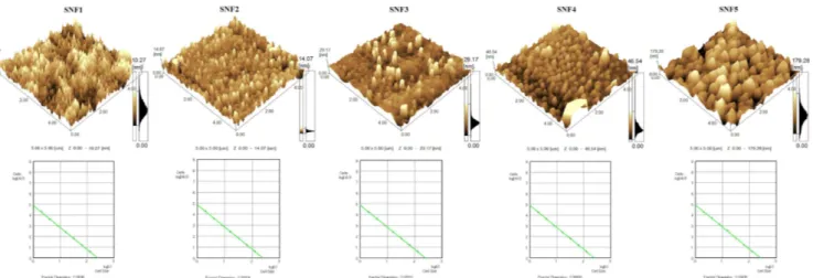
The influence of the amount of ammonia solution on the surface morphology of SNF is presented via three-dimensional (3D) AFM images in Fig. 1a. It is obvious
to see that all the films have a granular structure and consist of uniform, close-packed particle clusters. This figure can be interpreted that the SiO2particles are
uni-formly distributed over the whole surface of the films due to well-synthesized sol. Furthermore, according to the AFM images of SNF, one can observe that the par-ticle size increases when the amount of ammonia in sol increases. Here, it is worth mentioning that the parti-cle size monitored with AFM refers to the lateral feature size, namely lateral diameter of the agglomerated granu-lar structures. As a result, particle size is not correlated with the crystallite size due to amorphous structure of films heat treated at 450◦C. SPM Manager Program was used to evaluate the average particle size, roughness values, and fractal dimensions of the films. The calcu-lated average particle size values for films with different amount of ammonia are listed in Table I. Particle size is directly proportional to the amount of catalyst material, ammonia, in that the catalyst material lowers the activa-tion energy by altering the pathway and shortening the gelation time as mentioned in the literature, for both sil-ica [36–41] and other metal oxide nanostructures [42, 43]. As expected, z-range observations are in agreement with the particle size values.
Fig. 1. Three-dimensional (3D) AFM images of silica nanofilms (a) for different amounts of ammonia: 0.109, 0.174, 0.218, 0.436, 0.873 ml at heat treatment temperature of 450◦C, (b) the fractal dimensions of the films calculated based on the slope of the logarithmic plot of number of cells n(r) versus cell size r.
TABLE I AFM and spectrophotometer analysis results of nanostructured silica thin films with different volume ratio of ammonia/sol.
Film Ammonia/solvolume ratio Thickness [nm] size [nm]Particle Scatteringintensity
Fractal dimension verified by SPM Manager SNF1 1.45×10⁻³ 49 19 0.002 2.03041±0.0183 SNF2 2.32×10⁻³ 71 32 0.006 2.02154±0.0172 SNF3 2.91×10⁻³ 125 48 0.009 2.02520±0.0179 SNF4 5.82×10⁻³ 225 75 0.037 2.03600±0.0188 SNF5 1.16×10⁻² 340 158 0.168 2.03435±0.0185
Figure 1b represents the fractal dimensions of the films calculated based on the slope of the logarithmic plot of number of cells, n(r), versus cell size r. Fractal analy-sis tool of SPM Manager Program is based on the cube counting method [44, 45] derived from a definition of box-counting method to measure fractal dimension. The al-gorithm works on the z-surface by taking the number of cells that contain at least one pixel of the image. The slope of the plot of log(n(r)) versus log(r) gives the frac-tal dimension directly [46]. The measured fracfrac-tal dimen-sion values are given in Table I. They are very close to each other. The slight differences between them are only visible in the second digit after the decimal.
3.2. Fractal analysis via optical studies
In order to find the scattering response of the films, transmittance, reflectance and absorbance modulations were measured by the UV–vis spectrophotometer in a spectral range of 300–1000 nm. Figures 2 and 3 depict
Fig. 2. UV–vis (a) transmission, (b) absorption spec-tra of silica nanofilms for different ammonia volume ra-tios.
Fig. 3. The influence of ammonia/sol volume ratios on (a) reflection and (b) scattering response of silica nanofilms.
the effect of the ammonia solution’s amount on these measurements of the SNF with respect to wavelength. Transmittance values of the films were decreased with the increase of ammonia/sol ratios as shown in Fig. 2a. The decrease in the volume ratio of ammonia/sol produces a blue shift in the absorbance spectra of the films in Fig. 2b due to quantum size effect. Additionally, the difference in the absorption edge is related to the Burstein–Moss effect because of the increase of water concentration in the sol. Some part of the incident light absorbed by and some part of it transmitted through the film depended on its thickness. The scattering intensity of the films at 400 nm increases, when the ratio of ammonia/sol creases as seen in Fig. 3. The calculated scattering in-tensity and thickness values are provided in Table I.
The thickness values of the films increased with the increase of amount of ammonia in sol. This is not sur-prising because the increase in the thickness values of the films is in agreement with an increase of particle size observed by AFM measurements.
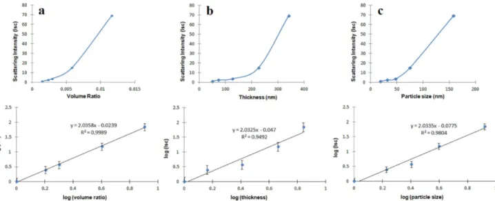
Fig. 4. The plots of scattering intensity versus (a) volume ratio, (b) thickness, and (c) particle size on linear and logarithmic scales.
The plots of scattering intensity versus volume ra-tio, thickness and particle size on linear and logarithmic scales are shown in Fig. 4a–c, respectively. The optical measurements for films display an increase in scattering by increasing these quantities.
It has been well established that fractals have self-similar structures that can be characterized by a single parameter, the fractal dimension D. If D = 2, the film has a planar [47] and smooth surface morphology like a substrate, as exemplified in [48] and [49]. The depen-dence of scattering intensity Iscon the ammonia/sol
vol-ume ratio V , the thickness T and the particle size S in the SNF films, can be suggested by the following equa-tions [50, 34]:
Isc= (V )D, Isc= (T )D, Isc= (S)D, (1 a, b, c)
where D is the fractal dimension of the film and Isc is
the scattering intensity of films. Here it is assumed that V , T , and S values are proportional to the scattering centers in the film presenting power law dependency as given in Eqs. (1a, b) and (c). It is obvious that Isc is
proportional to scattering centers of the films under con-sideration. In Fig. 4a–c log–log plot of normalized Isc
intensities are plotted versus V , T , and S, respectively. The fractal dimensions are produced from slopes of the logarithmic plots and given in Table II.
TABLE II Fractal dimensions of the nanostructured silica thin films measured by optical methods.
Method Fractal dimension
volume ratio V 2.0358
thickness T 2.0325
particle size S 2.0335
4. Conclusion
The nanoparticled silica thin films were synthesized by sol–gel spin coating process. Different particle sizes were obtained using various amounts (0.109, 0.174, 0.218, 0.436, 0.873 ml) of ammonia solutions at the same an-nealing temperature of 450◦C. The optical studies re-vealed that the scattering intensity of the films decreased with increase of wavelength. Moreover, the scattering in-tensity of SNF increased with the volume ratio of ammo-nia/sol. The absorption edge of the SNF shifted to longer wavelengths with an increase in amount of ammonia solu-tion due to the quantum confinement and the Burnstein– Moss effects of nanoparticles. Both optical measurements and SPM Manager Program results of SNF confirm the change of the particle size with the amount of ammo-nia. The fractal dimension D of the SNF measured by SPM was found to be around 2.03 which is very close to the D values (2.0358, 2.0325, and 2.0335) produced by optical method. These extremely small differences are within the error limits. The D values strongly support the structure of SNF film having planar and smooth sur-face morphology.
In summary, this study indicates that it is possible to determine the fractal dimension of the nanoparticled silica thin films precisely using both optical and surface analysis methods.
Acknowledgments
The authors would like to thank Materials Science Lab-oratory employees of SabancıUniversity for the morpho-logical analysis.
References
[1] H.D. Jang, H. Chang, Y. Suh, K. Okuyama, Curr. Appl. Phys. 6, e110 (2006).
[2] Y. Huang, J.E. Pemberton, Coll. Surf. A Physic-ochem. Eng. Asp. 360, 175 (2010).
[3] W. Stöber, A. Fink, J. Coll. Interface Sci. 26, 62 (1968).
[4] S.K. Park, K.D. Kim, H.T. Kim, Coll. Surf. A Physicochem. Eng. Asp. 197, 7 (2002).
[5] X.D. Wang, Z.X. Shen, T. Sang, X.B. Cheng, M.F. Li, L.Y. Chen, Z.S. Wang, J. Coll. Interface Sci. 341, 23 (2010).
[6] S. Santra, P. Zhang, K. Wang, R. Tapec, W. Tan,
Anal. Chem. 73, 4988 (2001).
[7] X. Lv, L. Zhang, F. Xing, H. Lin,Micropor. Mesopor. Mater. 225, 238 (2016).
[8] A.B. Corradi, F. Bondioli, A.M. Ferrari, B. Focher, C. Leonelli,Powder Technol. 167, 45 (2006). [9] S. Duhan, S. Devi, M. Singh,J. Rare Earths 27, 83
(2009).
[10] M. Marini, B. Pourabbas, F. Pilati, P. Fabbri,Coll. Surf. A Physicochem. Eng. Asp. 317, 473 (2008).
[11] M. Jafarzadeh, I.A. Rahman, C.S. Sipaut,J. Sol–Gel Sci. Technol. 50, 328 (2009).
[12] G.M. Pajonk,Coll. Polym. Sci. 281, 637 (2003). [13] K. Ishizaki, S. Komarneni, M. Nanko, in: Porous
Ma-terials, Vol. 4 of the series Materials Technology Se-ries, Springer, USA 1998, p. 67.
[14] S. Chang, M. Lee, W. Kim, J. Coll. Interface Sci. 286, 536 (2005).
[15] G.H. Bogush, C.F. Zukoski, J. Coll. Interface Sci. 142, 1 (1991).
[16] J. Li, L. Chen, Z. Zhang, C. Jiao, J. Wuhan Univ. Technol. — Mater. Sci. Ed. 29, 478 (2014). [17] K.S. Rao, K. El-Hami, T. Kodaki, K. Matsushige,
K. Makino,J. Coll. Interface Sci. 289, 125 (2005).
[18] Ö. Kesmez, E. Burunkaya, N. Kiraz, H.E. Çamurlu, M. Asiltürk, E. Arpaç, J. Non-Cryst. Solids 357, 3130 (2011).
[19] H.C. Wang, C.Y. Wu, C.C. Chung, M.H. Lai, T.W. Chung,Ind. Eng. Chem. Res. 45, 8043 (2006). [20] S.K. Park, K.D. Kim, H.T. Kim,Coll. Surf. A 197,
7 (2002).
[21] D.C.L. Vasconcelos, W.R. Campos, V. Vasconcelos,
Mater. Sci. Eng. A — Struct. Mater. Prop. Mi-crostruct. Process. 334, 53 (2002).
[22] W. Wang, B. Gu, J. Phys. Chem. B 109, 22175 (2005).
[23] I.A. Rahman, P. Vejayakumaran, C.S. Sipaut, J. Is-mail, M. Abu Bakar, R. Adnan, C.K. Chee, Coll. Surf. A294, 102 (2007).
[24] K.S. Kim, J.K. Kim, W.S. Kim,Ceram. Int. 28, 187 (2002).
[25] J.W. Yoo, D.S. Yun, H.J. Kim, J. Nanosci. Nan-otechnol. 6, 3343 (2006).
[26] J. Li, L.X. Chen, Z.M. Zhang, C.B. Jiao,Adv. Mater. Res. 560-561, 462 (2012).
[27] R. Watanabe, T. Yokoi, E. Kobayashi, Y. Otsuka, A. Shimojima, T. Okubo, T. Tatsumi,J. Coll. Inter-face Sci. 360, 1 (2011).
[28] C. Oh, S.B. Shim, Y.G. Lee, S.-G. Oh, Mater. Res. Bull. 46, 2064 (2011).
[29] N. Ibaseta, B. Biscans, Powder Technol. 203, 206 (2010).
[30] L.P. Singh, S.K. Bhattacharyya, R. Kumar, G. Mishra, U. Sharma, G. Singh, S. Ahalawat,
Adv. Coll. Interface Sci. 214, 17 (2014). [31] B.B. Mandelbrot,Phys. Scr. 32, 257 (1985).
[32] X. Zhang, Y. Xu, R.L. Jackson,Tribol. Int. 105, 94 (2017).
[33] B.M. Smirnov,Phys. Rep. 188, 1 (1990).
[34] J.E. Martin, A.J. Hurd,J. Appl. Crystallogr. 20, 61 (1987).
[35] B. Özuğur Uysal, F.Z. Tepehan,J. Sol-Gel Sci. Tech-nol. 63, 177 (2012).
[36] H.M. Lim, H.C. Shin, S.H. Huh, S.H. Lee,Solid State Phenom. 124-126, 667 (2007).
[37] V.I. Boev, A. Soloviev, C.J.R. Silva, M.J.M. Gomes, J. Pérez-Juste, I. Pastoriza-Santos, L.M. Liz-Marz, in: Nanostructured Materials for Advanced Techno-logical Applications, part of the series NATO Science for Peace and Security Series B: Physics and Bio-physics, Springer, Netherlands, 2009, p. 245.
[38] M. Fertani-Gmati, K. Brahim, I. Khattech, M. Jemal,
Thermochim. Acta 594, 58 (2014).
[39] M. Toki, T. Takeuchi, S. Miyasita, S. Kanbe,
J. Mater. Sci. 27, 2857 (1992).
[40] M. Darbandi, UV-VIS and Photoluminescence Spectroscopy for Nanomaterials Characterization, Springer, Berlin 2013, p. 431.
[41] G.-L. Davies, A. Barry, Y.K. Gunko, Chem. Phys. Lett. 468, 239 (2009).
[42] Z.-X. Tang, L.-E. Shi,Eclecita Quim. 33, 15 (2008). [43] L. Bian, S.P. Wang, X.B. Ma,Kinet. Catal. 55, 763
(2014).
[44] C. Douketis, Z. Wang, T.L. Haslett, M. Moskovits,
Phys. Rev. B 51, 11022 (1995).
[45] W. Zahn, A. Zösch,Fresenius J. Anal. Chem. 365, 168 (1999).
[46] W. Zahn, A. Zösch,Fresenius J. Anal. Chem. 358, 119 (1997).
[47] S. Sadi, A. Paulenova, P.R. Watson, W. Loveland,
Nucl. Instrum. Methods Phys. Res. A 655, 80 (2011). [48] A. Mannelqvist, M.R. Groth,Appl. Phys. A 73, 347
(2001).
[49] C.G. Sonwane, S.K. Bhatia, N.J. Calos,Langmuir 15, 4603 (1999).
[50] J. Bastide, L. Leibler, J. Prost, Macromolecules 23, 1821 (1990).

