Introduction
The family Lamiaceae has an important role as a source of medicinal and aromatic plants of commercial importance. The genus Hymenocrater Fisch. & C.A.Mey., which belongs to the family Lamiaceae, is represented in Turkey by only one species, H. bituminosus Fisch. & C.A.Mey. This plant is distributed in East Anatolia, Transcaucasia, north-west and central Iran, and Khorassan. It is an Irano-Turanian element and grows at high altitudes (1900-2100 m) in East Anatolia (Mill, 1982). The genus Hymenocrater is represented by 11 species in Flora Iranica and 2 species in Flora of the USSR (Gorshkova, 1976; Rechinger, 1982).
There are few studies on the essential oil of Hymenocrater species in Russia, Iran, and Turkey. It is reported in the Flora of the USSR that H. bituminosus has commercial value due to a lemon aroma (Gorshkova, 1976). The essential oil of H. incanus Bunge, which is endemic to Iran, was analysed by Mirza et al. (2001). Composition of the essential oil of H. calycinus (Boiss.) Benth. was investigated by Firouznia et al. (2005) and the essential oil of H. bituminosus in Turkey was analysed by Kürkçüo¤lu et al. (2005).
There are many studies of the anatomical and morphological characteristics of some species belonging to the family Lamiaceae in Turkey (Gönüz & Özörgücü,
Comparative Morphological and Anatomical Studies of
Hymenocrater bituminosus Fisch. & C.A.Mey. (Lamiaceae) in Turkey
Fatih SATIL1, Murat ÜNAL2, Ersin HOPA3
1
Bal›kesir University, Arts & Science Faculty, Department of Biology, 10145 Bal›kesir - TURKEY
2
Yüzüncü Y›l University, Arts & Science Faculty, Department of Biology, Van - TURKEY
3
Susurluk Anatolian High School, Bal›kesir - TURKEY
Received: 24.03.2006 Accepted: 28.03.2007
Abstract:Hymenocrater bituminosus Fisch. & C.A.Mey. is the only species of the genus Hymenocrater Fisch. & C.A.Mey. in Turkey. In this study, the morphological features of the species, such as stem, leaf, flower, and nutlet, are described in detail. The morphological results were compared to the Flora of Turkey, Flora Iranica, and Flora of the USSR. As a result of this study, the description of this species has been expanded, contributing to the knowledge of the flora of Turkey. In anatomical studies, transverse sections of stems and leaves were examined and are supported by illustrations and photographs. Furthermore, trichomes in stems, leaves, and calyces were investigated. Anatomical characters of the species were observed to be similar to the usual features of Lamiaceae anatomy.
Key Words: Anatomy, Hymenocrater, Lamiaceae, morphology
Türkiye’deki Hymenocrater bituminosus Fisch. & C.A.Mey. (Lamiaceae) Üzerinde Karflılafltırmalı Morfolojik ve Anatomik Çalıflmalar
Özet:Hymenocrater bituminosus Fisch. & C.A.Mey., Türkiye’deki Hymenocrater Fisch. & C.A.Mey. cinsinin tek türdür. Bu çalıflmada, türün gövde, yaprak, çiçek ve tohumunun morfolojik özellikleri detaylı bir flekilde incelenmifltir. Morfolojik sonuçlar, Türkiye Florası, ‹ran Florası ve Rus Florası ile karflılafltırılmıfltır. Çalıflma sonucunda türün deskripsiyonu geniflletilerek Türkiye Florası’na katkılar sa¤lanmıfltır. Anatomik çalıflmalarda, gövde ve yapraktan alınan enine kesitler incelenerek çizim ve foto¤raflarla desteklenmifltir. Ayrıca, gövde, yaprak ve kaliksteki tüy örtüsü incelenmifltir. Türün anatomik özelliklerinin, Lamiaceae familyasının genel anatomik yapısına benzer oldu¤u gözlenmifltir.
1999; Bafler et al., 2000; Kaya et al., 2000, 2002; Satıl et al., 2002; Kandemir, 2003); however, there has been no investigation of Hymenocrater species as yet.
Herein, the morphological and anatomical features of H. bituminosus were studied in order to provide more detailed descriptions of the species.
Materials and Methods
Plant material
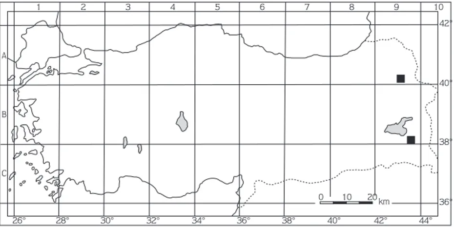
Specimens were collected from 2 different regions of Turkey (Figure 1): A9 Kars: 16 km from Ka¤ızman to Kars, 13 vi 2002, T. Dirmenci (1908!) VANF; B9 Van: 25 km from Gürpınar to Güzelsu, edge of Zernek Dam, 13 vi 2002, 1800-1900 m, F. Satıl (1035!) & M. Ünal (VANF).
Voucher specimens were deposited at the Herbarium of the Science and Arts Faculty of Yüzüncü Yıl University (VANF) and the Herbarium of the Science and Arts Faculty of Balıkesir University, Turkey.
Morphological and Anatomical study
Morphological characteristics were determined from fresh and herbarium materials. The taxonomic description of the plant was carried out according to Flora of Turkey and the East Aegean Islands (Mill, 1982) and was also confirmed by the herbarium samples of deposited species in VANF. An Olympus SZX12 stereomicroscope with a drawing tube was used for morphological study.
Anatomical studies were conducted on fully flowered fresh plants and herbarium material. Whole plants or portions of them were stored in 70% ethanol. Developed middle cauline leaves and stems from fully flowered plants were used for anatomical study. Transverse sections of leaves and stems were made manually.
Tissues were stained with Sartur reagent and embedded in glycerine jelly (Baytop, 1972). An Olympus BX50 phase contrast binocular microscope with a drawing tube was used for anatomical studies and drawings..
Results
Morphology
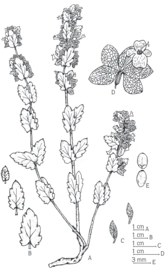
Perennial herbs or subshrubs, flowering stem 30-60 cm. Stem and branches rarely eglandular and glandular hairs. Leaves ovate or ovate-oblong, 10-30 × 6-24 mm, obtuse or subacute, cordate to truncate, crenate to dentate; petiole 1.5-11 mm. Bracts similar to leaves, but slightly smaller, oblong-lanceolate, 5-10 × 2.5-3 mm, sessile or shortly petiolate, 1-2 mm. Verticillasters 2-3-flowered, pedicel 0.5-1 mm. Calyx 10-20 mm, 5-lobed, pale green, papery, pubescent, densely glandular, calyx lobes 6-16 ×3-10 mm, obtuse or mucronate, calyx tube 3-5 mm, pubescent, glandular. Corolla 12-21 mm, violet, resupinate, corolla tube 12-18 mm, upper lip 4-5 mm and 2-lobed. Lower lips 3-lobed, lateral lobe similar to upper lips, the middle lobe is longer than the laterals. Stamens 4, 4-5 mm, exserted from lower lip. Style 1.5 mm, exserted from lower lip. Nutlets ovoid, 2.5-3 × 1.4-2 mm, brown, minutely verrucose (Figure 1.4-2).
1 2 3 4 5 6 7 8 9 42° 40° 38° 36° 26° 28° 30° 32° 34° 36° 38° 40° 42° 44° A B C 0 10 20 km 10
Flowering period: May-June
Ecology:H. bituminosus grows on bare shaly slopes, limestone scree, and on steppes, 1800-3000 m. It shares its habitat with Alopecurus arudinaceus Poiret, Sedum album L., Marrubium vanense Hub.-Mor., Dianthus crinitus Sm. subsp. crinitus, Arabis caucasica Willd subsp. caucasica, Salvia kronenburgii Rech.f., and Artemisia taurica Willd.
Anatomy
Stem: The epidermis is composed of a single layer. The upper and lower walls of the epidermis are covered with a thick cuticle and they are thicker than the lateral walls (Figures 3,4). Eglandular and glandular hairs are rare. Eglandular trichomes are generally 2-3-celled with cuticular micropapillae, unbranched. Glandular hairs are
quite simple in morphology. They have a short unicellular stalk and head (Figure 5).
The collenchyma tissue, located immediately under the epidermis, is 6-8-layered on the corners and 2-4-layered between the corners. They are irregular and fairly thick-walled. Parenchyma tissue is usually composed of squashed cells. This tissue is 3-4-layered on the corners
and 1-2-layered between the corners. The
sclerenchymatic tissue, located under the parenchyma, is 3-6-layered on the corners and 1-2-layered between the corners. Endodermis is indistinguishable.
The phloem is 3-5-layered and consists of irregular or rectangular cells. The cambium is not observed. The secondary xylem forms a large ring that comprises the trachea and tracheids. Trachea cells are round or ovoid, while tracheids are polyhedral. Rays are usually
D E C A 1 cm A 1 cm B 1 cm C 1 cm D 3 mm E B
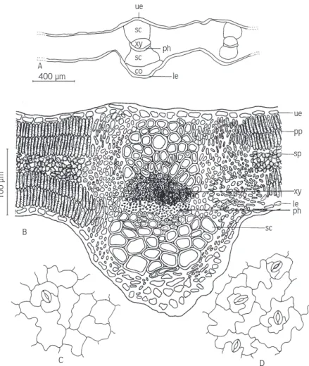
100 µm 200 µm A pi pi ep co pa sc ph xy B xy ph sc pa co ep
Figure 3. H. bituminosus. A-B: Cross-section of stem. ep: epidermis; co: collenchyma; pa: parenchyma; sc: sclerenchyma; ph: phloem; xy: xylem; pi: pith.
Figure 4. H. bituminosus. Cross-section of stem. ep: epidermis; co: collenchyma; pa: parenchyma; sc: sclerenchyma; ph: phloem; xy: xylem; pi: pith. Scale bar: 100 µm.
500 µm
A
C B
uniseriate. The pith consists of large hexagonal or polyhedral parenchymatous cells (Figures 3,4).
Leaf: The epidermis is composed of a single layer of cells, and the cells are rectangular or oval. Size of upper and lower epidermis is similar. Epidermal cells are covered with a thick cuticle. Stomata type is diacytic (Figure 6) and occurs on both surfaces (amphistomatic leaves). They are located almost on the same level as epidermis cells.
Eglandular trichomes are 2-4-celled with cuticular micropapillae, unbranched, and consist of elongated cells. Glandular trichomes are generally stalked and their head is unicellular (Figure 5). Trichomes are located on both surfaces of the leaf.
The leaf is isolateral. The mesophyll is differentiated into 2-seriate palisade and 1-4-seriate spongy parenchyma. The palisade tissue is under the upper and lower epidermis. The shape of the palisade parenchyma in transverse section is cylindrical. The spongy parenchyma cells, circular or ovoid, are located between the palisade tissues.
The midrib region is well developed and forms a projecting part towards the outside. Vascular bundles are collateral. The xylem faces towards the upper surface, while the phloem faces the lower epidermis. The upper and lower vascular bundles are covered with sclerenchymatous cells. There are collenchymatous cells, 2-3-layered, under the upper and lower epidermis in the midrib (Figures 6,7). 100 µm 400 µm B C D ue A sc xy sc co ph le ue pp sp xy le ph sc
Figure 6. H. bituminosus. A-B: cross-section of leaf; C: surface view of upper epidermis; D: surface view of lower epidermis. ue: upper epidermis; pp: palisade parenchyma; sp: spongy parenchyma; xy: xylem; le: lower epidermis; ph: phloem; sc: sclerenchyma.
Discussion
Hymenocrater bituminosus is a unique species in Turkey, which belongs to the genus Hymenocrater. It grows in Kars and Van provinces in East Anatolia. There have been no previous morphological or anatomical studies of the Hymenocrater species.
In this paper, we report the findings of a morphological study of H. bituminosus so as to improve the present knowledge of morphology for systematic purposes.
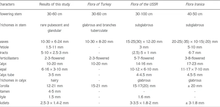
Our morphological results have been compared to those published in the Flora of Turkey (Mill, 1982), Flora Iranica (Rechinger, 1982), and Flora of the USSR (Gorshkova, 1976). The comparison of morphological characters is shown in Table 1.
The description of specimens in our study is different from the descriptions in the Flora of Turkey and the flora of neighbouring countries, in terms of some characteristics.
Petiole, bracts, sepal, calyx tube, trichomes in calyx, stamen, style, and nutlet sizes have been reported here for the first time. Some morphological variations have been observed in the indumentum and bract length characteristics (Table 1). The stem has simple hairs with sparsely glandular hairs. The indumentum in the calyx is
Figure 7. H. bituminosus. Cross-section of leaf midrib. ue: upper epidermis; pp: palisade parenchyma; xy: xylem; ph: phloem; sc: sclerenchyma. Scale bar: 100 µm.
Table 1. Morphological characteristics of H. bituminosus in Turkey compared to H. bituminosus in other Flora.
Characters Results of this study Flora of Turkey Flora of the USSR Flora Iranica
Flowering stem 30-60 cm 30-60 cm 30-100 cm 40-50 cm
Trichomes in stem rare pubescent and glabrous and branches subglabrous subglabrous glandular tuberculate
Leaves 10-30 ×6-24 mm 10-30 ×8-20 mm 15-25(30) ×12-20 mm 20-25(-35) ×10-15(-20) mm
Petiole 1.5-11 mm - 3 mm 5-10 mm
Bracts 5-10 ×2.5-3 mm - (2.5)-5 ×1 mm 6-7 mm
Verticillasters 2-3-flowered 2-3-flowered 5-7-flowered 3-8-flowered
Calyx 10-20 mm 10-20 mm 14-16 mm 17-23 mm
Sepal 6-16 ×3-10 mm - 10-12 ×6-10 mm 11-17 ×7-10 mm
Calyx tube 3-5 mm - 4-4.5 mm 4.5-5 mm
Trichomes in calyx hairy - glabrous glabrous
Corolla 12-21 mm 15-21 mm 15-17(20) mm ± 20 mm
Stamen 4-5 mm - -
-Style 1.5 mm - 1.6 mm
denser than the stem’s (Figure 5). Eglandular trichomes in the calyx are 1-4-celled and with cuticular micropapillae. In addition, there are glandular trichomes in the calyx. They are short, unicellular stalked, rarely bi-cellular, and head.
H. bituminosus is the most similar species to H. elegans Bunge, which is distributed in north-east Persia and Turkmenia. Both species are differentiated by their corolla colours; H. bituminosus has a reddish corolla, whereas H. elegans has a blue-whitish corolla.
Our research results are consistent with the representative taxa reported by Satıl et al. (2002) and Kaya et al. (2002). The usual features of Lamiaceae
anatomy (Metcalfe & Chalk, 1950) were observed in anatomical studies of the species. The stem is square in transverse section with well-defined groups of collenchyma in the corners. The midrib area is well developed in the leaf anatomy. The vascular bundle is covered with sclerenchymatous cells in the upper and lower sides, as is Sideritis gülendamiae (Kaya et al., 2002).
Acknowledgements
We would like to thank Dr. Tuncay Dirmenci for his helpful discussion concerning the description of the species.
References
Bafler KHC, Kaya A, Satıl F & Tümen G (2000). Thymus aznavourii Velen (Labiatae) Üzerinde Morfolojik ve Anatomik Çalıflmalar. Ot Sist Bot Derg 7: 95-105.
Baytop A (1972). Bitkisel Drogların Anatomik Yapısı. ‹stanbul: ‹stanbul Üniversitesi, Eczacılık Fakültesi. Yayın No: 829 (4. Baskı). Firouznia A, Rustaiyan A, Nadimi M, Masoudi S & Bigdeli M (2005).
Composition of the essential oil of Hymenocrater calycinus (Boiss.) Benth. from Iran. J Essent Oil Res 17: 527-529. Gorshkova SG (1976). Hymenocrater Fisch & CA Mey. In: Komarov VL
(ed.) Flora of the USSR, Labiatae Vol. XX: 488-490 (Translated from Russian). Israel Program for Scientific Translation, Jerusalem.
Gönüz A & Özörgücü B (1999). An investigation on the morphology, anatomy and ecology of Origanum onites L. Turk J Bot 23: 19-32.
Kandemir N (2003). The morphological, anatomical and karyological properties of Endemic Salvia hypargeia Fich. & Mey. (Lamiaceae) in Turkey. Pak J Bot 35: 219-236.
Kaya A, Satıl F, Bafler KHC & Tümen G (2000). Morphological and anatomical studies on Cyclotrichium origanifolium (Labill.) Maden & Scheng. (Labiatae). Turk J Bot 24: 273-278.
Kaya A, Satıl F, Tümen G & Bafler KHC (2002). Türkiye’de Yeni Bir Endemik Tür: Sideritis gülendamiae H. Duman & F.A. Karavelio¤ulları (Labiatae). In: Bafler KHC & Kırımer N (eds.) 14. Bitkisel ‹laç Hammaddeleri Toplantısı, pp. 490-496, Eskiflehir. Kürkçüo¤lu M, Arıkan fi & Bafler KHC (2005). The Essential Oil of
Hymenocrater bituminosus Fisch et Mey. from Turkey. 6th International Symposium on the Chemistry of Natural Compounds (SCNC), Ankara.
Metcalfe CR & Chalk L (1950). Anatomy of the Dicotyledons, Vol. 2. London: Oxford University Press.
Mill RR (1982). Hymenocrater Fisch & CA Mey. In: Davis PH (ed.) Flora of Turkey and the East Aegean Islands. Vol. 7: 293-294. Edinburgh: Edinburgh University Press.
Mirza M, Ahmadi L & Tayebikhorrami M (2001). Volative constituents of Hymenocrater incanus Bunge an Iranian endemic species. Flavour Fragr J 16: 239-240.
Rechinger KH (1982). Hymenocrater Fisch & CA Mey. In: Rechinger KH (ed.) Flora Iranica, Labiatae No 150: 239-250. Graz (Austria): Akademische Druck-u Verlagsanstalt.
Satıl F, Tümen G, Akçelik A & Bafler KHC (2002). Comparative morphological, anatomical, ecological and chemical studies on endemic Satureja parnassica subsp. sipylea from Turkey. Acta Bot Croat 61: 207-220.