INVESTIGATING THE EFFECTS OF
BIOACTIVE PEPTIDE NANOFIBERS ON THE
GROWTH AND DIFFERENTIATION
BEHAVIOUR OF NERVOUS SYSTEM CELLS
A THESIS SUBMITTED TO
THE GRADUATE SCHOOL OF ENGINEERING AND SCIENCE
OF BILKENT UNIVERSITY
IN PARTIAL FULFILLMENT OF THE REQUIREMENTS
FOR THE DEGREE OF MASTER OF SCIENCE
IN
NEUROSCIENCE
By
CANELİF YILMAZ
JULY 2017
INVESTIGATING THE EFFECTS OF BIOACTIVE PEPTIDE NANOFIBERS ON THE GROWTH AND DIFFERENTIATION BEHAVIOUR OF NERVOUS SYSTEM CELLS
By Canelif Yılmaz July 2017
We certify that we have read this thesis and that in our opinion it is fully adequate, in scope and in quality, as a thesis for the degree of Master of Science.
Ayşe Begüm Tekinay (Advisor)
Michelle Marie Adams
Çağdaş Devrim Son
Approved for the Graduate School of Engineering and Science:
ii
ABSTRACT
INVESTIGATING THE EFFECTS OF
BIOACTIVE PEPTIDE NANOFIBERS ON THE
GROWTH AND DIFFERENTIATION
BEHAVIOUR OF NERVOUS SYSTEM CELLS
Canelif Yılmaz M.S. in Neuroscience Advisor: Ayşe Begüm Tekinay
July 2017
Peripheral nerve regeneration is a tightly regulated process that entails the degeneration, proliferation, alignment and remyelination of Schwann cells. Tuning the bioactivity is important to support all of these processes and achieving successful regeneration. Extracellular matrix (ECM) proteins are important molecules for controlling cell behavior and differentiation. Mimicking the natural ECM proteins is a promising approach for promoting regeneration in peripheral nerve injury. In this study, we investigated the biocompatibility and bioactivity of two natural ECM mimicking peptide amphiphile (PA) molecules, heparan sulfate-mimicking PA (HM-PA) and laminin-mimicking PA (LN-PA), and showed that they self-assemble into ECM-like nanofibrous networks. These bioactive nanofibers promote the viability, proliferation and spreading of Schwann cells, and that the combination of the two bioactive epitopes supports both early and late neuroregenerative responses of Schwann
iii
cells. We have also shown that these nanofibers support the attachment and neurite extension of dorsal root ganglion neurons, and promotes neurite alignment and assembly in DRG-Schwann cell co-cultures.
KEYWORDS:
Peptides, Peptide nanofibers, biomimetic materials, peripheral nerve injury, nerve regeneration, Schwann cells, DRG.
iv
ÖZET
BİYOAKTİF PEPTİT NANOFİBERLERİN SİNİR
SİSTEMİ HÜCRELERİNİN BÜYÜME VE
FARKLILAŞMA DAVRANIŞLARI ÜZERINDEKİ
ETKİLERİNİN İNCELENMESİ
Canelif Yılmaz Nörobilim, Yüksek Lisans Tez Danışmanı: Ayşe Begüm Tekinay
Temmuz 2017
Periferal sinir rejenerasyonu, Schwann hücrelerinin Wallerian Dejenerasyonu, proliferasyonu, hizalanması ve remiyelinizasyonunu gerektiren ve sıkı bir şekilde düzenlenmiş bir süreçtir. Gerekli biyoaktivitenin sağlanması, bu süreçlerin tümünü desteklemek ve başarılı bir iyileşmeyi sağlamak için önemlidir. Ekstraselüler matris (ESM) proteinleri hücre davranışı ve farklılaşmasında önemli moleküllerdir. Doğal ESM proteinlerini taklit etmek, periferik sinir hasarında rejenerasyonun iyileştirilmesi için umut vadeden bir yaklaşımdır. Bu çalışmada, iki doğal ESM molekülünü taklit eden PA molekülleri heparan sülfat taklit eden PA (HM-PA) ve laminin-taklit eden PA (LN-PA) sentezlenmiş ve kendiliğinden bir araya gelmeyle ESM benzeri nanofiber yapılar oluşturdukları gösterilmiştir Ardından bu nanofiberlerin biyouyumluluk ve biyoaktivitesi araştırılmıştır. Nanofiber ağların, Schwann hücrelerinin canlılığını, çoğalmasını ve yayılmasını desteklediği ve iki biyoaktif molekülün kombinasyonunun
v
Schwann hücrelerinin hem erken hem de geç nörorejeneratif yanıtlarını desteklediği görülmüştür. Ayrıca bu nanofiberlerin DRG hücrelerinin yapışma ve nörit uzamasını desteklediği ve DRG-Schwann hücre ko-kültürlerinde nörit hizalanmasını ve toplanmasını desteklediği gösterilmiştir.
ANAHTAR KELİMELER:
Peptitler, Peptit nanofiberler, biyomimetik materyaller, periferik sinir yaralanması, sinir rejenerasyonu, Schwann hücreleri, DRG
Acknowledgem ents
Firstly, I would like to thank my Advisor Dr. Ayşe Begüm Tekinay, not only for her scientific guidance, but also for sharing countless examples of her life experience and helping me grow as a person. I would also like to thank my de-facto co-advisor Mustafa Özgur Güler, for his support and providing scientific insight during my thesis research.
I would like to thank my jury members Çağdaş Devrim Son and Michelle Marie Adams, for taking time to evaluate my thesis, and for their precious feedback. I would like to acknowledge financial support from TUBITAK BIDEB 2210-C scholarship, and TUBITAK ARDEB 113S959, and Bilkent University Interdisciplinary Neuroscience Graduate Program.
I would like to thank all members, past, present and future, of Nanobiotechnology Lab and Biomimetic Materials Lab, for welcoming me, for sharing an ever-growing amount of scientific expertise with me, and for providing a truly warm working environment. I have seen quite possibly the best times of both our groups, and I am proud to have worked alongside you guys.
I would like to thank Dr. Gülistan Tansık, Nuray Gündüz, Elif Arslan, Dr. Seher Üstün Yaylacı, Melike Sever, Dr. Ozlem Erol, Dr. Aref Khalily, Dr. Melis Şardan Ekiz and Dr. Göksu Çınar and Zeynep Erdoğan for their help with my technical questions.
I would like to thank Çağla Eren Çimenci for being a pillar of happiness and for the liters of coffee we consumed, İdil Uyan for our shared love of cats, and for dealing with impracticalities no one would bother to deal with, to Nurcan Haştar for being a constant source of cheer and pastry, to Gökhan Günay for always being there to hear me complain about things when I need to, to Merve Şen for being mostly unexpected and always Şen, to Zeynep Okur for convincing me every time that the cell isolation will work this time, to Fatih Yergöz for our endless quarrels, to Mustafa Beter for supplying the anti-capitalist spirit we
need, to Oğuz Tuncay for the teenaged girl in him (oh no I di’int!), to Özge Uysal for being the early bird and cheering us up, to Begüm Dikeçoğlu for always being helpful even when she’s busy, to Alper Devrim Özkan and Ahmet Emin Topal for being the most aweseome duo I’ve ever met, to Recep Erdem Ahan for always answering “no” to my utterly ridiculous scientific questions, to Egemen Deniz Eren for accompanying me day and night through my never-ending process of peptide synthesis, and finally to Oya İlke Şentürk for being my driving force to finish writing up my thesis.
I would also like to thank Seren Hamsici, Özüm Çalışkan, Ayşegül Dede, Göksemin Şengül, Zehra Tatlı Yıldırım, Aslı Ekin Doğan, Dr. Özlem Tufanlı, Dr. Begüm Kocatepe, Buket Gültekin, Behide Saltepe, Onur Apaydın, Side Selin Su Yirmibeşoğlu and Efe Işılak for making the life at UNAM 5. Kat quite more worth living.
I would like to thank the members of “AnKArA CreW”, Emin, Merve and Halil for our countless nights of partying, although our idea of partying slowly transformed into having dinner together and then studying on our own, to Erinç for being the best legal advisor, to Çağla for being my best clinician colleague, to Nazlı for always providing the best scientific support for each other without having the slightest idea about what the other one studies, to Güncel for her everlasting friendship and always being supercalifragilisticexpialidocious, to Ocan for the positive vibes, to Destan and Alican for constantly devising super-secret plans that we will never realize, to Emir for although having spent almost all of our friendship countries apart, always having exciting scientific news for me, to Canberk for being able to meet up only in Istanbul although we both live in Ankara, and finally to Barış for always being there to listen to my erratic worries and providing advice.
Finally, I would like to thank the members of my family, beginning with my mother Fatma Yaşar Yılmaz, my father Şükrü Önder Yılmaz, my aunts Ayşegül Zorlu, Hülya Varhan and Semiha Jensen for always supporting me through each decision I make, and raising me to become person I am. I hope to have become a person worthy of them.
Contents
1.1. Nervous system and peripheral nerve injuries ... 1
1.1.1. Anatomy and regenerative potential of the nervous system ... 1
1.1.2. Role of Schwann cells in peripheral nerve repair ... 5
1.1.3. Peripheral nerve injuries ... 7
1.1.4. Peripheral nerve repair ... 8
1.1.5. Usage of biomaterials for peripheral nerve repair ... 11
1.2. Natural ECM proteins and their mimetics ... 14
1.2.1. Laminin ... 15
1.2.2. Heparan Sulfate ... 16
1.2.3. Other proteins and epitopes... 17
1.3. Peptide amphiphile nanofibers and their applications in regenerative medicine ... 18
1.4. Objective of the study ... 20
2 Results and Discussion ... 21
2.1. Results ... 21
2.1.1. PA synthesis and characterizations ... 21
2.1.2. Schwann cell isolation and purity ... 28
2.1.3. Biocompatibility of PA nanofibers ... 29
3 Experimental ... 44
3.1. Materials ... 44
3.2. PA synthesis and characterizations... 45
3.2.1. PA synthesis and purification ... 45
3.2.2. Self-assembly and nanofiber formation ... 46
3.2.3. CD spectra of PA nanofibers ... 46
3.2.4. SEM imaging of PA nanofibers ... 47
3.2.5. AFM imaging of PA nanofibers ... 47
3.3. Primary cell isolation and culture ... 48
3.3.1. Schwann cell isolation and culture ... 48
3.3.2. DRG isolation and culture ... 49
3.3.3. DRG and Schwann Cell co-cultures: ... 50
3.4. Biocompatibility analyses ... 51
3.4.1. Preparation of cell culture surfaces ... 51
3.4.2. Viability of Schwann Cells ... 52
3.4.3. Proliferation of Schwann Cells ... 52
3.4.4. Spreading of Schwann Cells ... 53
3.5. Bioactivity analyses ... 54
3.5.1. Growth factor release from Schwann Cells ... 54
3.5.3. Immunocytochemistry of Schwann Cells and DRG cells ... 58
3.6. Statistical Analyses ... 58
4 Conclusions and Future Perspectives... 60
List of Figures
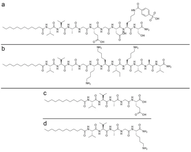
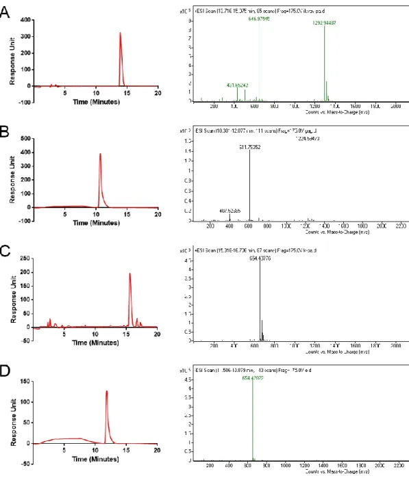
Figure 1.1: Transverse section of a peripheral nerve [6]. Figure reprinted with permission from Cold Spring Harbor Laboratory Press. ... 3 Figure 1.2: Anatomy of the peripheral nerve system. Figure reprinted under Creative Commons Attribution-Non-Commercial 3.0 Unported License. ... 4 Figure 1.3: PNI regeneration process [19] Figure reprinted under Creative Commons Attribution Non-Commercial license (CC-BY-NC) ... 7 Figure 1.4: Current treatment options for PNI [31]. Figure reprinted with permission from Elsevier. ... 10 Figure 1.5: Biomaterial use strategies for PNI regeneration [26]. Republished with permission of IOP Publishing. ... 12 Figure 1.6: Self-assembly of peptide amphiphiles into nanofibers [70]. Figure reprinted with permission from American Association for the Advancement of Science. ... 19 Figure 2.1 Self assembling PA molecules. Chemical structures of synthesized PA molecules HM-PA (a), LN-PA (b), E-PA (c), and K-PA (d). ... 22 Figure 2.3: Liquid chromatography and mass spectrometry analysis of synthesized PA molecules of LN- PA (A), HM-PA (B), K-PA (C) and E-PA (D). Mass spectrometry of LN-PA; [M+H]+ (calculated): 1292.93, [M+H]+ (observed): 1292.94, [M+2H]+2/2 (calculated): 646.96, [M+2H]+2/2(observed):
646.97, [M+3H]+3/3 (calculated): 431.64, [M+2H]+3/2(observed): 431.65. Mass spectrometry of HM-PA; [M-H]- (calculated): 1225.59, [M-H] -(observed):1224.59, [M-2H]-2/2 (calculated): 612.29, [M-2H]-2/2 (observed): 611.79, [M-3H]-3/3 (calculated): 407.86, [M-3H]-3/3(observed): 407.52. Mass spectrometry of K-PA; [M+H]+ (calculated): 654.48, [M+H]+ (observed): 654.49. Mass spectrometry of E-PA; [M-H]- (calculated): 654.42, [M-H] -(observed): 654.42. ... 23 Figure 2.4: Circular dichroism spectra of individual peptide amphiphiles ... 24 Figure 2.5: Circular dichroism spectra of nanofibers formed by mixing oppositely charged peptide amphiphile molecules. ... 25 Figure 2.6: Morphology of formed PA nanofiber networks. Gels prepared by mixing LN-PA/E-PA (A), LN-PA/HM-PA (B), PA/HM-PA (C) and K-PA/E-PA (D) as observed by SEM imaging. ... 26 Figure 2.7: Height map of the peptide nanofibers, as visualized with AFM, of LN-PA/E-PA (A), LN-PA/HM-PA (B), K-PA/HM-PA (C) and K-PA/E-PA (D). ... 27 Figure 2.8: Deflection map of the peptide nanofibers, as visualized with AFM, of LN-PA/E-PA (A), LN-PA/HM-PA (B), K-PA/HM-PA (C) and K-PA/E-PA (D). ... 28 Figure 2.9: Representative photomicrograph of immunostaining of Schwann cells against S100ß, scale bar = 50 µm ... 29
Figure 2.10: Representative photomicrographs of live-dead staining of Schwann cells cultured on PA nanofiber scaffolds and PLL for 24 and 48 h, scale bars = 100 µm. ... 30 Figure 2.11: Quantification of cellular viability of Schwann cells cultured on PA nanofiber scaffolds for 24 and 48 hours. No significant change was calculated. ... 31 Figure 2.12: Representative photomicrographs of spreading analysis performed by staining with Phalloidin (red) and immunostaining against Vinculin (green), of Schwann cells cultured on LN-PA/E-PA (A), LN-PA/HM-PA (B), K-PA/HM-PA (C), K-PA/E-PA (D) and PLL (E) coated surfaces for 48 h. Scale bars = 50 µm. Quantification of spreading of Schwann cells cultured on PA nanofiber scaffolds (F). No significant difference was calculated. ... 32 Figure 2.13: Proliferation rate of Schwann cells cultured on PA nanofiber scaffolds for 24 h, measured by BrdU ELISA, normalized to PLL. **: p<0.01 ***: p<0.001. Statistical analysis was performed with one-way-ANOVA with Bonferroni correction, n=3. ... 34 Figure 2.14: NGF release from Schwann cells cultured on PA nanofiber scaffolds for 24 and 48 h, quantified with sandwich ELISA. ***: p<0.001 ****: p<0.0001. Statistical analysis was performed with one-way-ANOVA with Bonferroni correction, n=3. ... 36
Figure 2.15: FGF release from Schwann cells cultured on PA nanofiber scaffolds for 24 and 48 h, quantified with sandwich ELISA. No significant difference was calculated. ... 36 Figure 2.16: Immunostaining of Schwann cells cultured for 7 days on PA nanofiber scaffolds, stained against MBP, p75-NTR or S100ß, the nuclei counterstained with TO-PRO-3. Scale bars = 50 µm. ... 37 Figure 2.17: Gene expression analysis of Schwann cells cultured on PA nanofiber scaffolds for 7 days. The expression level of each gene was normalized against the non-bioactive control K-PA/E-PA, and GAPDH and ß-Actin were used as internal controls. Error bars denote mean ± SEM, n=5, *p<0.05. ... 39 Figure 2.18: Immunostaining of DRG neurons cultured for 7 days on PA nanofiber scaffolds, stained against ß-III tubulin and Synaptophysin, the nuclei were counterstained with TO-PRO-3. Scale bars = 50 µm. ... 41 Figure 2.19: Immunostaining of DRG neurons and Schwann cells co-cultured for 10 days on PA nanofiber scaffolds, stained against ß-III tubulin and S100ß. Scale bars = 50 µm. ... 43
List of Tables
Table 3.1: Calculated mixing ratios for peptide nanofiber formation ... 46 Table 3.2: Primer sequences, elongation temperatures and efficiencies used for gene expression analysis with qRT-PCR reactions. ... 57
List of Abbreviations
AFM Atomic force microscopy AN OVA Analysis of variance
ASC Adipose derived stem cells CN S Central nervous system CN TF Ciliary neurotrophic factor DRG Dorsal root ganglion
ECM Extracellular matrix
ELISA Enzyme linked immunosorbent assay E-PA Non-bioactive (negatively charged) PA FG F-2 Basic fibroblast growth factor
G AG Glycosaminoglycan
G APDH Glyceraldehyde 3-phosphate dehydrogenase G DN F Glial derived neurotrophic factor
G F Growth factor
H BTU 2-(1H-benzotriazol-1-yl)-1,1,3,3-tetramethyluronium hexafluorophosphate
H M -PA Heparan sulfate-mimicking PA H S Heparan sulfate
ICC Immunocytochemistry
K-PA Non-bioactive (positively charged) PA LN -PA Laminin-mimicking PA
M SC Mesenchymal stem cell N G F Nerve growth factor PA Peptide amphiphile PBS Phosphate buffered saline PDL Poly-D-Lysine
PFA Paraformaldehyde
PLG A Poly lactic-co-glycolic-acid PLL Poly-L-Lysine
PN I Peripheral nerve injury PN S Peripheral nervous system
qRT-PCR Quantitative real-time polymerase chain reaction SEM Scanning electron microscopy
Chapter 1
1 Introduction
1.1. N ervous system and peripheral nerve injuries
1.1.1. Anatom y and regenerative potential of the nervous
system
The central nervous system (CNS) consists of the brain and the spinal cord. These structures are composed of neurons, that relay information; glia, that support the neurons; and the extracellular matrix (ECM). Neurons form an interconnected network to gather, process and conduct information via electrical signals. In the CNS, oligodendrocytes are the glial cells that insulate the axons to increase conduction speed [1], and microglia are cells that function as phagocytic cells that protects neurons and serve as an internal immune system [2]. Astrocytes are also glial cells with has many functions, such as neurotransmitter clearance, providing ionic balance, clearance and secretion of the interstitial fluid, maintenance of the blood brain barrier, and scar formation when an injury occurs [3].
Neurons are cells that relay information via electrochemical signals. The balance of ions, neurotransmitters, and neuromodulators are crucial for neural
conductivity. They form a large and extremely complex network by synaptic connectivity to function as an information processor. Injuries in the CNS cause this intricate balance to be lost, and cause disruption of the network. Not only the apoptotic neurons, but also their connections are affected, and this increases the effect of the injury. Damaged axons may grow over very limited lengths; however, regeneration in the CNS is virtually impossible [3]. This is due to neurons being terminally differentiated cells, which cannot replicate, and since the stem cell resources in the brain is scarce. In addition, the glial scar formation caused by protein deposition from astrocytes and endothelial cells poses a physical and molecular obstacle for CNS regeneration [4, 5].
The main cell types of the peripheral nerve system (PNS) are neurons and Schwann cells. Schwann cells myelinate the axons. The connective tissue around the axons are called the endoneurium, the vascular structure around the epineurium providing the flux of ions and macromolecules is called the perineurium, and the outermost connective tissue is called the epineurium (Figure 1.1).
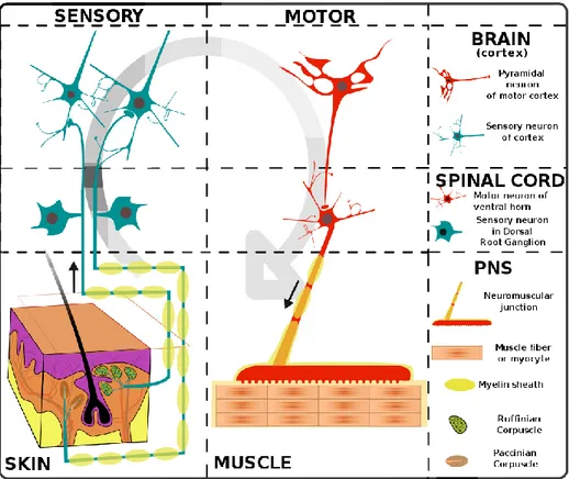
Figure 1.1: Transverse section of a peripheral nerve [6]. Figure reprinted with permission from Cold Spring Harbor Laboratory Press.
The neural cells of the PNS are classified as sensory, and motor nerves. Sensory neurons are bipolar nerves with cell bodies resting in the dorsal root ganglia (DRG), collecting information from the network of receptors around the body. Motor neurons are divided into two groups, somatic and autonomic nerves. The motor neurons’ cell soma lays in the brain, and they have long projections from the brain to the body. Somatic nerves relay signals to the skeletal muscles through neuromuscular junctions, and they control the voluntary movements. Autonomic nerves, on the other hand, control the involuntary movements such as breathing, peristalsis, and they also connect with the glands in the body (Figure 1.2).
Figure 1.2: Anatom y of the peripheral nerve system . Figure reprinted under Creative Com m ons Attribution-N on-Com m ercial 3.0 U nported License.
Compared to the CNS, the regeneration capacity of the PNS is higher. The regeneration in the PNS depends on the size of the injury, the location of the injury, and the biological condition of the patient [7]. Smaller injuries have better ability to regenerate. The ones further from the cell soma can regenerate better than the ones closer to the cell soma [8]. This difference is provided by the molecules in the interstitial fluid, the rapid clearance of the debris, and mainly, the Schwann cells’ ability to support the regeneration [9, 10]. Schwann cells can proliferate to replace the cells lost during the injury, but in the CNS oligodendrocytes cannot proliferate and compensate the cell loss [9]. The timing
of intervention is also critical because as time passes, Schwann cells may lose their supportive abilities [11].
1.1.2. Role of Schwann cells in peripheral nerve repair
Schwann cells are key players during the regeneration process. Following the injury, they undergo “Wallerian degeneration” by demyelinating the damaged axon, to assist in the clearance of cell debris from the injured axon [12, 13]. Macrophages infiltrate the distal stump of the damaged nerve and help the clearance of debris. Schwann cells that have detached from the axons, proliferate to form bands of Büngner by aligning inside the endoneurium [6, 14]. This alignment serves the purpose of a guidance tube for growth cones of regenerating axons. They do this by secreting ECM proteins such as laminin and fibronectin, proteins that have neural guidance properties [15] and they express surface markers that enable neural cell attachment, such as neural cell adhesion molecule, and cadherins, that also helps the neural guidance [16]. Another function of Schwann cells during peripheral nerve regeneration is to secrete growth factors (GFs) that accelerate regeneration, in response to the injury. These GFs include brain derived neurotrophic factor, ciliary neurotrophic factor (CNTF), glial derived neurotrophic factor (GDNF), nerve growth factor (NGF) and basic fibroblast growth factor (FGF-2) [17, 18]. These factors enable generation of growth cones and promote neurite extension, and
help growth towards the target organs. Finally, if successful regeneration is observed, they remyelinate the regenerated axons to provide insulation and improve signal conduction speed. However, the complete regeneration of a peripheral nerve injury (PNI) is only observed in small injuries, distant from the cell soma. Therefore, larger injuries closer to the cell soma often require medical intervention to ensure recovery (Figure 1.3).
Figure 1.3: PN I regeneration process [19] Figure reprinted under C reative Com m ons Attribution N on-Com m ercial license (CC -BY -N C)
1.1.3. Peripheral nerve injuries
PNIs are caused by blunt or penetrating traumas, and result in loss of autonomous, sensory or motor function in the affected areas. PNIs are a major
cause of disability in the modern world, with incidence rates estimated at 13-23 per 100,000 people per year in developed countries [20]. The PNS is composed of autonomic, sensory and motor neurons, and Schwann cells myelinating the axons of these neurons. The PNS has a better regeneration capacity compared to the CNS, which virtually has no ability to regenerate, however this capacity is still limited. The ability of the PNS to regenerate without external intervention depends on the size and location of the injury, as well as the proximity of the damaged axons to the cell soma. When injuries are large or close to the cell soma, regeneration is often inadequate, leading to muscular atrophy, loss of sensation and, depending on the location of the injury, irregularities in autonomous functions such as breathing, heart contraction or digestion.
1.1.4. Peripheral nerve repair
Several therapies such as direct suturing, allografts, autografts and natural or synthetic conduits are used for the treatment of PNIs to a certain level of success (Figure 1.4). Direct suturing is only successful in gaps shorter than 5 mm, and may introduce inflammation and impair recovery [21]. Autografts remain as the gold standard in PNI repair, however they cause additional damage to the patient through the nerves harvested for the autografting process [20]. Allografts are also effective in promoting regeneration, but they require the host
to receive immunosuppressants, which leaves the patient prone to infections and even tumor formation [22, 23]. Processed allografts manufactured from decellularized tissue [24] or natural proteins [25] are also used in proof of concept studies, but they are not fully suitable for proceeding into clinical use due to them being difficult and expensive to produce, and the rapid degradation of the proteins [19, 26, 27]. Conduits of synthetic polymers, on the other hand, despite being cheaper and easier to manufacture, often lack the required bioactivity and only serve as a guidance conduit rather than promoting regeneration [28]. They also hold the potential to cause foreign body reaction, causing inflammation and scar tissue formation [29]. In addition, due to them being rather large prosthetics, none of these therapies are applicable in the extremities, and their use is limited to injuries that occur on larger nerve bundles. Alternatively, injectable hydrogels may be injected directly to the wound site [26], and have also shown promise for PNI treatments. These hydrogels come forth as compelling materials since they are applicable in the extremities, as well as they can be utilized to fill the lumen of polymeric conduits to provide bioactivity [30].
Figure 1.4: C urrent treatm ent options for PN I [31]. Figure reprinted with perm ission from Elsevier.
A promising therapy in PNI repair is Schwann cell or stem cell transplant into the injury site. Although the isolation of Schwann cells shares similar downsides with autografts [32], mesenchymal stem cells (MSCs) and adipose-derived stem cells (ASCs) can be isolated in abundance with minimally invasive procedures. Recently, differentiation of ASCs into Schwann cell-like phenotypes was reported [33], and MSCs can be differentiated into neuronal phenotypes [34]. Alas, the viability after transplantation of these cells remains a challenge. Biomaterials providing mechanical and biochemical protection from the stress
caused by the transplantation process would improve the efficiency of these procedures.
1.1.5. U sage of biom aterials for peripheral nerve repair
A variety of biomaterials has been investigated to be used in PNI repair. These biomaterials include natural biomaterials, such as ECM proteins, synthetic biodegradable polymeric biomaterials or non-degradable polymers. The biomaterials used in PNI repair possess beneficial traits such as biocompatibility, biodegradability, ability to modify chemical and physical properties such as porosity and topography, and for synthetic materials, ease and low cost of production [35] (Figure 1.5).
Figure 1.5: Biom aterial use strategies for PN I regeneration [26]. Republished with perm ission of IOP Publishing.
Usage of natural proteins as biomaterials has been an interesting topic due to their high biocompatibility. Collagen and its various subtypes constitute the majority of the ECM; therefore, it has been used widely as a tissue engineering biomaterial. Reports indicate that conduits manufactured from bovine collagen functionalized with NGF can improve functional recovery of a rat sciatic nerve injury model [36]. Also, it has been shown that porcine collagen nerve guides enhance motor neurons’ axonal recovery in a 1 cm long defect in rats [37]. Hyaluronic acid, another natural polymer, composed of repeats of sugar units, has been shown to possess exceptional biocompatibility [38], and due to its
tunable viscoelasticity, it is able to support maturation of neurons [39], and can be used with laminin as a cell transplant scaffold to protect the neural stem cells from stress during transplantation [40]. While these materials show promising results in research, their rapid degradation rates prevent them from being translated into clinical practice [41].
Synthetic polymers, with the benefit of having received FDA approval, have also been widely investigated in PNI repair. Poly caprolactone/poly lactic acid copolymers provide significant permeability, which is crucial for the exchange of biomolecules at the wound site [42]. Poly lactic-co-glycolic-acid (PLGA) polymers functionalized with the cell binding epitope RGD has been shown to accelerate functional recovery on a rat sciatic nerve defect [43]. Since neurons are electrically active cells, electrically conductive polymers have also been suggested to be used as nerve guidance conduits. For example, an electrically conductive polyurethane has been shown to increase NGF release and expression of myelinating genes of Schwann cells [44]. In addition, NGF decorated electrically conductive polypyrrole has been shown to significantly increase neural phenotype and neurite extension of PC-12 cells [45].
These biomaterials can also be used for the local delivery of drugs, growth factors or cells into the injured area [26]. Crosslinked collagen hydrogels have been used for GDNF and NGF delivery in a 1 cm wide nerve gap, and improved functional recovery [46]. PLGA scaffolds decorated with microspheres that
release NGF have been shown to improve fasciculation and remyelination of a rat sciatic injury model [47].
1.2. N atural EC M proteins and their m im etics
The natural environment of the tissues is composed of a fibrous network of proteins that is called the ECM. The ECM is a porous network of different biomacromolecules such as laminins, collagens, tenascins, fibronectin and heparan sulfate proteoglycans. These molecules are important in cell adhesion, migration, and differentiation during development and regeneration. Schwann cell extracellular matrix is known to be especially rich in laminins, tenascins and glycosaminoglycans [48]. The use of natural extracellular matrix proteins is often an expensive and undesirable method, since high amounts of production and isolation are not easy. Protein resistance and disruption are also limiting factors in clinical practice. Identifying bioactive epitopes of these proteins and incorporating them into the structure of synthetic nanomaterials allow for the production of nanomaterials that are both more productive and more stable in terms of stability.
1.2.1. Lam inin
Laminin is a glycoprotein found on the basement membrane, which is known to promote neuronal cell adhesion and differentiation. Synthetic peptide chains derived from the alpha-1 chain of laminin were screened, and IKVAV pentapeptide was found to be the bioactive sequence regulating cell behavior [49]. Ever since its discovery in 1989, the IKVAV pentapeptide has been used in a large number of studies, both in vitro and in vivo, and has been shown to improve neural cell attachment, migration, neurite extension and neural stem cell differentiation.
In a study conducted by Hamsici et al., electro spun cyclodextrin nanofibers were functionalized with the IKVAV epitope, by synthesizing an adamantane conjugated IKVAV peptide, and utilizing the host-guest interaction between adamantane and cyclodextrin. Then, PC-12 cells were cultured on the functionalized nanofibers, and the functionalization was shown to improve cell attachment, and increase the expression of neural markers [50].
As another example of the use of short peptide sequences, Cheng et al. produced a nanofiber hydrogel functionalized with the IKVAV epitope. The self-assembling property of RADA16 peptides was exploited to produce hydrogels, and the IKVAV epitope was conjugated to RADA16 peptides in order to increase bioactivity. Firstly, neural stem cells were encapsulated inside these gels, and while the NSCs differentiated into glial lineage in RADA16 gels, in
RADA16-IKVAV gels, they were observed to commit into neural lineage. When NSCs encapsulated in these gels were injected to a brain injury model, and in a period of 6 weeks, they have been observed to proliferate and fill the injury site and differentiate into neural cells [51].
The amino acid sequences YIGSR from beta-1 and KDI from gamma-1 chain are also functional epitopes derived from laminin, and used as ECM mimetic epitopes. The YIGSR epitope is mainly functional in cell attachment through laminin receptors, and most commonly used in studies regarding endothelial cell attachment and regeneration [52, 53]. The epitope has also been used to decorate collagen scaffolds for improved nerve guidance [54]. The KDI epitope has been shown to reduce glutamate neurotoxicity by acting as a glutamate receptor antagonist, and there are also reports stating that it has neurite extension promoting and neurite guidance effects as well [55, 56].
1.2.2. H eparan Sulfate
Glycosaminoglycans (GAGs) are a large family of ECM proteins that are glycosylated and sulfated, and they function as a structural compound of mammalian tissues. Heparan sulfate (HS) is one of the major members of the GAG family, and is a highly sulfated GAG. HS has important functions in a large number of physiological processes: it can bind chemokines and GFs on heparin binding domains through electrostatic interactions, and functions in the
transport and internalization of these molecules [57]. It can also bind other ECM proteins to provide tissue stability, and cell surface receptors to promote adhesion and differentiation by anchoring growth factors in cell niches [58]. HS contains carboxyl, hydroxyl and sulfonate groups, which enable the electrostatic interactions that mediate the bioactivity of the molecule. The high amount of sulfonation on the molecule is thought to harbor the GF binding ability of the molecule [59].
In a study conducted by our group, GF binding ability of HS mimicking PA nanofibers was investigated, and it was shown that this molecule can bind GFs containing a heparin binding domain with high affinity, as opposed to GFs without heparin binding domains [60]. These nanofibers were also shown to improve neurite extension from PC-12 cells, and promote angiogenesis in vitro and in vivo [61, 62].
1.2.3. Other proteins and epitopes
Several other functional epitopes were identified from other ECM proteins and used in regenerative medicine studies. For example, DGEA epitope from Collagen-I is known to bind integrin receptors to signal osteogenic differentiation [63, 64]. REDV epitope derived from fibronectin is known to promote selective binding of endothelial cells, and has been used in functionalization of arterial stents [65]. RGDS epitope, again isolated from
fibronectin is a cell attachment epitope and has been used in a plethora of studies to functionalize biomaterials to become cell adhesive [66, 67].
1.3. Peptide am phiphile nanofibers and their
applications in regenerative m edicine
Peptide amphiphiles (PAs) are synthetic molecules that can self-assemble into nanofibrous networks that resemble the morphology of the natural ECM. When two oppositely charge PA molecules are mixed, they form beta sheet structures, accumulate into nanofibers, and form a soft, injectable, self-healing hydrogel through electrostatic interactions [68] (Figure 1.6). PAs are composed of naturally occurring peptides and fatty acids, and are often biocompatible and biodegradable with no potentially harmful degradation products. They can be easily synthesized in large amounts, and can be designed to include various bioactive molecules. Due to their tailorable biological and morphological properties, the use of peptide amphiphiles in regenerative medicine has been an increasingly studied topic in the recent years, with promising results in neural regeneration as well [51, 69].
Figure 1.6: Self-assem bly of peptide am phiphiles into nanofibers [70]. Figure reprinted with perm ission from Am erican Association for the Advancem ent of Science.
1.4. Objective of the study
In this study, the main objective was to investigate the individual and cooperative effects of two natural ECM-mimetic PA molecules; a heparan sulfate-mimicking PA (HM-PA) containing the functional groups found on heparan sulfate, carboxylate, hydroxylate and sulfonate; and laminin-mimicking PA (LN-PA), containing the IKVAV pentapeptide. For this purpose, we have synthesized these two molecules along with two non-bioactive molecules, K-PA and E-PA, to provide a non-bioactive negative control when mixed together, and to enable self-assembly through charge neutralization in LN-only and HM-only bioactive groups. Poly-L-Lysine (PLL) or poly-D-Lysine (PDL)-Laminin coated surfaces were used as a positive control, as PLL is a commonly used cell adhesion polymer and the recommended culture surface for Schwann cells. We have demonstrated that these molecules can self-assemble into a nanofibrous scaffold. We investigated the individual and cooperative effects of the two molecules on Schwann cell viability, spreading, proliferation and myelination, as well as NGF release. In addition, we have further shown that all peptide nanofiber combinations can support DRG neuron adhesion and neurite extension, and when DRG neurons are co-cultured with Schwann cells on the scaffolds, the bioactive nanofiber combinations enable neurite bundling and alignment which resembles axon formation.
Chapter 2
2 Results and Discussion
2.1. Results
2.1.1. PA synthesis and characterizations
In this study, the main objective was to investigate the individual and cooperative effects of two natural ECM-mimetic molecules, LN-PA, and HM-PA. For this purpose, the molecules shown on Figure 7 were synthesized. While LN-PA and HM-PA were the two bioactive molecules to be investigated, two non-bioactive molecules, K-PA (positively charged) and E-PA (negatively charged) were also synthesized to provide a non-bioactive negative control when mixed together, and to enable self-assembly through charge neutralization in LN-only and HM-only bioactive groups. The purity of the synthesized molecules was verified by LC-MS (Figure 8). the sharp peak on LC retention graph on Figure 8 indicated the purity of the crude product and the mass to charge counts matching with the theoretical molecular weights validated that the observed product was in fact the intended molecule.
Figure 2.1 Self assem bling PA m olecules. C hem ical structures of synthesized PA m olecules H M -PA (a), LN -PA (b), E-PA (c), and K -PA (d).
Figure 2.2: Liquid chrom atography and m ass spectrom etry analysis of synthesized PA m olecules of LN - PA (A), H M -PA (B), K -PA (C) and E-PA (D). M ass spectrom etry of LN -E-PA; [M + H ]+ (calculated): 1292.93, [M + H ]+ (observed): 1292.94, [M + 2H ]+2/2 (calculated): 646.96, [M + 2H ]+2/2(observed): 646.97, [M + 3H ]+3/3 (calculated): 431.64, [M + 2H ]+3/2(observed): 431.65. M ass spectrom etry of H M -PA; [M -H ] -(calculated): 1225.59, [M -H ]- (observed):1224.59, [M -2H ]-2/2 (calculated): 612.29, [M -2H ]-2/2 (observed): 611.79, [M -3H ]-3/3 (calculated): 407.86, [M -3H ]-3/3(observed): 407.52. M ass spectrom etry of K -PA; [M + H ]+ (calculated): 654.48, [M + H ]+ (observed): 654.49. M ass spectrom etry of E-PA; [M -H ]- (calculated): 654.42, [M -H ]- (observed): 654.42.
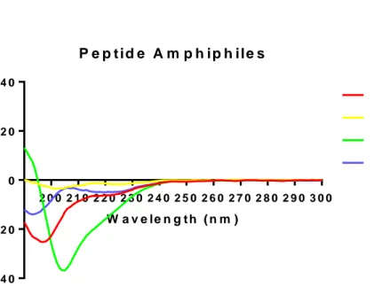
In order to investigate the secondary structure and beta sheet formation through self-assembly, CD spectra of molecules were recorded for PA only solutions and experimental nanofiber compositions. PA-only solutions predominantly showed random coil structures with local minima at around 195 nm, except for K-PA (Figure 9), which showed an alpha-helix like structure characterized by a local minimum between 200-210 nm and a local maximum at below 190 nm. P e p t id e A m p h ip h ile s W a v e le n g t h ( n m ) M o la r e ll ip ti c it y 2 0 0 2 1 0 2 2 0 2 3 0 2 4 0 2 5 0 2 6 0 2 7 0 2 8 0 2 9 0 3 0 0 - 4 0 - 2 0 0 2 0 4 0 L N - P A H M - P A K - P A E - P A
P e p t i d e A m p h ip h ile M ix t u r e s W a v e le n g t h ( n m ) M o la r e ll ip ti c it y 2 0 0 2 1 0 2 2 0 2 3 0 2 4 0 2 5 0 2 6 0 2 7 0 2 8 0 2 9 0 3 0 0 - 4 0 - 2 0 0 2 0 4 0 L N - P A /E - P A L N - P A /H M - P A K - P A /H M - P A K - P A / E - P A
Figure 2.4: Circular dichroism spectra of nanofibers form ed by m ixing oppositely charged peptide am phiphile m olecules .
When two oppositely charged molecules were mixed, self-assembly through electrostatic interactions was observed, and all combinations showed a beta sheet like structure characterized by local minima at around 220 nm and local maxima at around 200 nm (Figure 10).
Mimicking the morphology of the ECM is also important for functional recovery, as it is well-established that topographical cues positively affect the cell behavior, such as improving neurite length and alignment. Therefore, the morphology of the nanofiber networks formed through self-assembly was investigated by scanning electron microscopy (SEM) and atomic force microscopy (AFM) imaging. To observe the native 3D morphology of the gels, critical point dried samples were imaged using SEM (Figure 11). The structures
were observed to form a nanofibrous network resembling the natural ECM structure.
Figure 2.5: M orphology of form ed PA nanofiber networks. Gels prepared by m ixing LN -PA/E-PA (A), LN -PA/H M -PA (B), K -PA/H M -PA (C) and K -PA/E-PA (D ) as observed by SEM im aging.
For AFM imaging, nanofibers were formed on borosilicate coverslips, and dried overnight. The samples were prepared as they will be prepared as cell culture scaffolds. The AFM images of height (Figure 12) and deflection (Figure 13) maps that the nanofiber structures were preserved after drying of the substrates.
Figure 2.6: H eight m ap of the peptide nanofibers, as visualized with AFM , of LN PA/EPA (A), LN PA/H M PA (B), K PA/H M PA (C) and K -PA/E-PA (D).
Figure 2.7: Deflection m ap of the peptide nanofibers, as visualized with AFM , of LN -PA/E-PA (A), LN -PA/H M -PA (B), K -PA/H M -PA (C) and K -PA/E-PA (D).
2.1.2. Schwann cell isolation and purity
In order to verify the purity of the isolated Schwann cells, immunostaining against the Schwann cell marker S-100ß was performed at passage 2, before using the cells in further experiments. Immunostaining against S-100ß verified that the cells were Schwann cells, and the cell purity was calculated as 98.6% ±1.8 by dividing the S-100ß positive cells to the number of nuclei (i.e. the total number of cells) on randomly taken pictures (Figure 14).
Figure 2.8: Representative photom icrograph of im m unosta ining of Schwann cells against S100ß, scale bar = 50 µm
2.1.3. Biocom patibility of PA nanofibers
When designing and manufacturing biomaterials, it is important to ensure the biocompatibility of the product by investigating the short-term effects of the material on cell viability, morphology and proliferation. For analyzing the biocompatibility of the self-assembled peptide systems, the viability of the Schwann cells cultured on the nanofiber scaffolds was investigated by live-dead staining with Calcein and Ethidium Homodimer. Representative images at 24 hours and 48 hours are provided in Figure 16 and show few to no dead cells on all culture surfaces. The number of live and dead cells were counted from
pictures taken after live dead staining, and cellular viability was calculated (Figure 16). Quantification of the cellular viability showed no significant effect of any of the nanofiber scaffolds on cell survival, compared to positive control group PLL.
Figure 2.9: Representative photom icrographs of live-dead staining of Schwann cells cultured on PA nanofiber scaffolds and PLL for 24 and 48 h, scale bars = 100 µm .
% V ia b il it y 2 4 h 4 8 h 0 5 0 1 0 0 L N - P A /E - P A L N - P A /H M - P A K - P A /H M - P A K - P A / E - P A P L L
Figure 2.10: Quantification of cellular viability of Schwann cells cultured on PA nanofiber scaffolds for 24 and 48 hours. N o significant change was calculated.
The effect of the nanofibers on the cellular morphology of the Schwann cells was investigated by Phalloidin and Vinculin immunostaining, Phalloidin binds the actin cytoskeleton of the cells, and Vinculin is a protein found on cellular adhesion points. As shown in Figure 17, all scaffolds supported the adhesion of the cells, which were observed to exhibit similar morphology on all scaffolds, and actin cytoskeletons were intact with stress fibers. Cell areas were quantified for at least 30 cells in each group, from randomly taken pictures, and no significant difference in cell area was observed. Overall, the presence of stress fibers and focal adhesion points, as well as similar morphology and spreading areas, validate that the cells were able to fully interact with all experimental scaffolds as well as the positive control group PLL, and cells showed normal behavior in all conditions.
Figure 2.11: Representative photom icrographs of spreading analysis perform ed by staining with Phalloidin (red) and im m unostainin g against Vinculin (green), of Schwann cells cultured on LN PA/EPA (A), LN -PA/H M -PA (B), K --PA/H M -PA (C), K -PA/E-PA (D) and PLL (E) coated surfaces for 48 h. Scale bars = 50 µm . Quantification of spreading of Schwann cells cultured on PA nanofiber scaffold s (F). N o significant difference was calculated.
The effect of nanofiber scaffolds on Schwann cell proliferation was investigated with the help of a colorimetric BrdU cell proliferation enzyme linked immunosorbent assay (ELISA) kit (Figure 18). The cell proliferation on PLL surface was assumed to be 100% and other groups were normalized to this value. On LN-PA/E-PA scaffold, proliferation was observed to stay at the same level of the PLL surface, while on LN-PA/HM-PA and HM-PA/K-PA scaffolds proliferation decreased down to 85% and 84%, respectively, and proliferation on the K-PA/E-PA scaffold increased up to 123%. This change may be interpreted as an indirect measure of cell differentiation, as it is known that Schwann cell proliferation and myelination processes are regulated by opposing protein pathways. Even though the decrease on LN-PA/PA and HM-PA/K-PA is statistically significant, it is numerically low and cells still do proliferate close to their natural rate, so this decrease is not likely to interfere with the regeneration process.
S c h w a n n C e ll P r o lif e r a t io n % P r o li fe r a ti o n LN -PA /E-P A LN -P A/H M-P A K-P A/H M-P A K-P A/E -P A PL L 0 2 5 5 0 7 5 1 0 0 1 2 5 L N - P A /E - P A L N - P A /H M - P A K - P A /H M - P A K - P A / E - P A P L L *** **
Figure 2.12: Proliferation rate of Schwann cells cultured on PA nanofiber scaffolds for 24 h, m easured by BrdU ELISA, norm alized to PLL. **: p<0.01 ***: p<0.001. Statistical analysis was perform ed with one-way-AN OVA with Bonferroni correction, n= 3.
2.1.4. Bioactivity of PA nanofibers
To assess the regenerative potential of the PA nanofiber scaffolds, we have first investigated the amount of growth factors released from the Schwann cells cultured on these scaffolds, using sandwich ELISA method. Schwann cells are known to secrete NGF and FGF-2 during peripheral nerve regeneration, and both of them are known to be important for the healing process. The cells were cultured on PA nanofiber scaffolds for 24 or 48 h, the cell culture supernatants were collected and GF release was quantified (Figures 19,20). NGF release was significantly increased from Schwann cells cultured on LN-PA/HM-PA and K-PA/HM-PA on 48 h, and even though not statistically significant, the same
trend was observed at 24 h as well. In FGF-2 release, an overall increase on nanofibrous scaffolds was observed, which was up to 1.6-fold on LN-PA/HM-PA scaffold at 48 h, although this increase was statistically insignificant. However, in our previous work we have shown that HM-PA containing nanofibers can bind FGF-2 with high affinity through its heparin binding domain [60], and considering the released FGF-2 would be trapped on the PA-nanofiber scaffolds, results associated with FGF-2 release is likely to be underestimated due to limited growth factor recovery from the scaffolds. NGF and FGF-2 are growth factors that are known to be upregulated after PNIs, induce neurite extension, and support the survivability of nervous system cells. Although these growth factors can be delivered in soluble form to the injury site, they would diffuse and degrade rather quickly, and would require multiple injections to obtain effective results. Inducing higher levels of growth factor secretion from Schwann cells, and ensuring that the secreted growth factors are locally restrained through electrostatic interactions with the PA nanofiber scaffold, is an efficient strategy for providing a sustained presence of growth factor signaling, and would lead to better regeneration of PNIs. Hence, the inclusion of HM-PA in bioactive scaffolds can provide increased and prolonged presence of growth factors at the injury site, and makes HM-PA a favorable molecule to include in PNI regeneration.
N G F R e le a s e b y S c h w a n n C e ll s N G F ( n g /m L ) 24 h 48 h 0 5 1 0 1 5 2 0 *** ** *** * * L N - P A /E - P A L N - P A /H M - P A K - P A /H M - P A K - P A / E - P A P L L
Figure 2.13: N GF release from Schwann cells cultured on PA nanofiber scaffolds for 24 and 48 h, quantified with sandwich ELISA . *: p≤ 0.05, **: p≤0.01, **: p≤0.001 Statistical analysis was perform ed with one-way-AN OVA with Bonferroni correction, n= 3.
F G F - 2 R e le a s e b y S c h w a n n C e ll s A b s o r b a n c e 24 h 48 h 0 .0 0 0 .0 1 0 .0 2 0 .0 3 0 .0 4 L N - P A /E - P A L N - P A /H M - P A K - P A /H M - P A K - P A / E - P A P L L
Figure 2.14: FGF release from Schwann cells cultured on PA nanofib er scaffolds for 24 and 48 h, quantified with sandwich ELISA. N o significant difference was calculated.
We have then performed immunostaining against MBP, p75-NTR and S100ß (Figure 21) on Schwann cells cultured on PA nanofiber scaffolds for 7 days, and observed the stable expression of all Schwann cell markers, with similar morphology and subcellular localization in each experimental group. Although the cells on LN-PA/HM-PA and K-PA/HM-PA groups were observed to be bigger, we attribute this behavior to the reduced proliferation rate on these scaffolds resulting in lower confluency. This showed that the cells did not lose their characteristics, and the scaffolds were supportive of the long-term culture of Schwann cells.
Figure 2.15: Im m unostaining of Schwann c ells cultured for 7 days on PA nanofiber scaffolds, stained against M BP, p75-N TR or S100ß, the nuclei counterstained with TO-PRO-3. Scale bars = 50 µm .
To further assess the bioactivity of the scaffolds, we have investigated the gene expression profiles of Schwann cells cultured on PA nanofiber scaffolds for 7 days, using quantitative real-time polymerase chain reaction (qRT-PCR) (Figure 22). The non-myelinating Schwann cell marker p75-NTR was downregulated on LN-PA containing scaffolds, as well as the positive control PLL down to 0.7-0.6-fold, and upregulated to 1.2-fold on K-PA/HM-PA scaffold, compared to non-bioactive scaffold K-PA/E-PA, albeit insignificantly. For the premyelinating transcription factor Krox-20, we have observed a significant upregulation of 2-fold on LN-PA/E-PA scaffold compared to K-PA/HM-PA and K-PA/E-PA scaffolds. An insignificant increase in gene expression was also observed on LN-PA/HM-PA scaffold and PLL. For the myelinating Schwann cell marker MBP, an upregulation of 1.6 and 1.9-fold was observed on LN-PA/E-PA scaffold and PLL, respectively; however, these were also not statistically significant. The change in gene expression levels are not clearly defined possibly because the Schwann cells we used were primary cultures. In a recent study, suggesting the use of a polymeric electroactive biomaterial as conduits, both primary cultures and an immortalized Schwann cell line were used, and the fold-change in gene expression was much less defined in primary cultures as opposed to the immortalized cell line [44]. Given that we have used Schwann cells isolated from adult tissue, even a slight change in gene
expression would be clinically meaningful. From the gene expression profiles, we can see that the groups containing LN-PA are better in promoting myelination, and we can suggest that inclusion of LN-PA in nanofiber compositions is beneficial for the successful remyelination after PNI injuries.
Figure 2.16: Gene expression analysis of Schwann cells cultured on PA nanofiber scaffolds for 7 days. The expression level of each gene was norm alized against the non-bioactive control K -PA/E-PA, and GAPDH and ß-Actin were used as internal controls. Error bars denote m ean ± SEM , n=5, *p<0.05.
Although Schwann cells are important players in PNI regeneration, it is also important for a biomaterial to promote regeneration of the neurons as well. Therefore, we have further assessed the bioactivity of the scaffolds by culturing freshly isolated DRG neurons, and performing co-cultures of DRG neurons and Schwann cells on the scaffolds. Because attachment of the neurons is problematic, PDL-Laminin coated coverslips were used as a positive control in these experiments. The DRG neurons were seeded directly on the PA nanofiber scaffolds after isolation, and cultured for 7 days. Immunostaining was performed against ß-III tubulin, a cytoskeletal protein expressed in mature neurons, and Synaptophysin, the major synaptic vesicle protein at day 7 (Figure 23). As these are already differentiated neurons, both proteins were expressed along the neurites. All scaffolds supported the adhesion and viability of these cells, as exampled by the immunostaining experiment. When DRG neurons are cultured on the PA nanofiber scaffolds, they were able to extend long neurites on bioactive scaffolds. Another behavior observed on PA/E-PA and LN-PA/HM-PA scaffolds was the alignment and bundling of these neurites, an important process in maturation of functional nerves. On the non-bioactive scaffold, neurites were shorter and spread out. On PDL-Laminin coated surfaces, long but disorganized neurites were observed.
Figure 2.17: Im m unostaining of DRG neurons cultured for 7 days on PA nanofiber scaffolds, stained against ß-III tubulin and Synaptophysin, the nuclei were counterstained with TO-PRO-3. Scale bars = 50 µm .
The co-cultures of DRG neurons and Schwann cells were performed by seeding Schwann cells on top of DRG neurons cultured on PA nanofiber scaffolds for 8 days, and the co-cultures were maintained for 10 additional days before immunostaining against ß-III tubulin (DRG neurons) and S100ß (Schwann cells) (Figure 24). DRG neurons showed similar behavior on the scaffolds as they did in the individual cultures, they grew neurites that were able to align and bundle up on bioactive scaffolds, as well as on the PDL-Laminin coated positive control. On the non-bioactive K-PA/E-PA scaffold, however, the neurites were randomly oriented. Schwann cells migrated and colocalized with the growing neurites in all scaffolds, which is a required behavior for the initiation of myelination. The alignment of the neurites on the bioactive scaffolds is a promising result, considering that this behavior was observed even in the absence of a topographical pattern. The results obtained from cultures of DRG neurons on PA nanofiber scaffolds suggest that the inclusion of bioactive molecules in the nanofiber composition improves cell morphology and behavior, which makes these scaffolds promising biomaterials to be used in PNI regeneration.
Figure 2.18: Im m unostaining of DRG neurons and Schwann cells co-cultured for 10 days on PA nanofiber scaffolds, stained against ß -III tubulin and S100ß. Scale bars = 50 µm .
Chapter 3
3 Experim ental
3.1. M aterials
All protected amino acids, resins and 2-(1H-benzotriazol-1-yl)-1,1,3,3-tetramethyluronium hexafluorophosphate (HBTU) were purchased from NovaBiochem and ABCR. Cell culture plasticware were purchased from Corning or NEST Biotechnologies. Cell culture medium and supplements were purchased from PAN Biotech, Sigma-Aldrich, Abcam or Thermo Fischer. Antibodies and ELISA reagents were purchased from R&D systems and Invitrogen. Primary antibodies for immunocytochemistry were purchased from Abcam, and secondary antibodies from Chemicon-EMD Millipore. All other chemicals used in this study were from Sigma Aldrich, Alfa Aesar, or Thermo Fischer, and were at least molecular biology grade.
3.2. PA synthesis and characterizations
3.2.1. PA synthesis and purification
PA molecules were synthesized using solid phase peptide synthesis method, using either Wang or Rink Amide resins. Fmoc protected amino acids and lauric acid were coupled with 2 equivalents of amino acid, 1.95 equivalents of HBTU and 2 equivalents of DIEA in dissolved DMF, shaking for at least 3 h. Fmoc deprotection was performed with 20% piperidine in DMF for 20 min. The cleavage of the synthesized peptide was performed using a mixture of TFA:TIPS:water in the ratio of 95:2.5:2.5, for 2 h. Excess TFA was removed in a rotary evaporator, and the remainder was precipitated in ice-cold diethyl ether overnight. The precipitate was dissolved in distilled water and freeze-dried. The purity of the PA molecules was analysed using HPLC-MS, using an Agilent 6530 accurate-Mass Q-TOF LC/MS equipped with an Agilent 1200 HPLC, equipped with a Phenomenex Luna 3 μ C8 100A (50 × 3.00 mm) column for positively charged PA molecules, and Agilent Zorbax Extend-C18 (50 × 2.1 mm) column for negatively charged molecules. PA molecules with purity over 95% were used as is, and ones below 95% were purified using a water/acetonitrile gradient on an Agilent 1200 HPLC system with a Zorbax prepHT 300CB-C8 column for positively charged molecules, and with a Zorbax Extend C18 prep-HT column for negatively charged molecules. HCl treatment
was performed on positively charged PA molecules to remove residual TFA, by dissolving 100 mg of PA molecule in 5 mL of 1 mM HCl and freeze-drying afterwards.
3.2.2. Self-assem bly and nanofiber form ation
All PA molecules were dissolved in distilled water. The pH of the solutions was set to be between 6.8-7.8 using either NaOH or HCl, and were sonicated for 15 min to obtain a homogenous solution. Self-assembly was triggered by mixing equimolar solutions of oppositely charged PA molecules in different volume ratios, in order to obtain zero net charge. The theoretical charges of each PA molecule and mixing ratios for scaffolds are shown in the Table 3.1.
PA molecule\Gel LN-PA/E-PA LN- PA/HM-PA K-PA/HM-PA K-PA/E-PA LN-PA (Charge: +3) 2 eq 1 eq - - HM-PA (Charge: -3) - 1 eq 1 eq - K-PA (Charge: +1) - - 3 eq 2 eq E-PA (Charge: -2) 3 eq - - 1 eq
Table 3.1: Calculated m ixing ratios for peptide nanofiber form ation
3.2.3. CD spectra of PA nanofibers
0.25 mM solutions of PA molecules were mixed in the ratios given above, incubated overnight at +4℃ to enable self-assembly. CD spectra for individual
PA molecules and PA nanofibers were recorded using a J-815 CD spectrophotometer, between 190 and 300 nm with an interval of 1 nm, and scanning speed of 100 nm/min. Three sets of measurements were accumulated for each sample.
3.2.4. SEM im aging of PA nanofibers
10 mM solutions of PA molecules were mixed on silicon wafers in the ratios given above in a total volume of 60 µL. Samples were incubated at room temperature for 5 min to enable self-assembly, and dehydrated by immersing the samples in increasing concentrations of ethanol solutions. To preserve the nanofiber structure, samples were dried in a Tousimis Autosamdri-815B critical point drier. The samples were coated with 9 nm thickness of Au-Pd to increase electron conductivity during imaging. SEM imaging was performed with a FEI Quanta 200 FEG, using the GSED detector at ESEM mode.
3.2.5. AFM im aging of PA nanofibers
1 mM solutions of PA molecules were mixed on 9 mM borosilicate coverslips in the ratios given above, in a total volume of 50 μL and incubated at 37 ℃ for 30 min to enable self-assembly, followed by drying in a laminar flow hood overnight. AFM imaging was performed using an MFP-3D system under
contact mode using SiNi cantilevers. 1024 x 1024 images were taken over a 5 μm x 5 μm area.
3.3. Prim ary cell isolation and culture
3.3.1. Schwann cell isolation and culture
Schwann cells were isolated from 10-12 weeks old male Sprague-Dawley rats by following a method previously described by Kaewkhaw et al. [71]. All procedures on animals were approved by the Animal Ethics Committee of Gulhane Military Medical Academy. Briefly, the rats were sacrificed by cervical dislocation under anesthesia, and sciatic nerves were collected from both legs. Epineurium of the nerves were stripped under a stereomicroscope using fine forceps, remaining nerve fascicles were teased into fine fragments and enzymatic dissociation was performed with a filter sterilized 0.0625% (wt/vol) solution of collagenase in DMEM. 10 mL of collagenase solution was used per pair of nerves, and nerve fragments were incubated at 37 ℃ for 1 h. At the end of the incubation, the cell suspension was filtered through a 40 μm cell strainer to remove undigested debris, and cell suspension was centrifuged for 5 min at 500g for 5 min. The pellet was dissolved in Schwann cell medium, composed of high-glucose DMEM, containing D-Val instead of L-Val, 2 mM L-Glu, supplemented with 10% FBS,
1% Penicillin/Streptomycin, 1% N2 supplement, 20 μg/mL BPE, and 5 μM Forskolin. The cell suspension was seeded on a PLL-Laminin coated 25 cm2 tissue culture flask. The initial culture was maintained in a 37 ℃ humidified chamber with 5% CO2 atmosphere, and was allowed to proliferate for 21 days
with changing half of the culture medium with fresh medium every 2-3 days. At the end of 21 days, cells were detached from the tissue culture flask using 0.25% Trypsin-EDTA, and transferred to a 75 cm2 flask coated with PLL. From then on, cells were passaged every 5-7 days in 1:4 dilution, and used in experiments within 7 passages.
3.3.2. D RG isolation and culture
DRG cells were isolated from 8-12 weeks old male Sprague-Dawley rats according to a previously described method [72]. All procedures on animals were approved by Ankara University Animal Ethics Committee. Briefly, rats were sacrificed by CO2 asphyxiation, their spinal columns were dissected and washed
with ice-cold HBSS containing 1.6 μg/mL fungizone. Excess tissue around the spinal columns were trimmed, and the spinal columns were cut in half through the sagittal plane. The DRG were identified under a stereomicroscope, and collected in ice-cold HBSS by grasping and gently pulling up with fine forceps. The nerve trunks were trimmed using a surgical blade and HBSS was replaced with 3 mL of 0.125% solution of Collagenase in DRG culture medium, composed
of F-12 medium containing 10% horse serum, 1% Penicillin/Streptomycin, 2 μM L-Glutamine, supplemented with 50 μM 5-fluro-2′-deoxyuridine and 150 μM Uridine, 200 ng/mL NGF and 100 μg/mL normocin O.. Enzymatic digestion was allowed for 2 h at 37 ℃, with replacing the collagenase solution with a fresh aliquot at 1-h mark. At the end of the digestion, the DRG were collected in a sterile falcon tube with 3 mg of DNAse1 and centrifuged at 500 g for 1 min. The resulting DRG pellet was resuspended in 1.5 mL of DRG culture medium and cells were mechanically dissociated by pipetting until a homogenous suspension was obtained. The suspension was filtered through a 100 μm cell strainer and the filtrate was evenly distributed among PA nanofiber, or PDL-Laminin coated coverslips. DRG neuron culture was incubated for 7 days in a 37 ℃ humidified chamber with 3% CO2 atmosphere, while refreshing the cell
medium at days 3 and 5. At the end of 7 days, DRG cells were either paraformaldehyde (PFA) fixed for immunocytochemistry (ICC), or transferred into a 37 ℃ humidified chamber with 5% CO2 atmosphere, in DRG medium
without pyrimidine analogues for co-culturing with Schwann Cells.
3.3.3. D RG and Schwann Cell co-cultures:
DRG cells transferred to DRG medium without pyrimidine analogues were cultured for 24 h to allow adaptation to the new environment. After the adaptation period, trypsin dissociated Schwann cells were resuspended in
Schwann cell medium prepared with DMEM with L-valine, and seeded on top of DRG cells, at 6.25x10³ cells/well density, without removing the DRG medium. The co-culture was maintained in a 1:1 mixture of DRG medium without pyrimidine analogues and Schwann Cell medium with L-Valine for 10 days, supplemented with 50 μg/mL ascorbate starting from day 2. At the end of 10 days, the co-cultures were PFA fixed for ICC.
3.4. Biocom patibility analyses
3.4.1. Preparation of cell culture surfaces
1 mM solutions were mixed in the ratios given in Table 1, in multiwell tissue culture plates, or on 13 mM borosilicate coverslips in following total volumes: 80 μL for 96 well plates, 100 μL for 13 mM coverslips placed in 24 well plates, and 800 μL for 6 well plates. The plates were incubated at 37 ℃ for 30 min to allow self-assembly, and dried overnight in a laminar flow hood. The next day, plates were sterilized under UV light for at least 30 min, and cells were seeded on PA nanofiber coated surfaces. As a control, PLL coating was also performed, by adding a sterile 0.01% of PLL solution of the same volume used for PA solutions in each well, incubating in 37 ℃ for 30 min, removing the solution and drying overnight.
![Figure 1.1: Transverse section of a peripheral nerve [6]. Figure reprinted with permission from Cold Spring Harbor Laboratory Press](https://thumb-eu.123doks.com/thumbv2/9libnet/5841295.119730/20.892.189.759.133.390/figure-transverse-section-peripheral-figure-reprinted-permission-laboratory.webp)
![Figure 1.3: PN I regeneration process [19] Figure reprinted under C reative Com m ons Attribution N on-Com m ercial license (CC -BY -N C)](https://thumb-eu.123doks.com/thumbv2/9libnet/5841295.119730/24.892.171.667.105.807/figure-regeneration-process-figure-reprinted-reative-attribution-license.webp)
![Figure 1.4: C urrent treatm ent options for PN I [31]. Figure reprinted with perm ission from Elsevier](https://thumb-eu.123doks.com/thumbv2/9libnet/5841295.119730/27.892.316.626.109.559/figure-urrent-treatm-options-figure-reprinted-ission-elsevier.webp)
![Figure 1.5: Biom aterial use strategies for PN I regeneration [26]. Republished with perm ission of IOP Publishing](https://thumb-eu.123doks.com/thumbv2/9libnet/5841295.119730/29.892.254.693.103.538/figure-biom-aterial-strategies-regeneration-republished-ission-publishing.webp)
![Figure 1.6: Self-assem bly of peptide am phiphiles into nanofibers [70]. Figure reprinted with perm ission from Am erican Association for the Advancem ent of Science](https://thumb-eu.123doks.com/thumbv2/9libnet/5841295.119730/36.892.189.761.126.863/figure-peptide-phiphiles-nanofibers-reprinted-association-advancem-science.webp)