Ankara 06800, Turkey
∥
Neuroscience Graduate Program, Bilkent University, Ankara 06800, Turkey
§
Department of Chemistry
“Ugo Schiff”, University of Florence, Sesto Fiorentino, Florence 50019, Italy
⊥Institute for Molecular Engineering, University of Chicago, Chicago, Illinois 60637, United States
*
S Supporting InformationABSTRACT:
The ability of dendritic cells to coordinate innate and adaptive immune responses makes them essential targets for
vaccination strategies. Presentation of speci
fic antigens by dendritic cells is required for the activation of the immune system
against many pathogens and tumors, and nanoscale materials can be functionalized for active targeting of dendritic cells. In this
work, we integrated an immunogenic, carbohydrate melanoma-associated antigen-mimetic GM3-lactone molecule into
mannosylated peptide amphiphile nano
fibers to target dendritic cells through DC-SIGN receptor. Based on morphological
and functional analyses, when dendritic cells were treated with peptide nano
fiber carriers, they showed significant increase in
antigen internalization and a corresponding increase in the surface expression of the activation and maturation markers CD86,
CD83 and HLA-DR, in addition to exhibiting a general morphology consistent with dendritic cell maturation. These results
indicate that mannosylated peptide amphiphile nano
fiber carriers are promising candidates to target dendritic cells for antigen
delivery.
KEYWORDS:
cancer immunotherapy, cancer antigens, dendritic cells, carbohydrate melanoma associated antigen, peptide amphiphiles
■
INTRODUCTION
The immune system is a complex machinery that requires
regulatory interplay between its innate and adaptive
compart-ments to ensure an e
fficient surveillance against pathogens and
cancer cells. Generally, the immune system is not e
fficiently
induced against tumors, and cancer cells are tolerated because
of the evasion mechanisms they employ.
1Because cancer cells
are poor antigen-presenting cells (APC), a robust antitumor
immune response cannot be elicited. Cancer immunotherapy
approaches aim to induce a long-lasting and robust immunity,
enabling cancer patients to
fight against their own cancer. Thus,
understanding of cancer and immune system cross-interactions
holds a paramount importance for the improvement and
development of new cancer immunotherapy strategies.
2,3Dendritic cells (DCs)
4are promising candidates for cancer
immunotherapy, as they capture, process and present antigens
to T-cells for the initiation of adaptive immunity.
5,6T-cells
presented and activated with a tumor antigen initiate anticancer
responses. The activation of dendritic cells and their capability
to activate T cells depend on their maturation state,
7which is
determined by the upregulation of costimulatory molecules as
well as by morphological changes such as increased cell surface
area, which enables more connections with surrounding cells.
8Several attempts have been made to pulse DCs with speci
fic
Received: March 22, 2017Accepted: April 26, 2017 Published: April 26, 2017
antigens for the induction of anticancer immunity. The
targeting of C-type lectin receptors (CLR) is critical for the
pulsing and maturation of DCs.
9,10Among CLRs, dendritic cell
speci
fic intracellular adhesion molecule-3-grabbing nonintegrin
(DC-SIGN) is a receptor that is mainly expressed on immature
dendritic cells (iDCs),
11and modulates the uptake of speci
fic
pathogens through its interactions with mannosylated
struc-tures.
12−14Indeed, several studies reported that DC-SIGN
receptors can be targeted by using mannosylated structures.
15−17Discovery of human cancer antigens enabled scientists to
develop new immunotherapy strategies.
18,19Melanoma is often an
intractable cancer type and has been reported to be a promising
target for immunotherapy.
20GM3 ganglioside is a widespread
glycosphingolipid overexpressed in metastatic melanoma cells, and
several studies investigated its potential as a melanoma associated
antigen.
21A metabolite of GM3 ganglioside, GM3 lactone, is also
present in melanoma cells. Even though it is more immunogenic
than GM3 ganglioside, its amount is generally not su
fficient to
cause an e
fficient immune response because of its instability in
acidic tumor environment. Previously, it was shown that a
permannosylated dendron-containing hydrolytically stable mimetic
of GM3 lactone (antigenic GM3 lactone mimetic) is able to
induce DC activation through DC-SIGN receptor.
15Recently, antigens delivered through nanoscale materials have
been shown to provide considerable advantages over soluble
antigen administration, due to the stimulation of antigen uptake
and cellular activation by surface-functionalized delivery vectors.
Gold and PLGA nanoparticles, dendrimers and liposomes
have previously been functionalized for in vitro dendritic cell
targeting purposes.
15,22,23Molecular self-assembly is a process
by which molecules are organized into complex structures
through noncovalent interactions. Functional self-assembled
nanomaterials can be designed through a bottom-up fabrication
technique,
24,25which can be used in many biomedical
applica-tions such as drug delivery and regenerative medicine.
26,27Among these materials, self-assembling peptide amphiphiles
(PA) have been extensively used for their biocompatibility and
biodegradability.
28In addition, these molecules can be designed
to form nano
fibers and can be tailored with a variety of
func-tional groups and residues for cellular targeting.
29,30Here self-assembled mannosylated glycopeptide nano
fibers
were used for the delivery of an immunogenic mimetic of
the GM3 lactone antigen. Sugar speci
ficity was investigated
between mannose and glucose in terms of DC-SIGN receptor
targeting and antigen delivery into immature DCs. We showed
the activation and maturation of DCs in terms of phenotypic
expression of surface costimulatory molecules and
morpho-logical changes upon DC-SIGN targeting.
■
RESULTS AND DISCUSSION
Preparation of Fluorescent GM3-MIM Integrated
Glycopeptide Nano
fibers. A glycosylated amino acid residue
was
first synthesized in four steps prior to the synthesis of
glycopeptide PA molecules (
Scheme S1
). All compounds
synthesized in each step and the
final molecule, Fmoc-
L-Ser[
α-D-Man(OAc)
4]
−OH, were characterized by NMR and mass
spectrometry (
Figures S1
−S8
). Synthesized Fmoc-
L-Ser[
α-D-Man(OAc)
4]
−OH was used as the first amino acid residue of
the glycopeptide sequence during the elongation of the peptide
on solid support. After the resin cleavage, the amphiphilic
glycopeptide was obtained in protected form due to the
acetylated sugar hydroxyl groups, and deprotection reaction
was carried out in solution phase to prevent O-glycosidic
bond cleavage during acid treatment in solid phase peptide
synthesis.
31Although the mannosylated amino acid residue
existed at the C-terminus of the peptide segment, amphiphilic
character was obtained by the conjugation of a hydrocarbon tail
to the N-terminus of the peptide moiety (
Figure 1
). Here the
mannose residue was chosen because of its high a
ffinity to
DC-SIGN receptor, enabling the induction of DC activation.
In addition, an additional glycopeptide molecule, Glc-PA, was
synthesized by using a glucosylated serine amino acid in the
sequence to test the sugar speci
ficity of the glycosylated PA.
The chemical structures of the two amphiphilic glycopeptide
molecules, Man-PA and Glc-PA, were veri
fied by liquid
chromatography and mass spectrometry (
Figure S9
).
Before the integration of
fluorescent GM3-MIM into the
glycopeptide systems, the amphiphilic glycopeptides were
individually investigated in terms of their secondary structure
and morphological properties at physiological pH. Diluted
solutions of Man-PA and Glc-PA were studied by circular
dichroism (CD) spectroscopy and the results revealed that
both pure systems were oriented in a
β-sheet conformation,
exhibiting a positive peak at 202 nm and negative peak at
Figure 1.Chemical structures of the self-assembling molecules.
ACS Applied Materials & Interfaces
solutions were dissolved in slightly acidic water, GM3-MIM was
solubilized in a DCM:MeOH (7:1, v/v %) mixture. Prior to the
addition of the GM3-MIM solution, glycopeptide solutions were
separately sonicated at 50
°C at pH 6 to disrupt self-assembly
and generate single glycopeptide molecules. The glycopeptides
carry a positive net charge from their lysine residue, which
increases their water solubility and facilitates their integration
during self-assembly, as electrostatic interactions force the
hydrophobic tail of glyco-PAs within the nano
fiber structure,
whereas lysine and serine-conjugated glucose/mannose residues
are presented at the nano
fiber periphery. After addition of
the antigen solution, heating and sonication processes allow the
molecule into the water phase. The
fluorescence and UV spectra
of
fluorescent GM3-MIM integrated glycopeptide systems were
blue-shifted relative to GM3-MIM alone, which was also in good
agreement with visual observations (
Figures 2
B, C). UV spectra
results also showed that GM3-MIM exhibited comparable
absorbance values before and after its integration into
glyco-PAs, suggesting that it was successfully integrated into the PA
system and that integration was quantitative.
The successful transfer of GM3-MIM into the glycopeptide
solutions was followed by the investigation of the secondary
structure and morphology of glycopeptides in the presence
of the antigen by using CD and STEM, respectively. CD results
indicated that GM3-MIM integrated glycopeptide solutions
showed a
β-sheet composition (
Figure S13
). Although a higher
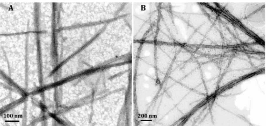
degree of bundling was observed after the integration in STEM
images, the PAs nevertheless self-assembled into nano
fibers
without any signi
ficant change in their diameters, indicating the
preservation of the PA packing process (
Figures 3
A, B).
More-over, because of the sulfur content of GM3-MIM molecule, it
was possible to test whether the antigen was successfully
integrated into the glycopeptide nano
fibers. Sulfur and oxygen
mapping was performed under TEM to con
firm that GM3-MIM
molecules were present in glycopeptide nano
fibers (
Figure S15
).
In addition, zeta potential measurements were performed in
order to investigate the surface charge of the glyco-PAs, which
was found to be around +27 mV (
Figure S16
).
Cell Viability Assay and iDC Di
fferentiation from THP-1
Monocytes. Cytotoxicity is another key factor when designing
materials for targeting purposes, and according to live
−dead
assay, 150
μM of glycopeptide nanofibers were found to
be nontoxic to cells (
Figure S17
). Immature dendritic cells
were obtained from THP-1 human monocytes as previously
reported.
32Cells di
fferentiation was monitored by quantifying
the surface expression of CD86, and
flow cytometry analyses
were performed to con
firm the de novo expression of this
marker (
Figure S18A
). In addition, the cells displayed extrusions
and protrusions that are consistent with the typical morphology
of immature dendritic cells (
Figure S18B
). As glyco-PAs are
biocompatible at 150
μM, and they formed nanofibers at this
concentration (as con
firmed by TEM and CD), subsequent
in vitro experiments were performed using this concentration.
Internalization of Fluorescent GM3-MIM Integrated
Glycopeptide Nano
fibers. Previous studies have reported
the internalization of mannosylated structures through
DC-SIGN receptors. Considering that iDCs strongly express
DC-SIGN on their surfaces,
11we investigated the
internal-ization of GM3-MIM integrated glycopeptide nano
fibers
through confocal microscopy imaging after obtaining iDCs
from THP-1 monocytes. The charge of a material has a critical
in
fluence over internalization, and it is feasible that different
Figure 2.(A) Image of solvent exchange (DCM:H2O) for GM3-MIM molecule. (B) Fluorescence and (C) UV spectra of GM3-MIM and GM3-MIM integrated glycopeptide solutions dissolved in DCM:MeOH (7:1) and water, respectively.
surface charges could alter the uptake of otherwise similar
PA structure.
33However, Zeta potential results indicated that
Man-PA and Glc-PA had similar charges at around +27 mV
(
Figure S16
), suggesting that any possible di
fference in the
uptake would not be caused by surface charge.
Cells were treated with Man-PA/GM3-MIM, Glc-PA/
GM3-MIM or control GM3-MIM for 24 h. BODIPY-conjugated
GM3-MIM was used for internalization analyses, and cells were
also stained with phalloidin and To-Pro for cytoskeletal and
nuclear staining, respectively. Confocal imaging indicated that,
under Man-PA/GM3-MIM treatment, antigen uptake was higher
compared to other groups (
Figure 4
A). This uptake can be
attributed to the high a
ffinity of mannose to DC-SIGN, and
induced maturation as observed by changes in cell morphology.
However, Glc-PA/GM3-MIM was internalized only to a lesser
extent, and could not promote an mDC-like morphology as
well as Man-PA/GM3-MIM (
Figure 4
B). However, the uptake
of Glc-PA/GM3-MIM was nevertheless higher compared to the
control, GM3-MIM (
Figure 4
C). Complementary results were
also obtained quantitatively through
flow cytometry analyses.
Man-PA/GM3-MIM showed 1.5-fold higher uptake compared
to Glc-PA/GM3-MIM and almost 7-fold higher uptake
compared to GM3-MIM (
Figure S19
). Glc-PA/GM3-MIM
showed 6-fold higher uptake compared to GM3-MIM
(
Figure S19
). This can be explained by the positive charge of
Man-PA and Glc-PA (GM3-MIM has no charge) and by the
ability of nano
fiber structure to promote endocytosis.
Surface Marker Analyses of Dendritic Cell Activation.
Activation and maturation state of DCs were measured by
analyzing the expression of CD86, CD83 and MHC-II receptors.
Increased expression of these molecules was previously shown
to be associated with the maturation and activation status of
dendritic cells.
15,34CD86 is a costimulatory molecule interacting
with CD28 on T cells and is required for proper activation.
35Man-PA/GM3-MIM group signi
ficantly increased the expression
of CD86 compared to other groups as measured by
flow
cytometry (
Figure 5
A). CD83 is another activation marker for
DCs and its expression is up-regulated during the transition
from immature state to mature state.
36,37Man-PA/GM3-MIM
also signi
ficantly increased the expression of CD83 compared to
the other compounds (
Figure 5
B), and the same pattern was
observed for the expression of MHC-II (
Figure 5
C), which is a
molecule essential for the proper presentation of antigens.
38Expression pro
files obtained by flow cytometry analysis were
compatible with
fluorescent imaging (
Figure 4
) and
flow
cytometry analyses (
Figure S19
) of GM3-MIM internalization.
In summary, considering the necessity of the up-regulation of
these molecules for DC activation and maturation, Man-PA/
GM3-MIM was shown to have a signi
ficant role in inducing the
activation and maturation of DCs in terms of surface expression
of CD86, CD83, and MHC-II.
Morphological Analyses of Dendritic Cell Activation.
Morphological analyses were carried out using scanning electron
microscopy (SEM) to investigate the changes in surface area
and cellular size. In agreement with
fluorescent imaging data
(
Figure 4
), iDCs treated with Man-PA/GM3-MIM increased
their surface area and morphologically became more mDC-like
(
Figure 6
A) compared to Glc-PA/GM3-MIM (
Figure 6
B) and
GM3-MIM (
Figure 6
C). It should be noted that increases in
surface area facilitate the interaction of DCs with surrounding
cells, and are therefore important for antigen presentation.
8Man-PA and Glc-PA nano
fibers lacking GM3-MIM integration were
also tested for morphological analyses to ensure that the changes
were due to GM3-MIM delivery. According to SEM images,
Figure 3.STEM images of (A) Man-PA/GM3-MIM and (B) Glc-PA/GM3-MIM.
Figure 4. Uptake of BODIPY conjugated GM3-MIM and morphological changes of iDCs when treated with (A) Man-PA/GM3-MIM, (B) Glc-PA/GM3-MIM, and (C) only GM3-MIM for 24 h. Nuclei were stained with To-Pro (blue), cytoskeletons were stained with Phalloidin (red) and BODIPY green (GM3-MIM). Scale bars: 20μm.
Man-PA (
Figure S20A
) and Glc-PA (
Figure S20B
) did not have any
e
ffect on morphological changes in iDCs, indicating the dominant
e
ffect of the antigen delivery through Man-PA/GM3-MIM.
■
CONCLUSION
In this study, peptide amphiphile molecules presenting mannose
moieties were used for the targeting of the DC-SIGN receptor
and the delivery of an immunogenic mimetic of GM3-lactone
antigen to induce DC maturation and activation. GM3-MIM
presented on Man-PA nano
fibers were more efficient over
Glc-PA/GM3-MIM and GM3-MIM in terms of antigen
inter-nalization, stimulation of CD86, CD83, and MHC-II expression
and the induction of a mature DC-like morphology, suggesting
that mannose/DC-SIGN interactions are primarily responsible
because of the bioactive epitopes expressed on their surfaces.
These epitope sequences can also be designed to interact with
speci
fic cell surface receptors, promoting entry into target cell
types. In the present study, the integration of GM3-MIM into
the peptide nano
fiber structure enabled efficient internalization
of the molecule. Considering that highly mannosylated
struc-tures are suitable candidates for DC-SIGN receptor targeting,
using sugar moieties for delivery purposes hold high potential
for future studies.
■
EXPERIMENTAL SECTION
Materials. 9-Fluorenylmethoxycarbonyl (Fmoc) protected amino acids, [4-[α-(2′,4′-dimethoxyphenyl) Fmoc aminomethyl] phenoxy] acetamidonorleucyl-MBHA resin (Rink amide MBHA resin) and 2-(1H-benzotriazol-1-yl)-1,1,3,3-tetramethyluronium hexafluorophos-phate (HBTU) were purchased from NovaBiochem. Fmoc-Ser[β-D-Glc(OAc)4]−OH was purchased from AAPPTec. Lauric acid and N,N- diisopropylethylamine (DIEA) were purchased from Merck. Other chemicals were purchased from Alfa Aesar or Sigma-Aldrich and used without any purification. Deionized water (resistance of 18 MΩ cm) was used during the experiments.
Synthesis of N-(9-Fluorenylmethoxycarbonyl)-L-serine allyl ester (3). Protection of the carboxylic acid of Fmoc-Ser-OH to produce Fmoc-L-Ser-OAll resulted in 86% yield. Fmoc-Ser-OH (1) (1.5 g, 4.5 mmol) was dissolved in DMF and mixed with allyl bromide (2.14 mL, 5.4 mmol) in the presence of K2CO3(0.93 g, 6.75 mmol). The reaction was allowed to stay for 24 h at room temperature. After CH2Cl2addition, the mixture was extracted with water and brine solution. Aqueous phase was discarded and Na2SO4was added into organic phase. The solution was concentrated with rotary evaporator and purified using flash column chromatography (1:1 nHex/EtOAc). 1H NMR (400 MHz, CDCl 3)δ (ppm) = 7.79 (d, J = 7.5 Hz, 2H), 7.63 (d, J = 6.7 Hz, 2H), 7.43 (t, J = 7.5 Hz, 2H), 7.34 (tt, J = 1.1, 7.5 Hz, 2H), 6.02−5.85 (m, 1H), 5.75 (d, J = 6.0 Hz, 1H), 5.37 (dd, J = 1.1, 17.2 Hz, 1H), 5.29 (dd, J = 1.1, 10.4 Hz, 1H), 4.72 (d, J = 2.3 Hz, 1H), 4.55−4.41 (m, 3H), 4.26 (t, J = 6.9 Hz, 1H), 4.10−3.92 (m, 2H).13C NMR (100 MHz, CDCl3)δ (ppm) = 170 0.12, 156.23, 143.69, 141.37, 141.33, 131.32, 127.75, 127.10, 125.06, 119.99, 119.04, 67.22, 66.36, 61.89, 56.13, 47.19. NMR data were in agreement with those reported Figure 5.Surface expression MFI values of (A) CD86, (B) CD83, and
(C) MHC-II under Man-PA/GM3-MIM, Glc-PA/GM3-MIM, and only GM3-MIM treatment for 24 h. Values represent mean± SEM (****p < 0.0001).
Figure 6. Morphological analyses of the differentiation in iDCs when treated with (A) Man-PA/GM3-MIM, (B) Glc-PA/GM3-MIM, and (C) GM3-MIM for 24 h. Scale bars are 30μm.
in the literature.39 ESI-TOF-HR-MS m/z calcd for C21H21NO5 [M + Na]+390.1420; found 390.1363, [2M+Na]+757.2814.
Synthesis of 1,2,3,4,6-Penta-O-acetyl-D-mannopyranose (4). Acetylation of D-mannose resulted in 93% yield. Three grams of D-mannose (2) (1 equiv.) was dissolved in pyridine (4 mL) and 15 mL of acetic anhydride (10 equiv.) was added into the solution. The reaction was kept at room temperature and stirred overnight. Pyridine was evaporated and kept under vacuum for 3−4 h. The product was purified by column chromatography (1:1 n-Hex:EtOAc, Rf = 0.45) 1H NMR (400 MHz, CDCl
3, mixture of both anomers): signals of β-anomer δ (ppm) = 6.11 (d, J = 1.8 Hz, 1H), 5.38−5.36 (m, 2H), 5.28 (d, J = 2.0 Hz, 1H), 4.30 (dd, J = 4.9, 12.4 Hz, 1H), 4.18−4.15 (m, 1H), 4.08 (dd, J = 2.4, 12.4 Hz, 1H), 2.20 (s, 3H), 2.19 (s, 3H), 2.11 (s, 3H), 2.07 (s, 3H), 2.03 (s, 3H); signals of α-anomer δ (ppm) = 5.87 (d, J = 1.2 Hz, 1H), 5.51 (dd, J = 1.1, 3.3 Hz, 1H), 5.31 (t, J = 10.0 Hz, 1H), 5.15 (dd, J = 3.3, 10.0 Hz, 1H), 4.32 (dd, J = 5.3, 12.4 Hz, 1H), 4.07 (dd, J = 2.4, 12.4 Hz, 1H), 3.83 (ddd, J = 2.4, 5.3, 9.8 Hz, 1H), 2.19 (s, 3H), 2.12 (s, 3H), 2.07 (s, 3H), 2.06 (s, 3H), 2.02 (s, 3H);13C NMR (100 MHz, CDCl 3, mixture of both anomers)δ (ppm) = 170.58, 170.14, 169.93, 169.68, 169.53, 169.49, 168.00, 90.64, 90.44, 73.34, 70.65, 68.75, 68.36, 68.19, 65.81, 65.61, 65.48, 62.13, 62.09, 21.00, 20.81, 20.71, 20.70, 20.67, 20.66, 20.62, 20.61, 20.59, 20.49. NMR data were in agreement with those reported in the literature.40 ESI-TOF-HRMS m/z calculated for C16H22O11 [M + Na]+413.1162; found 413.1064.
Synthesis of N-(9-Fluorenylmethoxycarbonyl)-3-O-(2,3,4,6-tetra-O-acetyl-α-D-mannopyranosyl)-L-serine Allyl Ester (5). D-Mannose pentaacetate coupling to Fmoc-L-Ser-O. All to produce Fmoc-L -Ser[α-D-Man(OAc4)]-O. All resulted in 40% yield. The Lewis acid, BF3·Et2O (8 mL, 0.3 mmol), was added to the mannose pentaacetate (4) (1.3 equiv., 4 g) and N-α-Fmoc-Ser-OAll (3) (1 equiv., 2.9 g) in dry CH2Cl2under argon atmosphere. The reaction was cooled to 0°C for Lewis acid addition, after 5−10 min reaction was allowed to stay at room temperature for 12 h. The progress of the glycosylation reaction was monitored by TLC (2:3 nHex/EtOAc). The solution was diluted with CH2Cl2and extraction was done with water (3 times). Na2SO4 was added to remove trace amounts of water and the solution was concentrated in a rotary evaporator. Compound was purified with column chromatography (2:3 nHex/EtOAc, Rf = 0.3). 1H NMR (400 MHz, CDCl3)δ (ppm) = 7.79 (d, J = 7.5 Hz, 2H), 7.65 (d, J = 7.3 Hz, 2H), 7.42 (t, J = 7.3 Hz, 2H), 7.34 (t, J = 7.4 Hz, 2H), 6.01− 5.92 (m, 1H), 5.90 (d, J = 7.8 Hz, 1H), 5.39−5.36 (m, 1H), 5.34−5.27 (m, 3H), 5.22 (brs, 1H), 4.82 (s, 1H), 4.77−4.69 (m, 2H), 4.65−4.60 (m, 1H), 4.43 (d, J = 7.1 Hz, 2H), 4.29−4.23 (m, 2H), 4.15−4.12 (m, 1H), 4.11−4.07 (m, 1H), 4.04−3.97 (m, 2H), 2.19 (s, 3H), 2.09 (s, 3H), 2.04 (s, 3H), 2.01 (s, 3H). 13C NMR (100 MHz, CDCl 3) δ (ppm) = 170.57, 169.91, 169.75, 169.69, 155.84, 148.98, 148.28, 143.77, 141.30, 131.24, 127.92, 127.76, 127.11, 125.15, 120.32, 120.01, 119.55, 98.58, 90.62, 70.63, 69.68, 69.19, 68.78, 68.35, 66.62, 65.98, 62.35, 62.12, 60.37, 54.43, 47.11, 20.82, 20.68, 20.65, 20.63. NMR data were in agreement with those reported in the literature.41 ESI-TOF-HRMS m/z calculated for C35H39NO14 [M + H]+ 698.2371; found 698.2002, [M + Na]+720.1802, [M+K]+736.1543.
Synthesis of N-(9-Fluorenylmethoxycarbonyl)-3-O-(2,3,4,6-tetra-O-acetyl-α-D-mannopyranosyl)-L-serine (6). Deprotection of allyl group for production of Fmoc-L-Ser[α-Man(OAc)4]−OH resulted in 72% yield. Glycosylated amino acid (5) (4.02 mmol, 2.8 g), N-methylaniline (40.68 mmol, 4.40 mL), and Pd(PPh3)4(0.048 mmol, 55 mg) were dissolved in THF. The reaction took place at room temperature under argon overnight and monitored with TLC (98:2, DCM:AcOH). The compound was concentrated with rotary evaporator and the product was purified with column chromatography (1:1 nHex/EtOAc).1H NMR (400 MHz, CDCl 3)δ (ppm) = 7.78 (d, J = 7.5 Hz, 2H), 7.63 (t, J = 6.7 Hz, 2H), 7.41 (t, J = 7.4 Hz, 2H), 7.32 (t, J= 7.4 Hz, 2H), 6.46 (d, J = 7.7 Hz, 1H), 5.45 (dd, J = 3.0, 10.0 Hz, 1H), 5.33−5.25 (m, 2H), 4.88 (brs, 1H), 4.71 (d, J = 8.0 Hz, 1H), 4.45 (dd, J = 7.5, 10.4 Hz, 1H), 4.39−4.33 (m, 1H), 4.29 (d, J = 5.5 Hz, 1H), 4.27−4.21 (m, 2H), 4.18−4.12 (m, 2H), 4.10−4.06 (m, 2H), 2.18 (s, 3H), 2.08 (s, 3H), 2.04 (s, 3H), 2.00 (s, 3H)).13C NMR (100 MHz, CDCl3) δ (ppm) = 171.82, 170.73, 170.68, 170.20, 169.82, 156.03, 143.81, 143.76, 141.30, 141.28, 127.93, 127.74, 127.09, 125.22, 125.15, 121.48, 120.33, 120.00, 98.13, 69.40, 69.34, 69.02, 68.93, 67.34, 66.16, 62.33, 54.05, 47.12, 20.85, 20.79, 20.67, 20.62. NMR data were in agreement with those reported in the literature.41 ESI-TOF-HRMS m/z calculated for C32H35NO14 [M-H]−656.2058; found 656.1897, [2M-H]−1313.3849.
Synthesis of Amphiphilic Glycopeptides. Protected glycopep-tides (Man-PA and Glc-PA) were constructed on MHBA Rink Amide resin. All amino acid couplings were performed with 2 equiv of Fmoc protected amino acid, 1.95 equiv of HBTU and 3 equiv of N,N-diisopropylethylamine (DIEA) in DMF for 3 h. Fmoc removals were performed with 20% piperidine/dimethylformamide (DMF) solution for 20 min. Cleavage of the peptides from the resin and deprotection of acid labile protecting groups were carried out with a mixture of trifluoroacetic acid (TFA):triisopropyl silane (TIS):water in the ratio of 95:2.5:2.5 for 2 h. Excess TFA was removed by rotary evaporation. The remaining residue was triturated with ice-cold diethyl ether and the resulting white pellet was freeze-dried. Deacetylation reaction was carried out in solution. For the cleavage of acetyl groups, 210 mg of protected glycopeptide (1 equiv.) was dissolved in 105 mL of anhydrous methanol. Two moles of NaOMe (4.4 equiv.) was dissolved in methanol and poured into the solution. After adjusting pH to 8−8.5, the reaction was carried out at room temperature for 2−3 h. The solution was neutralized with a few drops of acetic acid in order to terminate the reaction. The solvent was removed by vacuum. After water addition, it was frozen at−80 °C and freeze-dried. Before and after the deacetylation reaction, glycopeptides were identified and analyzed by reverse phase HPLC on an Agilent 6530 accurate-Mass Q-TOF LC/MS equipped with an Agilent 1200 HPLC. A Phenomenex Luna C8 100A (50× 3.00 mm) column as stationary phase and water/acetonitrile gradient with 0.1% formic acid as mobile phase were used to identify the peptide amphiphile. They were purified on Agilent 1200 by using a Zorbax prepHT 300CB-C8 column with a water−acetonitrile (0.1% TFA) gradient.
Synthesis of Fluorescent GM3-MIM ® (GM3-MIM-BODIPY). The alkyl derivative of GM3-MIM was prepared according to the previously reported procedure.42 For the synthesis of thefluorescent
derivative of GM3-MIM, the procedure is described below: To a stirred solution of amino alkyl MIM-GM3 (17.8 mg, 0.037 mmol) in a mixture of DMF:H2O (4:1, 1 mL) were added NaHCO3 (4.3 mg, 0.052 mmol) and BODIPY-NHS ester (20.2 mg, 0.052 mmol). The reaction mixture was stirred for 18 h at room temperature, then concentrated to dryness. The crude was purified by flash chromatog-raphy on silica gel (eluent: CH2Cl2:MeOH 5:1) to afford GM3-MIM-BODIPY as a glassy brown solid (24.0 mg, 0.032 mmol, 86%). [α]D22= −54.3 (c 0.41, CH3OH);1H NMR (500 MHz, CD3OD)δ 7.44 (s, H-b8), 7.03 (d, J = 3.9 Hz, H-b2), 6.34 (d, J = 3.9 Hz, H-b3), 6.23 (s, H-b6), 4.98 (s, H-a), 4.18−4.15 (m, H-3), 4.10−4.08 (m, H-e), 4.04 (ad, J = 2.2 Hz, H-4), 3.96 (ad, J = 9.6 Hz, H-5), 3.83−3−64 (m, 7H, H-xa, H-f, H-d, H-6, H-7), 3.52−3.47 (m, 1H, H-xb), 3.24 (t, J = 7.5 Hz, 2H, H-w), 3.19 (t, J = 6.8 Hz, 2H, H-y), 3.04−3.02 (part A of an AB system, JA‑B = 12.8 Hz, H-1’a), 3.0−2.97 (part B of an AB system, JB‑A= 12.8 Hz, H-1’b), 2.62 (t, J = 7.5 Hz, 2H, H-z), 2.53 (s, 3H, CH3), 2.30 (s, 3H, CH3), 1.95−1.93 (m, 2H, H-2), 1.63−1.30 (m, 8H, CH2).13C NMR (125 MHz, CD3OD)δ 173.1 (Cq), 159.9 (Cq), 157.1 (Cq), 144.4 (Cq), 141.8 (Cq), 135.1 (Cq), 133.5 (Cq), 128.2 (C-b1), 124.4 (C-b8), 119.9 (C-b6), 116.3 (C-b2), 106.4 (Cq), 96.3 (C-a), 92.3 (C-1), 72.5 (C-e), 70.9 (C-5), 68.4 (C-d), 67.8 (C-x), 66.4 (C-4), 66.2 (C-3), 64.5 (C-6), 62.8 (C-7), 61.0 (C-f), 39.0 (C-y), 36.2 (C-2), 34.7 (C-w), 33.2 (C-1’), 29.1, 28.9, 26.3, 25.6 (CH2), 24.3 (C-z), 13.5, 9.8 (CH3); HRMS: m/z calcd for C34H47O11N3BF2S [M-H]−753.30274, found 753.30342. Characterizations offluorescent GM3-MIM (GM3-MIM-BODIPY) are shown inFigures S10−S12.
Preparation of Fluorescent GM3-MIM Integrated Glycopep-tide Nanofibers. One mM of Man-PA and Glc-PA (2 mL) were dissolved in water and heated up to 50°C during sonication. 1.33 mM of GM3-MIM was dissolved in DCM:MeOH mixture (7:1 v/v %, 610 μL) and added into each glycopeptide solution. They were sonicated and vortexed for an hour at 50°C. Although the mixture was initially turbid, it became transparent as the solvents evaporated.
ACS Applied Materials & Interfaces
μL of the sample was transferred into a 1 mm quartz cuvette and spectra were obtained at room temperature from 300 to 190 nm with a data interval of 1 nm and a scanning speed of 100 nm/min. TEM images were obtained with a FEI Tecnai G2 F30 TEM at 200 kV. A high-angle annular darkfield (HAADF) detector was used for images taken in STEM mode. One mM of Glc-PA, Man-PA and GM3-MIM integrated glycopeptide nanofiber systems were first diluted to 150 μM and then dropped on 300-mesh carbon TEM grids. Samples were allowed to stay at room temperature for 3 min, stained by 2 wt % uranyl-acetate staining for another 1−2 min and air-dried prior to STEM imaging. Zeta potential of the nanofibers was measured by ZetaSizer. A Malvern Nanosizer/ZetaSizer Nano-ZS ZEN 3600 (Malvern Instruments, USA) instrument was used for the analysis. Measurements were per-formed in quartz cuvettes and repeated at least three times. Samples were prepared by dissolving each component in water at a concentra-tion of 0.25 mM.
Cellular Viability. THP-1 human monocytes were kindly provided by Prof. E. Erbay of Bilkent University. Cells were seeded onto 96 well plates at a density of 5000 cells/well within media containing Man-PA and Glc-PA. Viability was assessed at 150μM and 250 μM for both peptide molecules. At the end of 24 h, cells were washed with 1× PBS and were stained with Calcein-AM and ethidium homodimer for 30 min at room temperature in dark. Then, images of the cells were taken and quantified by using ImageJ program.
Differentiation of THP-1 Human Monocytes into Immature Dendritic Cells. THP-1 human monocyte cell line was differentiated into immature dendritic cells in RPMI medium containing 10% FBS, rhIL-4 (100 ng/mL), and rhGM-CSF (100 ng/mL) for 5 days. Medium was exchanged with fresh cytokines after 2 days.
Internalization of Fluorescent GM3-MIM Integrated Glyco-peptide Nanofibers. Thirteen mm glass coverslips were placed in 24-well plates and 5× 10⁴THP-1 cells were seeded and differentiated into immature dendritic cells as previously stated. After 5 days, the PA molecules were administered and cells were incubated for 24 h. After incubation, cells were washed with PBS two times,fixed with 4% paraformaldehyde, and permeabilized with 3% Triton-X and stained with phalloidin and TO-PRO-3. Cells were visualized by using Laser Scanning Confocal Microscope (LSM 510, Zeiss).
Surface Marker Analyses of Dendritic Cell Activation. iDCs were obtained from THP-1 monocytes as mentioned above. At the end of 5 days, cells were treated with Man-PA, Glc-PA, Man-PA/ GM3-MIM, Glc-PA/GM3-MIM, and only GM3-MIM for 24 h. Total peptide concentration for each group was 150μM and GM3-MIM amount was 40μg. At the end of 24 h, media were discarded, cells were centrifuged at 2500 rpm for 5 min and washed with 1× PBS and then centrifuged again. Then, pellet was dissolved in 1× PBS and cells were stained with CD86 (PerCP), CD83 (Phycoerythrin) and HLA-DR (MHC-II) (PE/Cy5) antibodies for 15 min at room temperature. 10 000 cells were recorded for each condition duringflow cytometry analyses.
Scanning Electron Microscopy (SEM) Imaging. SEM imaging was carried out in order to evaluate the morphological changes during dendritic cell maturation. 5 ×10⁴ THP-1 cells were seeded onto coverslips in 24-well plates and differentiated into immature DCs as previously stated. Treatment was carried out for 24 h and at the end
Synthesis details,
H NMR and
C NMR spectra,
HR-MS spectra, liquid chromatograms and mass spectra
(LC-MS) of amphiphilic glycopeptides, CD spectra of
amphiphilic glycopeptides, STEM images of glyco-PAs,
TEM mapping of
fluorescent GM3-MIM integration,
zeta potentials of glyco-PAs, Live
−Dead assay for
glyco-PAs, THP-1 di
fferentiation into iDC, flow cytometry,
SEM images (
)
■
AUTHOR INFORMATION
Corresponding Authors
*E-mail:
[email protected]
(M.O.G.).
*E-mail:
[email protected]
(A.B.T.).
*E-mail:
cristina.nativi@uni
fi.it
(C.N.).
ORCID
Cristina Nativi:
0000-0002-6312-3230Ayse B. Tekinay:
0000-0002-4453-814XMustafa O. Guler:
0000-0003-1168-202XAuthor Contributions
†
G.G. and M.S.E. contributed equally to this work.
Funding
G.G. acknowledge support from TUBITAK-BIDEB 2210-C
fellowship. This work was partially supported by TUBITAK,
TUBA, and by AIRC (Italy).
Notes
The authors declare no competing
financial interest.
■
ACKNOWLEDGMENTS
The authors thank Dr. A. Shaikh for the help in glycosylated
amino acid synthesis, M. Guler for help with TEM imaging,
Dr. Hamid Muhammed Syed for help in THP-1 culturing, and
Alper D. Ozkan for scientific discussion.
■
REFERENCES
(1) Rabinovich, G. A.; Gabrilovich, D.; Sotomayor, E. M. Immunosuppressive Strategies That Are Mediated by Tumor Cells. Annu. Rev. Immunol. 2007, 25, 267.
(2) Pardoll, D. M. The Blockade of Immune Checkpoints in Cancer Immunotherapy. Nat. Rev. Cancer 2012, 12 (4), 252−264.
(3) Croci, D. O.; Fluck, M. F. Z.; Rico, M. J.; Matar, P.; Rabinovich, G. A.; Scharovsky, O. G. Dynamic Cross-Talk between Tumor and Immune Cells in Orchestrating the Immunosuppressive Network at the Tumor Microenvironment. Cancer Immunol. Immunother. 2007, 56 (11), 1687−1700.
(4) Steinman, R. M.; Cohn, Z. A. Identification of a Novel Cell Type in Peripheral Lymphoid Organs of Mice I. Morphology, Quantitation, Tissue Distribution. J. Exp. Med. 1973, 137 (5), 1142−1162.
(5) Comabella, M.; Montalban, X.; Münz, C.; Lünemann, J. D. Targeting Dendritic Cells to Treat Multiple Sclerosis. Nat. Rev. Neurol. 2010, 6 (9), 499−507.
(6) Banchereau, J.; Steinman, R. M. Dendritic Cells and the Control of Immunity. Nature 1998, 392 (6673), 245−252.
(7) Bretscher, P. The Two-Signal Model of Lymphocyte Activation Twenty-One Years Later. Immunol. Today 1992, 13 (2), 74−76.
(8) Stockwin, L. H.; McGONAGLE, D.; Martin, I. G.; Blair, G. E. Dendritic Cells: Immunological Sentinels with a Central Role in Health and Disease. Immunol. Cell Biol. 2000, 78 (2), 91−102.
(9) Zelensky, A. N.; Gready, J. GE. The C-Type Lectin-Like Domain Superfamily. FEBS J. 2005, 272 (24), 6179−6217.
(10) Park, C. G. Vaccine Strategies Utilizing C-Type Lectin Receptors on Dendritic Cells in Vivo. Clin. Exp. Vaccine Res. 2014, 3 (2), 149−154.
(11) Soilleux, E. J.; Morris, L. S.; Leslie, G.; Chehimi, J.; Luo, Q.; Levroney, E.; Trowsdale, J.; Montaner, L. J.; Doms, R. W.; Weissman, D. Constitutive and Induced Expression of DC-SIGN on Dendritic Cell and Macrophage Subpopulations in Situ and in Vitro. J. Leukoc. Biol. 2002, 71 (3), 445−457.
(12) Figdor, C. G.; van Kooyk, Y.; Adema, G. J. C-Type Lectin Receptors on Dendritic Cells and Langerhans Cells. Nat. Rev. Immunol. 2002, 2 (2), 77−84.
(13) Mitchell, D. A.; Fadden, A. J.; Drickamer, K. A Novel Mechanism of Carbohydrate Recognition by the C-Type Lectins DC-SIGN and DC-SIGNR Subunit Organization and Binding to Multivalent Ligands. J. Biol. Chem. 2001, 276 (31), 28939−28945.
(14) Hong, P. W.-P.; Flummerfelt, K. B.; de Parseval, A.; Gurney, K.; Elder, J. H.; Lee, B. Human Immunodeficiency Virus Envelope (Gp120) Binding to DC-SIGN and Primary Dendritic Cells Is Carbohydrate Dependent but Does Not Involve 2G12 or Cyanovirin Binding Sites: Implications for Structural Analyses of Gp120-DC-SIGN Binding. J. Virol. 2002, 76 (24), 12855−12865.
(15) Ribeiro-Viana, R.; Bonechi, E.; Rojo, J.; Ballerini, C.; Comito, G.; Richichi, B.; Nativi, C. Human Dendritic Cell Activation Induced by a Permannosylated Dendron Containing an Antigenic GM3-Lactone Mimetic. Beilstein J. Org. Chem. 2014, 10 (1), 1317−1324.
(16) Varga, N.; Sutkeviciute, I.; Guzzi, C.; McGeagh, J.; Petit-Haertlein, I.; Gugliotta, S.; Weiser, J.; Angulo, J.; Fieschi, F.; Bernardi, A. Selective Targeting of Dendritic Cell-Specific Intercellular Adhesion Molecule-3-Grabbing Nonintegrin (DC-SIGN) with Mannose-Based Glycomimetics: Synthesis and Interaction Studies of Bis (Benzyla-mide) Derivatives of a Pseudomannobioside. Chem. - Eur. J. 2013, 19 (15), 4786−4797.
(17) van Liempt, E.; Bank, C.; Mehta, P.; Kawar, Z. S.; Geyer, R.; Alvarez, R. A.; Cummings, R. D.; Kooyk, Y. v.; van Die, I. Specificity of DC-SIGN for Mannose-and Fucose-Containing Glycans. FEBS Lett. 2006, 580 (26), 6123−6131.
(18) Renkvist, N.; Castelli, C.; Robbins, P. F.; Parmiani, G. A Listing of Human Tumor Antigens Recognized by T Cells. Cancer Immunol. Immunother. 2001, 50 (1), 3−15.
(19) Wang, R.-F. Tumor Antigens Discovery: Perspectives for Cancer Therapy. Mol. Med. 1997, 3 (11), 716.
(20) Mansh, M. Ipilimumab and Cancer Immunotherapy: A New Hope for Advanced Stage Melanoma. Yale J. Biol. Med. 2011, 84 (4), 381−389.
(21) Mazorra, Z.; Mesa, C.; Fernández, L. E. GM3 Ganglioside: A Novel Target for the Therapy against Melanoma. Biotecnol. Apl. 2009, 26 (3), 256−259.
(22) Cruz, L. J.; Rueda, F.; Cordobilla, B.; Simón, L.; Hosta, L.; Albericio, F.; Domingo, J. C. Targeting Nanosystems to Human DCs Via Fc Receptor as an Effective Strategy to Deliver Antigen for Immunotherapy. Mol. Pharmaceutics 2011, 8 (1), 104−116.
(23) Cruz, L. J.; Tacken, P. J.; Fokkink, R.; Figdor, C. G. The Influence of Peg Chain Length and Targeting Moiety on Antibody-Mediated Delivery of Nanoparticle Vaccines to Human Dendritic Cells. Biomaterials 2011, 32 (28), 6791−6803.
(24) Petkau-Milroy, K.; Brunsveld, L. Supramolecular Chemical Biology; Bioactive Synthetic Self-Assemblies. Org. Biomol. Chem. 2013, 11 (2), 219−232.
(25) Stupp, S. I.; Pralle, M. U.; Tew, G. N.; Li, L.; Sayar, M.; Zubarev, E. R. Self-Assembly of Organic Nano-Objects into Functional Materials. MRS Bull. 2000, 25 (04), 42−48.
(26) Hosseinkhani, H.; Hong, P.-D.; Yu, D.-S. Self-Assembled Proteins and Peptides for Regenerative Medicine. Chem. Rev. 2013, 113 (7), 4837−4861.
(27) Cui, H.; Webber, M. J.; Stupp, S. I. Self-Assembly of Peptide Amphiphiles: From Molecules to Nanostructures to Biomaterials. Biopolymers 2010, 94 (1), 1−18.
(28) Matson, J. B.; Stupp, S. I. Self-Assembling Peptide Scaffolds for Regenerative Medicine. Chem. Commun. 2012, 48 (1), 26−33.
(29) Mumcuoglu, D.; Sardan Ekiz, M.; Gunay, G.; Tekinay, T.; Tekinay, A. B.; Guler, M. O. Cellular Internalization of Therapeutic Oligonucleotides by Peptide Amphiphile Nanofibers and Nano-spheres. ACS Appl. Mater. Interfaces 2016, 8 (18), 11280−11287.
(30) Webber, M. J.; Tongers, J.; Renault, M.-A.; Roncalli, J. G.; Losordo, D. W.; Stupp, S. I. Development of Bioactive Peptide Amphiphiles for Therapeutic Cell Delivery. Acta Biomater. 2010, 6 (1), 3−11.
(31) Liu, M.; Barany, G.; Live, D. Parallel Solid-Phase Synthesis of Mucin-Like Glycopeptides. Carbohydr. Res. 2005, 340 (13), 2111− 2122.
(32) Berges, C.; Naujokat, C.; Tinapp, S.; Wieczorek, H.; Höh, A.; Sadeghi, M.; Opelz, G.; Daniel, V. A Cell Line Model for the Differentiation of Human Dendritic Cells. Biochem. Biophys. Res. Commun. 2005, 333 (3), 896−907.
(33) Wischke, C.; Borchert, H.-H.; Zimmermann, J.; Siebenbrodt, I.; Lorenzen, D. R. Stable Cationic Microparticles for Enhanced Model Antigen Delivery to Dendritic Cells. J. Controlled Release 2006, 114 (3), 359−368.
(34) Shariat, A.; Karimi, M. H.; Mokhtariazad, T.; Moazzeni, S. M.; Geramizadeh, B.; Hosseini, S. A. M.; Yaghobi, R. Maturation State and Function of Monocyte Derived Dendritic Cells in Liver Transplant Recipients. Iran J. Immunol. 2014, 11 (3), 153.
(35) Pagán, A. J.; Pepper, M.; Chu, H. H.; Green, J. M.; Jenkins, M. K. CD28 Promotes CD4+ T Cell Clonal Expansion During Infection Independently of Its Ymnm and Pyap Motifs. J. Immunol. 2012, 189 (6), 2909−2917.
(36) Zhou, L.-J.; Tedder, T. F. A Distinct Pattern of Cytokine Gene Expression by Human CD83+ Blood Dendritic Cells. Blood 1995, 86 (9), 3295−3301.
(37) Zhou, L.-J.; Tedder, T. F. CD14+ Blood Monocytes Can Differentiate into Functionally Mature CD83+ Dendritic Cells. Proc. Natl. Acad. Sci. U. S. A. 1996, 93 (6), 2588−2592.
(38) Engering, A.; Geijtenbeek, T. B.; van Vliet, S. J.; Wijers, M.; van Liempt, E.; Demaurex, N.; Lanzavecchia, A.; Fransen, J.; Figdor, C. G.; Piguet, V. The Dendritic Cell-Specific Adhesion Receptor DC-SIGN Internalizes Antigen for Presentation to T Cells. J. Immunol. 2002, 168 (5), 2118−2126.
(39) Brimble, M. A.; Kowalczyk, R.; Harris, P. W.; Dunbar, P. R.; Muir, V. J. Synthesis of Fluorescein-Labelled O-Mannosylated Peptides as Components for Synthetic Vaccines: Comparison of Two Synthetic Strategies. Org. Biomol. Chem. 2008, 6 (1), 112−121.
(40) Šardzík, R.; Noble, G. T.; Weissenborn, M. J.; Martin, A.; Webb, S. J.; Flitsch, S. L. Preparation of Aminoethyl Glycosides for Glycoconjugation. Beilstein J. Org. Chem. 2010, 6 (1), 699−703.
(41) Motiei, L.; Rahimipour, S.; Thayer, D. A.; Wong, C.-H.; Ghadiri, M. R. Antibacterial Cyclic D, L-A-Glycopeptides. Chem. Commun. 2009, 25, 3693−3695.
(42) Arcangeli, A.; Toma, L.; Contiero, L.; Crociani, O.; Legnani, L.; Lunghi, C.; Nesti, E.; Moneti, G.; Richichi, B.; Nativi, C. Stable GM3 Lactone Mimetic Raises Antibodies Specific for the Antigens Expressed on Melanoma Cells. Bioconjugate Chem. 2010, 21 (8), 1432−1438.