POLR2A/RPB1 SUBUNIT OF RNA POLYMERASE II INTERACTS
WITH NTD-MED14 CONTAINING CORE MEDIATOR COMPLEX
TO FACILITATE BASAL AND ACTIVATOR DRIVEN
TRANSCRIPTION
A THESIS SUBMITTED TO
THE GRADUATE SCHOOL OF ENGINEERING AND SCIENCE OF BILKENT UNIVERSITY
IN PARTIAL FULFILLMENT OF THE REQUIREMENTS FOR THE DEGREE OF
MASTER OF SCIENCE IN
MOLECULAR BIOLOGY AND GENETICS
By Javaid Jabbar
POLR2A/RPB1 SUBUNIT OF RNA POLYMERASE II INTERACTS WITH NTD-MED14 CONTAINING CORE MEDIATOR COMPLEX TO FACILITATE BASAL AND ACTIVATOR DRIVEN TRANSCRIPTION
By Javaid Jabbar June 2020
We certify that we have read this thesis and in our opinion it is fully adequate, in scope and in quality, as a thesis for the degree of Master of Science.
Murat Alper Cevher (Advisor)
Serkan İsmail Göktuna
Ayşe Elif Erson Bensan
Approved for Graduate School of Engineering and Science:
Ezhan Karaşan
ABSTRACT
POLR2A/RPB1 SUBUNIT OF RNA POLYMERASE II INTERACTS WITH NTD-MED14 CONTAINING CORE MEDIATOR COMPLEX TO FACILITATE BASAL
AND ACTIVATOR DRIVEN TRANSCRIPTION
Javaid Jabbar
M.Sc in Molecular Biology and Genetics Advisor: Murat Alper Cevher
June 2020
The Metazoan Mediator is a 2-MDa protein complex that consist of 30 subunits, most of which are evolutionarily conserved from yeast to humans8. The maintenance and
regulation of the cell is dependent on spatiotemporal control of RNA polymerase II (Pol II) mediated transcription as a result of intrinsic and extrinsic signals. Perturbations caused by the environment and genetics can alter the fate of the cells and can lead to many diseases such as cancer. The role of Mediator is critical in maintaining the cellular environment as it relays signal to RNA polymerase II to regulate homeostasis, cell growth, cell differentiation and development. Thus, it is essential to understand the mechanism by which Mediator regulates the expression of Pol II genes. We have utilized Multibac expression system to synthesize recombinant protein subcomplexes of Mediator and Pol II subunits to elucidate the interaction surface between core Mediator complex and RNA Polymerase II. Our data indicates that POLR2A (RPB1) subunit of Pol II interacts with ~84 kDa N terminal region of Med14 (NTD-Med14) containing core Mediator complex. Furthermore, we also show that other subunits of Pol II including POLR2C (RPB3), POLR2D (RPB4), POLR2E (RPB5), POLR2F (RPB6), POLR2G (RPB7), POLR2H (RPB8), POLR2I (RPB9) POLR2L (RPB10), POLR2J (RPB11) and POLR2K (RPB12) does not interact with core Mediator complex.
The binding assay also demonstrates that the recombinant RPB1 subunit competes with endogenous Pol II for the interaction with core Mediator, forming a stable RPB1-core
Mediator protein complex. The interaction between RPB1 subunit and NTD-Med14 containing core Mediator complex is independent of Med26. We propose a model for Pol II recruitment to the promoter by core Mediator complex which demonstrates that NTD-Med14 of Core Mediator complex interacts with RPB1 subunit of RNA
polymerase II and recruits it to the promoter to facilitate basal and activated transcription.
Key words: Mediator complex, Multibac expression system, RNA polymerase II, RPB1, NTD-Med14, transcription.
ÖZET
RNA POLİMERAZ II'YE AİT POLR2A/RPB1 ALT BİRİMİNİN NTD-MED14 İÇEREN KOR MEDİATÖR KOMPLEKSİ İLE ETKİLEŞİME GİREREK BAZAL VE
AKTİVATÖR BAĞIMLI TRANSKRİPSİYONU KOLAYLAŞTIRMASI
Javaid Jabbar
Moleküler Biyoloji ve Genetik Yüksek Lisans Tez Danışmanı: Murat Alper Cevher
Haziran 2020
Metazoana özgü Meditör, çoğu alt birimi mayadan insana evrimsel süreçte korunmuş olan ve toplamda 30 alt birimden oluşan 2-MDa moleküler ağırlığında bir protein kompleksidir.
Hücrenin kendini idame ettirmesi ve düzenlenmesi, içsel ve dışsal sinyaller sonucunda gerçekleşen RNA polimeraz II (Pol II) aracılı transkripsiyonun zamansal ve uzamsal kontrolüne bağlıdır. Çevresel koşulların ve diğer genetik ifadelerin neden olduğu bozulmalar hücrelerin kaderini değiştirebilir ve kanser gibi birçok
hastalığa yol açabilir. Mediatör; hücre büyümesini ve farklılaşmasını, homeostazı ve gelişimi düzenlenmek için RNA polimeraz II’ye sinyal gönderiminden sorumlu olması nedeniyle hücresel ortamın korunmasında kritik bir rol oynar. Bu nedenle, Mediatör’ün Pol II genlerinin ekspresyonunu hangi mekanizmalarla düzenlediğinin anlaşılması önemlidir.
Kor Mediatör kompleksi ve RNA Polimeraz II arasındaki etkileşim
yüzeyinin aydınlatılması için, Multibac ekspresyon sistemini kullanarak Mediatör ve Pol II alt birimlerini içeren rekombinant protein alt komplekslerini sentezledik.
Sonuçlarımız, Pol II'ye ait POLR2A (RPB1) alt biriminin 84 kDa büyüklüğündeki Med14’ü içeren Kor Mediatör kompleksi ile etkileşime girdiğini ve bu etkileşimin Med14 alt biriminin N terminal bölgesi (NTD-Med14) aracılığıyla gerçekleştiğini göstermektedir.
Ayrıca, Pol II'ye ait POLR2C (RPB3), POLR2D (RPB4), POLR2E (RPB5), POLR2F (RPB6), POLR2G (RPB7), POLR2H (RPB8), POLR2I (RPB9) POLR2L (RPB10), POLR2J (RPB11) ve POLR2K (RPB12) gibi diğer alt birimlerin Kor Mediatör kompleksi ile etkileşmediği gösterilmiştir.
Yaptığımız bağlanma analizi, rekombinant RPB1 alt biriminin, Kor Meditör ile
etkileşime girebilmek için endojen Pol II ile rekabet ettiğini ve böylece stabil bir RPB1-Kor Mediatör protein kompleksi oluşturduğunu da göstermiştir. RPB1 alt birimi ile NTD-Med14’ü içeren Kor Mediatör kompleksi arasındaki etkileşim Med26'dan bağımsız olarak gerçekleşmektedir. Bizim modelimiz Pol II’nin Kor Mediatör
kompleksi aracılığıyla promotor bölgesine getirildiğini önermektedir. Bu modele göre NTD-Med14 içeren Kor Mediatör kompleksi, RNA polimeraz II'nin RPB1 alt birimi ile etkileşime girmekte ve bazal ve aktivatöre bağımlı transkripsiyonu kolaylaştırmak için promotor bölgesine toplanmasını sağlamaktadır.
Anahtar Kelimeler: Mediatör kompleks, Multibac ekspresyon sistemi, RNA polimeraz II, RPB1, NTD-Med14, transkripsiyon.
Contents
Abstract...iii Özet………...………..v Contents………...……….vii Acknowledgements………..………..x List of Figures………..……….xi List of Tables………..……….xii Abbreviations………...………..….xiii Chapter 1 Introduction………...……….11.1 The Mediator Complex………...…………..1
1.1.1 The Head Module………..…………..3
1.1.2 The Middle Module………..…………...4
1.1.3 The Tail Module………..………4
1.1.4 The Kinase Module………...………...6
1.1.5 The Core Mediator………...………....7
1.2 The Role of Mediator Complex in Human Diseases……….7
1.3 RNA Polymerases………...……….11
1.3.1 RNA Polymerase I………..…...11
1.3.2 RNA Polymerase II………...……….12
1.3.3 RNA Polymerase III………...…………16
1.4 Baculovirus Expression System………...……17
1.5 The aim of the study………...………..19
Chapter 2 Materials and Reagents………...………….20
2.1 Materials for Cell culture, buffers, reagents and glassware………...………..20
2.2 Buffers for protein extraction and purification………...……….21
2.3 Buffers for SDS-PAGE, Western blot analysis and Coomassie blue analysis...….21
2.4 Materials used for Immobilize template recruitment assay…………...…………..22
2.5 Antibodies used in Immunoprecipitation and immunoblotting………...……23
2.6 Kits utilized during the experiments………...…….24
Chapter 3 Methods………...………25
3.1.1 Primer Design for Polymerase Chain Reaction………..…...25
3.1.2 cDNA synthesis………..……….…..25
3.1.3 Protocol for Polymerase Chain Reaction (PCR)………...…26
3.1.4 Digestion of Vector and PCR products………..…...26
3.1.5 Ligation………..……...27
3.2 Preparation of competent DH5α cells………..…...28
3.3 Preparation of competent DH10b cells……….………..28
3.4 Transformation of competent cells (DH5α and DH10b)……….…...29
3.5 Recombinant bacmid Isolation from transformed DH10b cells………...29
3.6 Transfection of Sf9 cells with recombinant bacmids……….…....30
3.7 Purification of recombinant proteins using anti-flag M2 agarose beads………....30
3.8 Immunoprecipitation (IP) using Med30 antibody……….….31
3.9 Immunoprecipitation (IP) using anti-flag M2 agarose beads……….……32
3.10 SDS-PAGE analysis, western blot analysis, Coomassie staining and silver stain analysis………...32
3.11 Cell culture………....33
3.12 Nuclear extract preparation from HEK 293T, MCF7 and B9b cells……….34
3.13 UV treatment of HEK 293T cells………...…………...34
3.14 Immobilize template recruitment assay………...35
3.15 Competition assay using anti-flag M2 agarose beads………...36
3.16 Crosslinking using disuccinimidyl sulfoxide (DSSO)………...37
3.17 Gel filtration for protein purification………37
Chapter 4 Results……….38
4.1 Purification of baculovirus expressed reconstituted human Mediator sub-complexes………...38
4.2 The interaction surface between RNA polymerase II and Mediator complex lies between RPB1 subunit of Pol II and NTD Med14 of core-Mediator complex………..39
4.3 RPB1 is the only subunit that interacts with core-Mediator complex……….43
4.4 Characterisation of human Mediator subunits in HEK 293T cells upon UV damage………...46
4.5 Characterisation of human Mediator subunits in MCF-7 wild type and Tamoxifen
resistant cells and their recruitment to the ERE-promoter………..48
Chapter 5 Discussion………..…….51
Chapter 6 Future Perspectives………..…...55
Bibliography………...………….57
Acknowledgements
I would like to express my gratitude to my advisor and mentor Assistant professor Murat Alper Cevher for his continued guidance and support throughout the course of the project. His encouragement, scientific expertise and active participation in the lab has enabled me to learn a lot and prepared me to tackle hurdles that might be in my way of academic career. Science is not a one way road and there are many times when
experiments do not work out, his undedicated support and guidance helped me to overcome the disappointments of failed experiments and encouraged me to come up with better ways to improve my results. I am blessed to be a member of Cevher lab and would like to thank Dr. Cevher for giving me the opportunity to contribute to his research and enhance my research skills to succeed in the field of biochemistry.
I would also like to extend my gratitude to my family members as they have provided love and support throughout my life. I am very grateful to past and current member of Cevher lab for their help to accomplish the goals of this study. I would like to extend my special regard to Beste Uygur for her undedicated moral support and standing by me through ups and downs during this period. We have accomplished so much even during this Covid-19 pandemic and it would not be possible without the help and support of everyone. I would like to express my gratitude to other faculty members of Molecular Biology and Genetics department at Bilkent University for their support, the support staff for ensuring the experiments run smooth and lastly, a special thanks to Marzana Ishraq, Hazal Beril Çatalak, Melike Dinççelik Aslan and Beste Uygur for the quality time spent during the incubation times of the experiments. I would also like to thank the undergraduate students who participated in this project.
Lastly, I would like to thank the department of Molecular Biology and Genetics at Bilkent University for allowing me to conduct this research and European Molecular Biology Organization (EMBO) for granting the funds for this study.
List of Figures
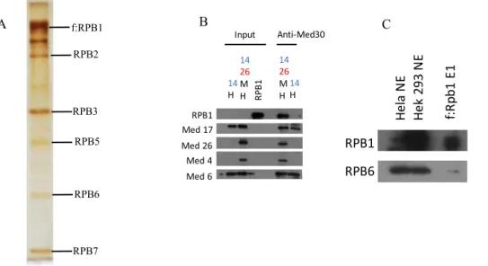
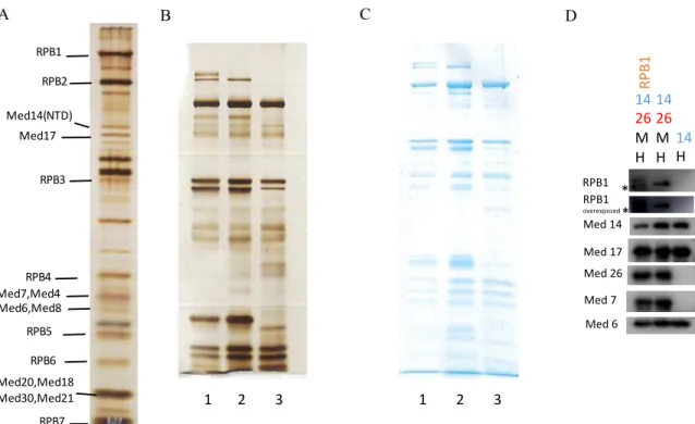
Figure 1: The Mediator complex organised within modules and its interactions with various factors adapted from Malik et al……….………..….2 Figure 2: The Mediator acts as a transmission hub, transmitting activator information to the transcription machinery adapted from Toth-Petroczy et al..………6 Figure 3:Reconstituted human Mediator sub-complexes and purified human RNA polymerase II………..……….39 Figure 4: Purification of RPB1 subunit of human RNA polymerase II (Pol II) transiently expressed in HEK 293T cells and its immunoprecipitation (IP) with Mediator
sub-complex………40 Figure 5: Immunoprecipitation of reconstituted Mediator sub-complexes with Pol II and His-RPB1………...………..41 Figure 6: Immunoprecipitation (IP) of His-RPB1 subunit of human RNA polymerase II (Pol II) and natural Pol II with different modules of Mediator complex…………...…..44 Figure 7: Purification of baculovirus expressed recombinant His-RPB1 subunit of Pol II………...45 Figure 8: Immunoprecipitation (IP) of human RNA polymerase II subunits with purified core Mediator complex and Head module of Mediator complex………...………..47 Figure 9: Screening of Mediator complex subunits in HEK 293T cells upon UV
treatment………...48 Figure 10: Characterisation and Immobilised template recruitment of Mediator complex subunits in wild-type and tamoxifen resistant MCF7 cells………..……49 Figure 11: A proposed model for Pol II recruitment to promoter by core-Mediator complex………52 Figure 12: Purification of His-RPB1-core Mediator sub complex………...……..55
List of Tables
Table 1: List of items used for insect and mammalian cell culture………...…………..20
Table 2: Buffers used for protein extraction from insect cells………21
Table 3: Buffers used for protein extraction from mammalian cells………...21
Table 4: Products used for Immuno-precipitation and protein purification…………....21
Table 5: Buffers used for SDS-PAGE, Western blot analysis and Coomassie staining and their contents………….……….22
Table 6: Materials and Buffers used in immobilize template recruitment assay and their contents………...………..22
Table 7: Antibodies used in immunoprecipitation and western blot analysis………...24
Table 8: Kits and reagents used throughout the experiments………...…...24
Table 9: Contents of a PCR reaction using 2x HF Phusion Master Mix………...26
Table 10: Conditions for PCR………..……...26
Table 11: Schematics for double digestion of PCR product and pFBDM vector…..….27
Abbreviations
AcMNPV Autographa californica nucleopolyhedrovirus
BEVS Baculovirus expression vector system CDK8 Cyclin dependent kinase 8
cMed Core Mediator complex CTD Carboxyl terminal domain CycC Cyclin C
DNA Deoxyribonucleic acid
DPE Downstream promoter element DSSO Disuccinimidyl sulfoxide E.coli Escherichia coli
EM Electron microscopy ERα Estrogen receptor alpha ERβ Estrogen receptor beta GTFs General transcription factors kDa Kilo Dalton
MDa Mega Dalton mRNA Messenger RNA MTE Motif ten element
NMR Nuclear magnetic resonance NPV Nucleopolyhedrovirus NTD N terminal Domain PIC Preinitiation complex Pol I RNA polymerase I Pol II RNA polymerase II Pol III RNA polymerase III RNA Ribonucleic acid RPB RNA Polymerase B rRNA Ribosomal RNA
S. pombe Saccharomyces pombe
Srb Suppressor of RNA polymerase B TAFs Tata binding protein associated factors TBP Tata binding protein
TGA Transposition of the great arteries tRNA Transfer RNA
CHAPTER 1
Introduction
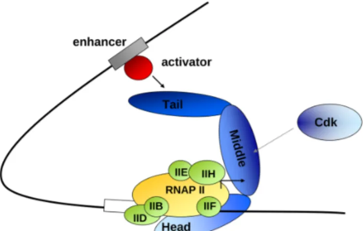
The Central dogma of molecular biology dictates the flow of genetic information from DNA to RNA through a process known as transcription. The information is translated into proteins that perform distinct functions1. The Eukaryotic transcription begins with the formation of the pre-initiation complex (PIC) that consists of RNA Polymerase II, general transcription factors (GTFs) such as Transcription Factor IIA (TFIIA),
Transcription Factor IIB (TFIIB), Transcription Factor IID (TFIID), Transcription Factor IIE (TFIIE), Transcription Factor IIF (TFIIF) and Transcription Factor IIH (TFIIH) and the Mediator Complex. Mediator complex acts as a centralised hub, transmitting the activator/repressor signal to the RNA polymerase II that helps to regulate various functions including transcription and long term epigenetic silencing2.
1.1 The Mediator Complex
In early 1990s, the Mediator Complex was first identified in yeast independently by two groups; Kornberg and Young3. The Kornberg and colleagues used crude yeast fractions that stimulated activator dependent transcription in vitro to isolate Mediator complex4,5. On the other hand, the Young and colleagues identified first Mediator genes using yeast genetic screens and coined them as suppressor of truncation of RNA polymerase II Carboxyl-terminal domain (CTD). These genes were termed suppressor of RNA
polymerase B (Srb) and the four dominant suppressors; Srb2, Srb4, Srb5 and Srb64 were found to be a part of a large multi-subunit complex that was tightly bound to RNA Polymerase II (RNA Pol II)6. Later, it was shown that the 20-subunit protein complex containing Srb2, Srb4, Srb5 and Srb6 stimulated in vitro transcription7.
The Metazoan Mediator is a 2-MDa protein complex that consist of 30 subunits, most of which are evolutionarily conserved from yeast to humans8. Since the metazoan
and Med31 to a substantially weaker relationship for the other remaining subunits8,9. Moreover, the metazoan Mediator complex contains metazoan specific subunits such as Med26 and Med30. For both humans and yeast, the subunits of the Mediator Complex are organised into four distinct modules namely head, middle tail and the kinase module (Figure 1)2. The components of the head and middle modules are known to be tightly associated with each other to form a stable core complex and interact with RNA Pol II machinery. On the other hand, the subunits of the tail module are loosely associated with each other and are target for many activators10,11. The subunits in the kinase module are mobile and associate reversibly with the Mediator complex enabling repressive
properties of the protein complex11.
Over the past years, many studies have been conducted to determine the structure and architecture of the Mediator Complex. However, the large size, heterogeneity and multiple subunit configuration makes it very challenging to determine its’ structure at high resolution. Early electron microscopy (EM) studies laid out a low resolution
architecture of Mediator complex as well as the Mediator-Pol II holoenzyme complex. It also identified the modules within the yeast Mediator complex; Head, Middle and Tail 22-24.
Figure 1: The Mediator complex organised within modules and its interactions with various factors adapted from Malik et al2.
1.1.1 The Head Module
The head module of Saccharomyces cerevisiae Mediator consists of seven subunits; Med6, Med8, Med11, Med17, Med18, Med20 and Med22. Together with the middle module, it is known to play an essential role in the assembly of preinitiation complex12. In 2006, the seven subunit head module from Saccharomyces cerevisiae was
recombinantly expressed in insect cells13 that enabled first negative stain analysis by EM14. Later after five years, the crystal structure of head module of Saccharomyces
cerevisiae was determined at 4.3 Å resolution15. This structure resembled a wrench
containing three major domains that were named as neck, fixed jaw and movable jaws 13-15. The structure of the head module of S. pombe at 3.4 Å resolution confirmed the wrench like organisation of the subunits of the head module16. The head module is stabilised by the neck domain formed by five subunits; Med6, Med8, Med11, Med17 and Med2217. Moreover, these studies suggest that upon recombinant expression of the individual subunits, the proteins tend to be insoluble but constitute a soluble head module when co-expressed together18-22.
The structure determination of the head module was the first milestone towards the characterisation of the Mediator at high resolution. The year 2014 was a very important year for the Mediator studies as two important studies were published which reported the cryo-electron microscopy (cryo-EM) data on Mediator complex, completely redefining its’ modular organisation23,24. Previously, the head module was allocated on one side and the middle and tail folded on top of each other to form the opposite side of the Mediator structure25. These studies revealed the cryo-EM structure of the Mediator Complex at 20-40 Å resolution showing that the head and middle modules were forming the
structure on the Mediator complex that was previously thought to be the middle and the tail module. Moreover, the large opposite domain that was thought to be the head module corresponded to the tail module of the Mediator23,24. A low resolution structure of human Mediator complex was also obtained that confirmed the similarity between human and yeast Mediator despite their evolutionary differences. Furthermore, the unassigned subunits of the Mediator complex that were metazoan specific such as
Med27, Med 28, Med29 and Med30 showed multiple contacts with the head module whereas, Med26 associated with the middle module of the Mediator complex23. For the very first time, a recombinant head module of the human Mediator complex was recombinantly expressed in the insect cells with an addition of a metazoan specific subunit Med30, making it a 8 subunit head module. Moreover, a functional 15 subunit core-Mediator was reconstituted that consists of Head and middle module being held together by Med1410.
1.1.2 The Middle Module
The yeast middle module consists of 8 subunits that includes Med1, Med4, Med7, Med9, Med10, Med19, Med21 and Med31. In metazoan, there is an additional subunit Med26 that is a part of the middle module. Until 2017, the structure of the middle module was unknown and the information was limited to two small individual sub-complexes known as Med7N/Med3126 and Med7C/Med2119. In 2010, Cramer and colleagues used the heterologous co-expression strategy to purify a 7-subunit middle module that lacked Med19 similar to the previous 7 subunit endogenous middle module from Δmed19 strain27. The protein interaction map formed at that time was very limited and solely based on the previous published data28-31 and pulldown experiments27. Later, cross-linking experiments of six subunit middle module lacking Med1 was co-expressed in bacteria that enabled a three-dimensional model of the middle module of Mediator complex32. In 2014, middle module of human Mediator complex was reconstituted using insect cells that consist of five subunits; Med4, Med7, Med10, Med21 and Med31 respectively10. These studies did not reveal three dimensional structural data of middle module of the human Mediator complex. The available structural data of middle module of human Mediator is limited to Med26 N terminal domain serving as a an overlapping docking site for ELL/EAF family containing super-elongation complexes and TFIID33.
The least conserved subunits of the Mediator complex between yeast and human lie in the tail module. Tail module of S. cerevisiae is composed of Med2, Med3, Med5, Med15 and Med16. Among these subunits, Med15 and Med16 are the most conserved subunits. In metazoan module, three more subunits including Med23, Med24 and Med25 exists among which Med24 is known to be a divergent ortholog of Med5 of the yeast Mediator Complex. Moreover, there is evidence which indicates that Med 27 and Med29 are also distant orthologs of Med3 and Med2 of yeast Mediator complex, respectively9. Thus, these subunits are referred as Med2/29, Med3/27 and Med5/24 in the literature. More studies are required for their specific assignment alongside with the other
metazoan specific subunit Med28 and Med30. Med27, Med28, Med29 and Med30 has previously shown to make numerous contact with head module. Whereas, in S.pombe, Med27 shows a tail connection34. Med14 plays a key role in the architectural backbone of the Mediator as it makes numerous contact with tail as well as head and middle module10,23,35.
The heterogeneity in the conformation of the tail module has rendered the structure to be unresolved36. One of the study reported a rotation in the tail module of S. cerevisiae in the presence of transcription factor Gcn423. It has been hypothesised that these structural transitions take place, making a stable association of Mediator with the transcription machinery37. Thus, it can be extrapolated that such transcription factors that are capable of inducing changes in the conformation of Mediator might have a critical role in regulating the transcription process. The first structure of the largest subunit of tail module of Mediator was fully characterised in 2018. The crystal structure at 2.8 Å resolution paved the way for better understanding of Mediator with activators38. As reported previously, the truncations in the C terminus of Med14 resulted in loss of interaction between the tail subunits of Mediator23,35. The precise organisation of metazoan tail subunits is not yet obtained despite the fact that Med16, Med23 and Med24 has been reported to form a stable sub-complex39,40. Many studies have reported the interaction of various activators with the tail module of the Mediator41. The nuclear magnetic resonance (NMR) analysis has identified the interactions between the activator binding domain of the tail subunits of Mediator with transactivation domains of several
transcription factors. These include VP16/Med2542,43, sterol regulatory element binding protein/Med1544, Gcn4/Med1545,46, and ATF6α/Med2547. A detailed structural
architecture of the tail module is required to have a deeper insight into activator mediated transcription of Mediator complex. This would enable the development of specific drugs to interfere the interaction surface of the Mediator-Activators48 and cure diseases such as cancer.
Figure 2: The Mediator acts as a transmission hub, transmitting activator information to the transcription machinery adapted from Toth-Petroczy et al49.
1.1.4 The Kinase Module
The four subunit kinase module comprising of CycC, CDK8, Med12 and Med13 dissociates reversibly from the head, middle and tail of the Mediator complex50. The kinase module is primarily known for its repressive function but some suggest that it can take part in activation of transcription as well52. The dissociation of kinase module is essential for the PIC assembly such that Mediator can join the PIC complex52.The structure of two subunit of kinase module of human Mediator has been determined; CDK8 and CycC, respectively53. However, no structural data regarding Med12 and Med13 exists. The basic structure of yeast kinase module was determined at 15 Å using cryo-EM analysis in 2013. It showed that Med12 is situated at the centre forming a central lobe, connecting Med13, CDK8 and CycC on the opposite end54-55. Structural data available at the moment suggests that Mediator with kinase module does not interact with the RNA polymerase II56. High resolution cryo-EM analysis is required to
determine the inhibitory surface that prevents the Mediator from interacting with RNA Polymerase II.
1.1.5 The Core Mediator
The term Core Mediator has been used in a variety of context in the past. However, it is dedicated to the minimum number of Mediator subunits required that are essential to induce transcription. These subunits belong to head and middle modules with an
addition of Med1410,57. One of the largest subunit of the Mediator complex, Med14 was characterised to be a part of middle or the tail module58-60. Subsequent studies revealed that Med14 had numerous contacts with various subunits of middle module that includes Med1, Med4, Med7, Med9 and Med21 and for the tail module including Med2, Med3, Med15 and Med16. As a result, it was hypothesized that the role of Med14 is very central in connection to different modules of the Mediator. It was confirmed in 2014 when a study demonstrating Cryo-EM analysis of the yeast Mediator at 18 Å resolution revealed the role of Med14 as a scaffold protein23,24. It was also found that the critical role of Med14 as a scaffold protein exist in human Mediator as well thus, it is a conserved function10. At 3.4 Å resolution, the Cramer laboratory reported the crystal structure of S.pombe 15-subunit core Mediator complex (cMed) in 2017. The core structure was divided into 13 submodules; 8 submodules in the head and 5 in the middle module. It was also shown that N terminus of Med17 and C terminus of Med6 interacts with Med14 that brings the head and middle module together61. As a result of these studies, our understanding of the Mediator structure has improved. However, The
organisation of Med1 within the Mediator could not be confirmed and remains a mystery to be resolved10,61.
1.2 The Role of Mediator Complex in Human Diseases
The maintenance and regulation of the cell is dependent on spatiotemporal control of RNA polymerase II mediated transcription as a result of intrinsic and extrinsic signals62. Perturbations caused by the environment and other genetic comments can alter the fate of the cells and can lead to many diseases such as cancer. The role of Mediator is critical
in maintaining the cellular environment as it relays signal to RNA polymerase II (Figure 2) to regulate homeostasis, cell growth, cell differentiation and development2,64. The aberration in Mediator complex subunits has known to cause many diseases such as cancer, neurodevelopmental disorders, and cardiovascular diseases62. Charcot-Marie tooth disease also known as hereditary motor and sensory neuropathy is a peripheral neuropathy that is commonly inherited64. A missense mutation in MED25 gene (A335V) has been implicated as a likely cause of this disease. As a result, it disrupts the Med25-Mediator interface necessary for transcriptional regulation of genes pertaining to function of peripheral nervous system62. Another missense mutation L371P in MED17 gene is associated with infantile cerebral and cerebellar atrophy65. This mutation was identified as a result of screening of 79 individuals of Caucasus Jewish origin. However, none of the 113 Arab Muslim or 110 Ashkenazi Jewish carried this mutation, signifying that it is a founder mutation in Caucasus Jewish community66. The missense mutations R961W and N1007S in Med12 gene results in syndromal X-linked mental retardation including FG and Lujan syndromes67,68.
Approximately 1% of the live births exhibit congenital heart disease, making it the most common birth defect in humans. The most common cyanotic heart defect in new-borns is the transposition of the great arteries (TGA) that account for 7% of all the congenital heart diseases69.With regard to the disease, MED13L, a paralog of MED13 was
identified in a patient with TGA. Analysis of 97 additional patients revealed 3 mutations in MED13L that were categorised as missense. These mutations are E251G, R1972H and D2023G that were absent in 400 control samples70. DiGeorge syndrome occurs in approximately 1 in every 3000 live births and is categorised as the most recurrent multiple congenital anomaly syndrome71. It results in cardiac defects, immune dysfunction, psychoses, schizophrenia, palatal anomalies, characteristic facial dysmorphism, and hypocalcaemia72. This disease is caused by the deletion of approximately 3Mb region on chromosome 22 that contains 60 genes including the MED15 gene73. Med15 in humans is categorised as transducer of SREBP1α and TGFβ-activated SMAD2/3 that regulates the metabolism of lipid and developmental
indicates that 22q11.2 deletion syndrome/DiGeorge syndrome can be the result of reduced expression of Med1562.
The Mediator complex plays a wide role in several signalling pathways regulating growth, differentiation and development. Many developments have been made recently that linked its’ subunits to variety of cancers. The Med1 subunit of the Mediator was among the first subunits to have a clear link established with the breast cancer. Breast cancer is the most common type of cancer diagnosed in women and is the leading cause of cancer related death worldwide among women75. Animal studies in the past have revealed that steroid hormone estrogen (17-β-estradiol; E2) induces and promotes breast cancer therefore, using drugs to oppose these affects or using estrogen ablation therapy can reduce the severity of breast cancer76,77. The effects of E2 are mediated through two receptors that are similar in structure; estrogen receptor α (ERα) and β (ERβ)
respectively78. It is estimated that 70% of the breast cancer patients are scored positive for ERα upon diagnosis79, hence a valuable target for breast cancer therapy. In vivo chromatin association studies and in vitro transcription based assays identified the recruitment of Mediator by ERα to promote the formation of pre-initiation complex and transcription of ERα target genes80-83. Moreover, mutant mice for defective Med1-ERα-binding resulted in mammary gland development defects and reduced expression of ERα responsive genes84. This indicates that Med1 of the Mediator complex is a critical co-activator of ERα. The study by Zhu et al, investigated the role of Med1 in ERα positive breast tumours and found that Med1 mRNA was overexpressed in more than 50% of the breast tumour85. Another study by Vijayvargia et al determined that Med1 is
overexpressed in approximately 50% prostate cancer, indicating its’ role in the progression of prostate cancer86. Apart from Med1, Med28 has also been found to be associated with breast cancer. Med28 expression analysis on samples from breast cancer patients based on clinicopathological variables, histopathological subtypes and disease outcome exhibited an increase in protein level in ductal and invasive ductal carcinoma of the breast tissue as compared to non-malignant breast epithelium of ductal and glandular origin. Furthermore, the expression level of Med28 protein also correlated with the disease outcome signifying a higher expression of the protein with elevated risk of death
in early and late stage of breast cancer87, demonstrating its role as a critical prognostic marker.
Colon cancer is among the leading causes of cancer related death. It is the second most common type of cancer in women and third most common among men75. Colon tumours originate in the intestinal crypts, where the progenitor derived epithelial cells start differentiation and ascend towards the intestinal villi88. The homeostasis of crypt progenitor phenotype depends on gene expression of Wnt/β-catenin pathway. When the pathway becomes constitutively active, it provides the driving force for the proliferation of intestinal epithelial cells that results in colon cancer88-91. The kinase module of the Mediator was thought to be involved in Wnt/β-catenin signalling as the C terminal transactivation domain of β-catenin targets Med12 in Mediator complex to activate transcription92. The architecture of the Mediator complex links Cdk8/CyclinC to the Med12 since the depletion of Med12 leads to reduced level of CDK8/CyclinC
incorporation to the Mediator complex93. The biochemical assays suggests that Med12 activates the Cdk8 kinase94. The study by Firestein et al demonstrates the important modulators of β-catenin pathway for colon cancer. Based on the high throughput RNAi, loss of function screens were conducted to determine kinases and phosphatases essential for the transactivation of β-catenin and the proliferation of colon cancer cells. The study found 9 candidate genes that were critical for both functions. However, only Cdk8 was found to be residing in the region of copy number gain in a notable proportion of colon cancer. It was also found that Cdk8 activity was required for colon cancer proliferation, β-catenin mediated gene transcription and cell transformation95. As shown earlier, β-catenin does not bind to CylinC or Cdk8 but instead binds directly to Med1292. Thus, it is very likely that the transduction of β-catenin signalling is mediated through Med12 to Cdk8. Another study by Kapoor et al revealed Cdk8 role in melanoma progression96. Although not very common, melanoma is categorised as the most deadly type of skin cancer with approximately 4% of skin cancer cases and 75% skin cancer related deaths97. The identification of macroH2A (mH2A) knockdown cell lines gene
expression revealed Cdk8 as a mH2A repressed gene, which indicates that it can be a possible mediator of melanoma malignancy96. Further investigations are required to fully
understand the role of individual Mediator subunits in order to develop therapeutic to treat wide range of human diseases.
1.3 RNA Polymerases
The expression of protein coding genes is pivotal for the biological processes carried out within the cell which is regulated predominantly at the level of transcription. Sam Weiss is the first discoverer of the RNA polymerase activity as he showed the incorporation of all four nucleoside triphosphate into a nucleotide in rat liver nuclei in the year 195998. In 1961, Jacob and Monod published the paper on lac operon that explained the landmark of mechanism and regulation of transcription99. Although many studies about bacterial RNA polymerase were being conducted in the late 1960, RNA polymerase was purified to homogeneity first time in 1969 by Richard Burgess who later showed that the
dissociable sigma subunit was regulating the initiation of transcription100. During that time, the understanding of eukaryotic transcription was very limited. The biggest milestone in understanding the eukaryotic mechanism of transcription was reached in 1969 with the discovery of three distinct classes of RNA polymerases by Roeder and colleagues101.
1.3.1 RNA Polymerase I
The RNA polymerase I (Pol I) synthesises approximately 60% cellular RNA as it transcribes many copies of ribosomal RNA (rRNA) genes. A microscopic structure that depicts a ‘Christmas tree’ is produced as each gene of rRNA is produced by many Pol I enzymes102. RNA polymerase I regulates the levels of ribosomal components and cell growth103. Furthermore, deregulation of Pol I results in several diseases that includes cancer104. Pol I is a 590 kDa enzyme that consists of 14 subunits105. Several attempts had been made to solve the structure of Pol I. In 2013, Christoph et al deciphered the crystal structure of yeast Saccharomyces cerevisiae Pol I at 2.8 Å resolution. The purified Pol I was active in both DNA templated RNA extension and cleavage. The structure consist of a Pol I dimer that contains 8681 amino acids, lacking the mobile A49 WH domain
and some other surface loops105. The Core of Pol I consist of 10 subunits; A190, A135, AC40, AC19, A12.2, Rpb5, Rpb6, Rpb8, Rpb10, and Rpb12106. Furthermore, RNA polymerase I and III contain a TFIIF like sub-complex which stabilises Pol II. In addition to that, Pol I and II also use TFIIB like factors and contain TFIIE related domains. This indicates that the core of transcription limitations of Pol I, II and III is conserved both functionally and structurally106.
1.3.2 RNA Polymerase II
The RNA polymerase II (Pol II) is a 12 subunit enzyme that transcribes mRNA from the protein coding genes. It regulates many cellular processes such as cell differentiation, homeostasis, and maintenance of cell identity. These regulations occur throughout the transcription process with most important one being the initiation stage of transcription. It is fundamental to understand the structure of the Pol II initiation complex to be able to understand the mechanism by which Pol II regulates gene transcription107. The general transcription factors TFIIB, TFIID, TFIIE, TFIIF, and TFIIH, along with the Pol II and Mediator complex assemble at the Promoter DNA to form pre-initiation complex (PIC)2.
About two decades ago, the structural analysis of PIC started. The free form structures of Tata binding protein (TBP)108,109 and DNA bound110 demonstrated that TBP is a molecule shaped like a saddle that binds to the minor groove of DNA and bends it by 90 degrees. Further investigations revealed that TFIIA and TFIIB transcription factors flank the DNA-TBP complexes111,112. The complete structure of 12 subunit TFIIB bound Pol II was elucidated in 2009 which highlighted the location of TFIIB on Pol II wall. Consequently, a model of Pol II-TFIIB-TBP-DNA complex in an open and closed promoter DNA conformation was established113. In 2013, TFIIE, TFIIF, and TFIIH were added to Pol II, TFIIB, TBP, DNA and TFIIA containing human PIC and visualised using electron microscope114. The structure appeared to be quite similar to the existing yeast core initiation complex. Moreover, it showed the TFIIE location on the clamp domain of Pol II, which was coherent with the homologous yeast factor location115,116.
The specific binding of the Tata-binding protein to TATA box is the first step in the canonical assembly of PIC. In humans, the consensus sequence of the TATA box is located approximately 30 base pair upstream of the transcription start site and is denoted by TATAWAWR117. The consensus sequence of the TATA box is conserved from yeast to humans as the evidence suggests118. A variety of promoters lack the TATA boxes and are referred as TATA-less promoters. In yeast, TBP along with other general factors are known to be bound throughout its genome, suggesting that the architecture of the PIC overall is similar for both TATA box containing and TATA-less promoters119-120. The mode of binding of TBP in yeast121, plant110 and human122 is highly conserved as exhibited by the structures formed by TBP bound to the TATA box containing DNA. The factor TFIIA stabilises the TBP-DNA complexes but is not essential for basal transcription123. It is a Pol II specific transcription factor that enables constitutive and activator driven transcription124. The structure of human125 and yeast18 TFIIA-TBP-DNA complexes showed a boot shaped TFIIA heterodimer which does not change the structure of TBP-DNA112. There are two conserved domains in TFIIA; a 4-helix bundle and 12 stranded β-barrel that enables TFIIA to bind underside of TBP saddle and the upstream region of TATA box, stabilising the TBP-DNA complex126.
One of the fundamental factor required for the recruitment of Pol II to the promoter is the transcription factor TFIIB127,128. It also promotes the binding of TBP to DNA and enables the bending of DNA54. The N and C terminal domains of TFIIB are responsible for the recruitment of Pol II and the interaction of TBP with Promoter,
respectively129.TFB is a TFIIB homologue in archaea. TFB and TBP are the only required initiation factors for the transcription system in archaea130. The Pol II -TFIIB complex structure elucidated the role of B-ribbon of TFIIB in binding to the docking domain of Pol II in order to recruit it to the promoter131. These structure also reveal the region of TFIIB that B-ribbon and B-core domains transversing the cleft of Pol II, forming B-reader and B-linker113. If you follow the N-terminus of B-ribbon, the polypeptide chain goes along with the exit channel of RNA on Pol II and continues towards the opposite direction of exiting RNA all the way to the cleft and forms a B-reader and B-linker helix. The B-linker helix enables opening of DNA and maintenance
of the transcription bubble113. The B-reader binds to the template DNA strand and position it for synthesis of RNA chain, and it helps with the recognition of the initiator sequence on the DNA132. In general, TFIIB initiates the Pol II mediated RNA synthesis and stabilises the initiation complex that contains a five nucleotide RNA strand131.
TFIIF is another transcription factor that was previously identified in mammalian cells due to its interaction with Pol II133. It is a heterodimer consisting of TFIIFα and TFIIFβ also known as RAP74 and RAP30, respectively134. In yeast, Tfg1 and Tfg2 are
homologous pairs of TFIIFα and TFIIFβ, respectively135.Furthermore, yeast contains an additional TFIIF element Tfg3 that is not required for transcription136. In yeast, about 50% of Pol II is coupled to TFIIF137. TFIIF stabilises PIC138 and prevents the non-specific interaction of Pol II with DNA139. Moreover, it also plays a role in the selection of TSS140, prevents Pol II pausing94, promotes formation of phosphodiester bond and early synthesis of RNA141. TFIIF also stabilises the transcription bubble and plays a very important role in transcription as it can be initiated to an extent in the absence of TFIIE and TFIIH in vitro, but it cannot in the absence of TFIIF142. TFIIF has several domains and most of their structures have been determined. The N terminal region of both TFIIFα and TFIIFβ dimerises, forming a triple barrel fold and a β-hairpin that is termed as an ‘arm’143. The winged helix domain is located at the C terminal end of both
subunits144. The winged helix domain connect to the dimerization module using a charged region on TFIIFα and linker region on TFIIFβ145,146. The structural studies elucidated that the dimerization module of TFIIF anchors it to Pol II and binds to the lobe of Pol II on one side of the cleft116,145,147. The winged helix domain of Tfg2 is found upstream of DNA148.This domain is located close to DNA in the initiation complex and is mobile in Pol II- TFIIF complex145. Nevertheless, the concurrent data does not support the existing EM model of Pol II-TFIIF complex149, thus, more studies are required to get a better understanding of Pol II-TFIIF complex.
TFIIE and TFIIH are among other essential factors for PIC formation and are critical for the opening of promoter DNA. The TFIIH factor encompasses DNA dependent ATPase activity that is critical for the initiation of transcription150-151. Furthermore, studies
involving transcription assays using linear, supercoiled or mismatched DNA
demonstrated that TFIIH is involved in the opening and escape of Promoter DNA152-153. The TFIIE factor acts like a bridge between TFIIH and Pol II as it helps in to recruit TFIIH to initiation complex153,154. TFIIE is a heterodimer comprising of two subunits TFIIEα and TFIIEβ155. These subunits are distant homologs of bacteria initiation factor sigma156. The N terminus of TFIIEα contains winged helix domain and a zinc finger domain, which is enough to interact with TFIIEβ to facilitate transcription157. The cross-linking experiments pertaining to TFIIEβ suggests that it is located upstream in the proximity of transcription start site158.TFIIE binds to the clamp domain of Pol II116, which is similar to archaea homolog TFE159. The 10 subunits transcription factor TFIIH comprise of a six subunit core module; ATPase XPD, p62, p52, p34, p8, and p44 which are known as Rad3, Tfb1, Tfb2, Tfb4, Tfb5, and Ssl1 in yeast, respectively. ATPase XPB which is known as Ssl2 in yeast and a kinase module consisting of three subunits CDK7, cyclin H and MAT1 known as Kin28, Ccl1 and Tfb3 in yeast160,161. For
transcription to occur, a complete 10 subunit TFIIH is required but the DNA repair does not require the kinase module of TFIIH162. The mutations in the catalytic ATPase subunits XPB and XPD are associated with diseases such as Cockayne syndrome, xeroderma pigmentosum and trichothiodystrophy163. While XPB is required for the opening of promoter during transcription in vitro and in vivo164,165, XPD is essential for opening of DNA during DNA repair166. There are many subunits of TFIIH for which the structures are available. These include cyclin H, CDK7, a part of MAT1, and the
homologs of XPB and XPD in archaea167-169. The structural information identified the location of TFIIH within the PIC and it demonstrated the mechanism by which TFIIH opens the DNA. TFIIH rotates the DNA from a fixed protein complex at the TATA box acting like a ‘wrench’, hence it creates sufficient torque to melt the DNA170.
Lastly, the transcription factor TFIID is multifunctional transcription factor that is conserved and plays a role in the promoter recognition, assembly of PIC and chromatin remodelling171. TFIID is approximately 1.2MDa protein complex, consisting of TBP and 13-14 TBP associated factors (TAFs), among which 13 are conserved throughout yeast and humans172. Studies using yeast have demonstrated that TFIID contributes
significantly to most of the genes transcribed by Pol II173. The primary role of TFIID is the recognition of core promoter sequence. Previously, it was thought that the
recruitment of TFIID to Pol II promoters was through TATA binding activity of TBP. However, only 10-20% of human and yeast promoters contain a TATA box174.
Investigation of core promoter sequence identified many core promoter elements that are being recognised by TAFs175, including motif ten element (MTE) and downstream promoter element (DPE) which were discovered in Drosophila melanogaster and were later attributed as human promoters176-178. The TFIID can fine tune its binding to DNA using different composition of elements. The structural studies for holo-TFIID has been very challenging due to large size, low amount of intact complex and heterogeneity in subunit composition. A recent study was published that recombinantly expressed core TFIID complex179 and enabled the structure to be resolved at 12 Å resolution. The gaps in the structure were filled with the existing data about the TFIID structure107. All in all, the transcription of Pol II genes is a complex process that relies on a variety of proteins and protein complexes that work in a cooperative manner to locate the TSS and induce transcription.
1.3.3 RNA Polymerase III
The RNA polymerase III (Pol III) is responsible for transcription of majority of RNAs in the eukaryotic cells that includes transfer RNAs (tRNAs) and many other noncoding RNA that are associated with ribosomes and protein synthesis180. RNA polymerase III consists of 10 subunit core that is a common feature of all nuclear RNA polymerases. The transcription of tRNA genes via Pol III requires TFIIIB; a 3 subunit initiation factor that consist of TBP and TFIIB paralog Brf1 and Bdp1, and TFIIIC; a six subunit
assembly factor that binds to intragenic A and B block promoter elements. The 17 subunit Pol III is recruited to stably assembled TFIIIB upstream of TSS181. Transcription of ribosomal RNA requires a different approach as the assembly of PIC on 5S RNA gene recruits an additional factor TFIIIA that guides the binding of TFIIIC. In metazoans, a third class of Pol III promoters are found that do not rely on intragenic sequence elements thus, the assembly of initiation complex relies on SNAPc instead of
TFIIIA and TFIIIC. Transcription of SNAPc mediated genes uses Brf2 (different paralog of TFIIB) in the initiation factor182183. Since the late 1970, many studies had been focused on the ability of yeast to respond to nutrient level by altering ribosome and tRNA synthesis184,185. It became more clear upon the identification of Maf1 which is a master regulator of transcription in-response to variable nutrition, cellular stress and change in environment186,187. Although Maf1 knockout yeast cells are viable, its’ ability to repress Pol III mediated gene transcription was hindered drastically. This process is conserved in higher eukaryotes as MAF1 is essential for Pol III mediated transcription repression in mammalian cells upon serum starvation, treatment with DNA damaging agents and TORC1 inhibitors188,189. There is abundant evidence that deregulation of Pol III transcription machinery can causes complex diseases including cancer. Since the tumour cells proliferate faster as compared to normal cells, up-regulation of tRNA levels and its’ iso-acceptors is observed in breast cancer and myeloma cell lines when
measured using microarray190,191. It is important to study nuclear RNA polymerases as they play a vital role in regulating critical processes within the cell and understanding the structure would shed light into the mode and mechanism by which these enzymes carry out their function in a cooperative manner.
1.4 Baculovirus Expression System
The baculoviruses are also known as nucleopolyhedrovirus (NPV) as they form inclusions in the nucleus of the infected cells. These viruses are widely used in pest control and production of recombinant proteins. The baculovirus expression vector system also known as BEVS was developed by two laboratories in the early 1980192,193. A mixture of viral DNA were co-transfected along with a donor vector to Sf21 cells to replace the polyhedrin gene from Autographa californica nucleopolyhedrovirus (AcMNPV) to generate recombinant viruses which were isolated from the viral
plaques192. The technology has improved significantly since then as many improvements have been made to obtain high yield of expressed proteins.
The most important breakthrough in the generation of recombinant baculoviruses was made in 1993 upon development of a vector that contained the entire genome of AcMNPV (referred as bacmid) and could be propagated in Escherichia coli (E.coli)194. This is known as Bac-to-Bac® system which is now commercialised by Invitrogen. In this system, the shuttle vector containing gene of interest is introduced into the genome of bacmid in the host bacteria using the transposition of shuttle vector via
Tn7-recombinase. There are many advantages of this system such as selection and isolation of bacmids is easier and high yield of recombinant baculovirus195. Moreover, more than one protein could be expressed using this technique. Previously, co-infection of multiple baculoviruses was required to express multiple proteins196. However, this method was very inefficient since simultaneous infection of all baculovirus cannot be guaranteed197. The problem was overcome by development of new shuttle vectors that can accept more than one gene. These vectors include pFastBacDual (Invitrogen) that are used to express two protein and the Multibac system that can facilitate the expression of multi-subunit protein complexes198. Other vectors include flashBAC™ system; a combination of bacmid technology and insect homologous recombination for the generation of baculoviruses199, OmniBac vector that can use both homologous recombination and bacmid technology to generate baculoviruses200, Bac-2-the-Future that has the essential features of Bac-to-Bac system but it reduces the time and labour to generate viruses201, and biGBac202 that utilises Gibson assembly method202 to assemble many DNA
fragments to generate baculoviruses.
The recombinant proteins expression can be attenuated by promoters, enhancers or regulatory cis elements. Most widely used promoters used for production of recombinant proteins using BEVS are polh (polyhedrin) and p10. These two promoters are very strong corresponding to high rate of transcription but start expressing proteins at a later stage of infection203. More recently, a combination of two promoters is being utilised to increase the yields of protein. One such example would be combination of polh with
vp39 or pSeL204,205. There are many insect cell lines that are used for the expression of
recombinant proteins. The most widely used ones are IPLB-SF21-AE (known as Sf21) derived from pupal ovarian tissue of Spodoptera frugiperda206, Sf9 which is a subclone
of Sf21 and BT1-Tn5B1-4 (commonly known as Hi5) derived from adult ovarian tissue of Trichoplusia ni 207. The choice of cell line depends on the type of protein that is going to be expressed. For example, Sf21 and Sf9 are highly vulnerable to viral infection therefore high titre of virus can be obtained. These cells can be grown as monolayers without CO2 at 27oC or as a suspension and are adaptable to serum free media208. The Sf9 cells as compared to Sf21 cells grow faster and in high density, are more tolerant to sheer stress, pH and osmotic changes209. The Hi5 cells are usually used for protein production as they are more resistant to nutrient stress and expresses recombinant proteins in high level due to its large size. A limitation of using this cell line is that as compared to Sf9 and Sf21, it produces three times more proteases which results in degradation of the target protein210. New improvements in the engineering of BEVS and host cell lines are being made to enhance the quality and quantity of target proteins. Advancements in genome editing techniques such as CRISPR-Cas9 will enable induction of specific mutations, deletions and insertions to improve the expression of recombinant proteins using baculovirus expression system211.
1.5 The aim of the study
The primary aim of the study was to identify the interaction surface between human Mediator complex and RNA polymerase II in order to elucidate the mechanism of transcription of RNA polymerase II mediated gene transcription. Multibac expression system was utilised to express the proteins and perform biochemical assays to
demonstrate the interaction between core Mediator complex and critical subunits of RNA polymerase II responsible for the interaction with the Mediator. Consequently, minimum subunits of Pol II-core Mediator has been identified that might be facilitating transcription hence, highlighting the critical role of Mediator in Pol II mediated gene transcription.
CHAPTER 2
Materials and Reagents
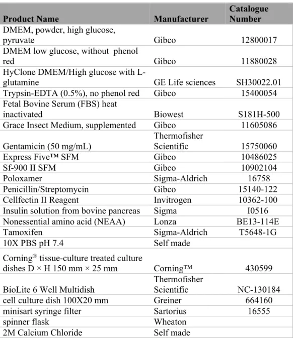
2.1 Materials for Cell culture, buffers, reagents and glassware
Product Name Manufacturer Catalogue Number DMEM, powder, high glucose,
pyruvate Gibco 12800017
DMEM low glucose, without phenol
red Gibco 11880028
HyClone DMEM/High glucose with
L-glutamine GE Life sciences SH30022.01
Trypsin-EDTA (0.5%), no phenol red Gibco 15400054 Fetal Bovine Serum (FBS) heat
inactivated Biowest S181H-500
Grace Insect Medium, supplemented Gibco 11605086 Gentamicin (50 mg/mL)
Thermofisher
Scientific 15750060
Express Five™ SFM Gibco 10486025
Sf-900 II SFM Gibco 10902104
Poloxamer Sigma-Aldrich 16758
Penicillin/Streptomycin Gibco 15140-122
Cellfectin II Reagent Invitrogen 10362-100
Insulin solution from bovine pancreas Sigma I0516 Nonessential amino acid (NEAA) Lonza BE13-114E
Tamoxifen Sigma-Aldrich T5648-1G
10X PBS pH 7.4 Self made
Corning® tissue-culture treated culture
dishes D × H 150 mm × 25 mm Corning™ 430599 BioLite 6 Well Multidish
Thermofisher
Scientific NC-130184
cell culture dish 100X20 mm Greiner 664160
minisart syringe filter Sartorius 16555
spinner flask Wheaton
2M Calcium Chloride Self made
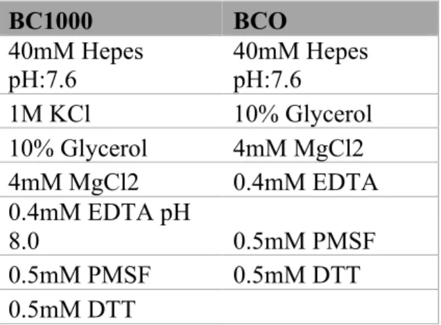
2.2 Buffers for protein extraction and purification. BC1000 BCO 40mM Hepes pH:7.6 40mM Hepes pH:7.6 1M KCl 10% Glycerol 10% Glycerol 4mM MgCl2 4mM MgCl2 0.4mM EDTA 0.4mM EDTA pH 8.0 0.5mM PMSF 0.5mM PMSF 0.5mM DTT 0.5mM DTT
Table 2: Buffers used for protein extraction from insect cells. Buffer I Buffer II 40mM Hepes pH:7.6 40mM Hepes pH:7.6 1.5mM MgCl2 1.5mM MgCl2 10mM KCl 0.2mM EDTA pH 8.0 0.5mM PMSF 300mM NaCl 0.5mM DTT 25% Glycerol 0.5mM PMSF 0.5mM DTT
Table 3: Buffers used for protein extraction from mammalian cells.
Product Name Manufacturer Catalogue Number
Anti-flag M2 Affinity Agarose beads Sigma-Aldrich A4596
Flag Peptide Sigma-Aldrich F3290
Dynabeads™ His-Tag Isolation and Pulldown
Thermofisher
Scientific 10103D
Superose 6 Prep Grade GE life sciences 17048901
Table 4: Products used for Immuno-precipitation and protein purification.
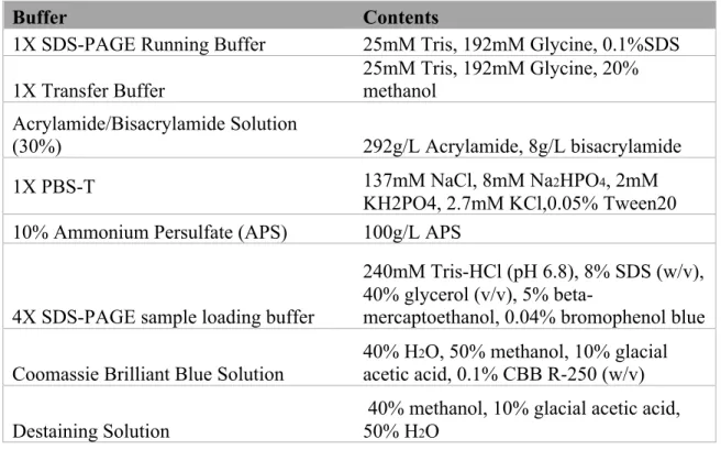
Buffer Contents
1X SDS-PAGE Running Buffer 25mM Tris, 192mM Glycine, 0.1%SDS 1X Transfer Buffer 25mM Tris, 192mM Glycine, 20% methanol Acrylamide/Bisacrylamide Solution
(30%) 292g/L Acrylamide, 8g/L bisacrylamide
1X PBS-T 137mM NaCl, 8mM Na2HPO4, 2mM
KH2PO4, 2.7mM KCl,0.05% Tween20 10% Ammonium Persulfate (APS) 100g/L APS
4X SDS-PAGE sample loading buffer
240mM Tris-HCl (pH 6.8), 8% SDS (w/v), 40% glycerol (v/v), 5%
beta-mercaptoethanol, 0.04% bromophenol blue Coomassie Brilliant Blue Solution 40% H
2O, 50% methanol, 10% glacial acetic acid, 0.1% CBB R-250 (w/v)
Destaining Solution
40% methanol, 10% glacial acetic acid, 50% H2O
Table 5: Buffers used for SDS-PAGE, Western blot analysis and Coomassie staining and their contents.
2.4 Materials used for Immobilize template recruitment assay. Dynabeads M-280 Streptavidin Invitrogen Cat no: 11205D
10X Assay Mix 50mM MgCl2, 200mM HEPES-KOH (pH
8.2)
Blocking Buffer 1X Assay Mix, BSA 5mg/ml, 0.03% NP-40, 12.5mM DTT, PVP 5mg/ml
2X B&W before incubation 10mM Tris-HCl (pH:7.5), 2M NaCl, 1mM EDTA
2X B&W after incubation 10mM Tris-HCl (pH:7.5), 2M NaCl, 1mM EDTA, BSA 1mg/ml, 0.006% NP-40
Wash Buffer 40mM Hepes (pH: 7.5) 150 mM KCl, 4mM
MgCl2, 4mM DTT, 0.1% NP-40
Table 6: Materials and Buffers used in immobilize template recruitment assay and their contents.
2.5 Antibodies used in Immunoprecipitation and immunoblotting.
Antibody Manufacturer Catalogue Number Dilution
Pol II (N-20) Santa cruz Biotechnology sc-899 1:1000
Pol II (A-10) Santa cruz Biotechnology sc-17798 1:1000 Pol II (8WG16)
R.G.R Lab at Rockefeller
University Home made 1:1000
Med14 rabbit pAb
R.G.R Lab at Rockefeller
University Home made 1:1000
Med6 rabbit pAb
R.G.R Lab at Rockefeller
University Home made 1:1000
Med7 rabbit pAb
R.G.R Lab at Rockefeller
University Home made 1:1000
Med8 (A-5) Santa cruz Biotechnology sc-365960 1:1000
His-Tag Antibody Cell Signaling Technology 2365s 1:1000
ANTI-FLAG M2 Sigma-Aldrich F1804 1:1000
ANTI-FLAG M2 Sigma-Aldrich F7425 1:1000
HA-Tag R.G.R Lab at Rockefeller University Home made 1:1000 Med1 rabbit pAb R.G.R Lab at Rockefeller University Home made 1:1000 Med12 rabbit pAb R.G.R Lab at Rockefeller University Home made 1:1000 Med13 rabbit pAb
R.G.R Lab at Rockefeller
University Home made 1:1000
Med15 rabbit pAb Proteintech 115661AP 1:1000
Med17 rabbit pAb
R.G.R Lab at Rockefeller
University Home made 1:1000
Med19 rabbit pAb
R.G.R Lab at Rockefeller
University Home made 1:1000
Med23
R.G.R Lab at Rockefeller
University Home made 1:1000
Med24
R.G.R Lab at Rockefeller
University Home made 1:1000
Med25 (A-7) Santa cruz Biotechnology sc-393759 1:1000 Med26 rabbit pAb
R.G.R Lab at Rockefeller
University Home made 1:1000
Med27 (B-7) Santa cruz Biotechnology sc-390296 1:1000
Med28 rabbit pAb
R.G.R Lab at Rockefeller
University Home made 1:1000
Med29 (B-1) Santa cruz Biotechnology sc-393800 1:1000
Med30 rabbit pAb
R.G.R Lab at Rockefeller
Cyclin C Abcam ab85927 1:1000 CDK8
R.G.R Lab at Rockefeller
University Home made 1:1000
Rpb5 rabbit pAb
R.G.R Lab at Rockefeller
University Home made 1:1000
Rpb6 rabbit pAb R.G.R Lab at Rockefeller University Home made 1:1000 Anti-rabbit IgG,
HRP linked antibody Cell Signaling Technology 7074S 1:1000 Anti-mouse IgG,
HRP linked antibody Cell Signaling Technology 7076S 1:1000 Estrogen Receptor α
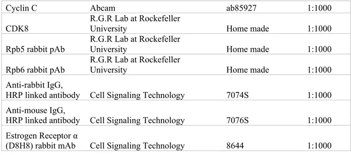
(D8H8) rabbit mAb Cell Signaling Technology 8644 1:1000 Table 7: The antibodies used in immunoprecipitation and western blot analysis.
2.6 Kits utilized during the experiments.
Product Name Manufacturer
Catalogue Number GeneJET™ Gel Extraction Kit Thermofisher Scientific K0691 GeneJET™ Plasmid Miniprep
Kit Thermofisher Scientific K0503
Clean-Blot™ IP Detection Kit
(HRP) Thermofisher Scientific 21232
Pierce Silver Stain Kit, 1L kit Thermofisher Scientific 24612 BCA protein assay kit 500ml kit Thermofisher Scientific 23227
CHAPTER 3
Methods
3.1 Construction of Plasmids.
3.1.1 Primer Design for Polymerase Chain Reaction.
The majority of the plasmids used in these experiments were already constructed by former students or Dr. Cevher himself. However, some of them had to be constructed to fulfil the aim of the project. The DNA coding sequence of His-Med14 (141-2420) was amplified using forward 5’ GC GAA TTC ATG CAT CAC CAT CAC CAT CAC GCA GCC CCA GTG CAG 3’ and reverse 5’ GC TCT AGA CTA TCT ACC ACC AAC AGG 3’ primer containing EcoRI and XbaI restriction sites, respectively. The DNA coding sequence of His-Med14 (141-1280) was amplified using forward 5’ GC GAA TTC ATG CAT CAC CAT CAC CAT CAC GCA GCC CCA GTG CAG 3’ and reverse 5’ GC TCT AGA CTA AGC TGG CAA AGG AGG 3’ primer containing EcoRI and XbaI restriction sites, respectively. The DNA coding sequence of His-Med14 (1341-2420) was amplified using forward 5’ GC GAA TTC ATG CAT CAC CAT CAC CAT CAC GCA CTG ATT GAC AGT GTC 3’ and reverse 5’ GC TCT AGA CTA TCT ACC ACC AAC AGG 3’ primer containing EcoRI and XbaI restriction sites,
respectively. The DNA coding sequence of His-Med14 (2403-3455) was amplified using forward 5’ GC GCT AGC ATG CAT CAC CAT CAC CAT CAC GCA GAG CCT GTT GGT GGT 3’ and reverse 5’ GC GGT ACC CTA TCG TCC ACT TGG TGA 3’ primer containing NheI and KpnI restriction sites, respectively. The DNA coding sequence of His-RPB2 was amplified using forward 5’ GC CTC GAG ATG CAT CAC CAT CAC CAT CAC GCA TAC GAC GCG GAT GAG 3’ and reverse 5’ GC GGT ACC CTA CTA AAC ACT CAT CAT TCG 3’ containing XhoI and KpnI restriction sites,
respectively. There were many more plasmids that were constructed but they are being used for other projects therefore, they are excluded from this project.
The total mRNA of HEK 293T cells was used to synthesize cDNA according to the manufacturer’s instructions for RevertAid First Strand cDNA Synthesis Kit (Thermo Fisher Scientific Cat no: K1622).
3.1.3 Protocol for Polymerase Chain Reaction (PCR).
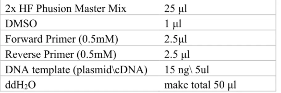
The coding DNA for all of the inserts was amplified using Phusion High-Fidelity PCR Master Mix from Thermofisher Scientific (Cat no: F-5315). The reactions were designed as follows.
2x HF Phusion Master Mix 25 μl
DMSO 1 μl
Forward Primer (0.5mM) 2.5μl Reverse Primer (0.5mM) 2.5 μl DNA template (plasmid\cDNA) 15 ng\ 5ul
ddH2O make total 50 μl
Table 9: Contents of a PCR reaction using 2x HF Phusion Master Mix.
Table 10: Conditions for PCR.
The PCR amplified DNA fragments were ran on agarose gel (percentage depending upon the size of fragments) and after visualization, the fragments were excised using GeneJET™ Gel Extraction Kit.
Step Temperature Time Cycle
Initial denaturation 98°C 30 sec 1
Denaturation 98°C 10 sec
Annealing variable 30 sec
Extension 72°C variable
Final Extension 72°C 7 mins 1
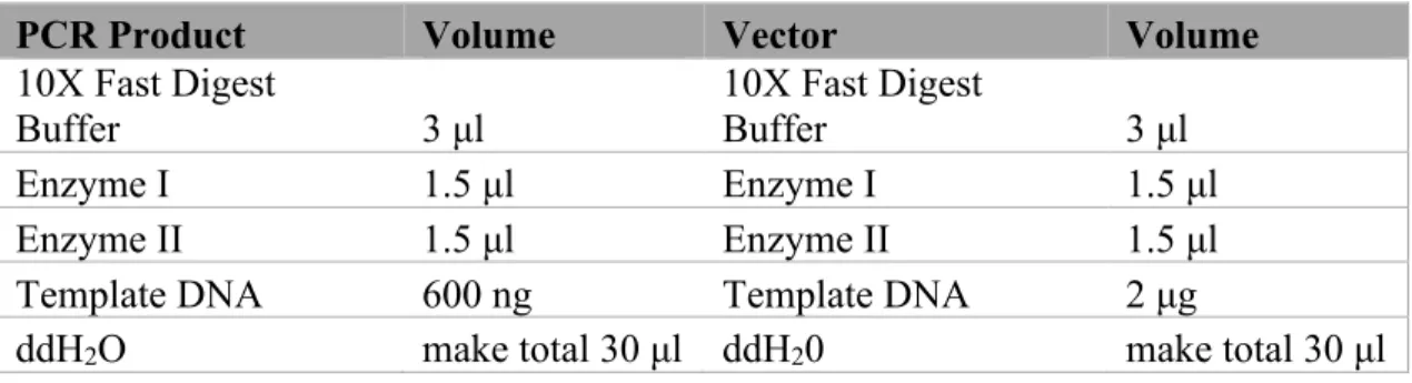
FastDigest value pack kit (Cat no: K1991) manufactured by Thermofisher Scientific was used in digestion of PCR amplified inserts and pFBDM vector. The Pack contains most of the commonly used enzymes. FastDigest NheI (Cat no: FD0973) from Thermofisher Scientific was also used for the respective cleavage sites. The reactions were set up as shown in table 11. Afterwards, the samples were incubated at 37oC for 30 minutes.
PCR Product Volume Vector Volume
10X Fast Digest
Buffer 3 μl 10X Fast Digest Buffer 3 μl
Enzyme I 1.5 μl Enzyme I 1.5 μl
Enzyme II 1.5 μl Enzyme II 1.5 μl
Template DNA 600 ng Template DNA 2 μg
ddH2O make total 30 μl ddH20 make total 30 μl
Table 11: Schematics for double digestion of PCR product and pFBDM vector.
The double digested vector and insert were purified using GeneJET™ Gel Extraction Kit after running on the agarose gel. The double digested vector was dephosphorylated using Quick CIP (NEB cat no: M0525S) as shown in table 12. The sample was incubated at 37oC for 10 minutes. After dephosphorylation, the sample was purified using
GeneJET™ Gel Extraction Kit.
Template DNA 1 pmol CutSmart Buffer
10X 2 μl
Quick CIP 1 μl
ddH2O make total 20 μl
Table 12: Recipe for dephosphorylation using Quick CIP.