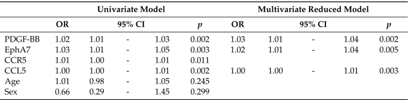
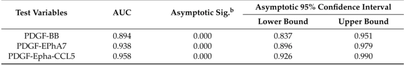
The Diagnostic Significance of PDGF, EphA7, CCR5, and CCL5 Levels in Colorectal Cancer
Tam metin
Şekil

Benzer Belgeler
Association be- tween brain-derived neurotrophic factor and von Willebrand factor levels in patients with stable coronary artery disease. Evaluation of serum sST2 and sCD40L values
Görülüyor ki Ticari İşlemlerde Taşınır Rehni Kanunu’nda, taşınır varlık üzerinde birden fazla alacaklının rehin hakkı söz konusu olacağı haller için, hem teslime
Bırakma motivasyonu, tanı ölçütleri, anksiyete düzeyi ve güvensiz davranışın cezaevinde kalan kişilerde şiddetli madde isteğini yordadığı saptanmıştır (Tablo
Yazar, Türkiye’nin üye olmak için bir talepte bulunma- ması ve ŞİÖ’den de bu yönde bir teklif gelmemesini, Türkiye için kaçırıl- mış büyük bir fırsat
Results: BMI, WHR, Ferriman eGallwey score, and triglyceride, LDL, total cholesterol, estradiol, dehydroepiandrosterone sulfate, free testosterone, luteinizing hormone,
Taking both Moss-Burstein effect and band gap renormalization into account, the corresponding quantity for the absorption edge is the imaginary part of the.. The Dirac delta term in
In conclusion, significantly higher MPV in patients with HcV infection may be responsible for the viral effect that causes platelet dysfunction, supporting the association between
In a 5-year follow up study, they showed that increased total homocysteine levels are linearly associated with CRP elevation and they found that higher total
