A Contribution to the Age-Class Determination of Martes foina Erxleben,
1777 from Turkey (Mammalia: Carnivora)
‹rfan ALBAYRAK1, Ahmet Selçuk ÖZEN2, Andrew Charles KITCHENER3
1University of K›r›kkale, Faculty of Sciences and Arts, Department of Biology, 71450, K›r›kkale-TURKEY 2
University of Dumlup›nar, Faculty of Sciences and Arts, Department of Biology, 43000, Kütahya-TURKEY
3Department of Geology and Zoology, National Museums of Scotland, Edinburgh EH1 1JF, UNITED KINGDOM
Received: 07.11.2006
Abstract: This study was based on 57 beech marten (Martes foina) specimens collected in Turkey between 1994 and 1998. The specimens were divided into 3 age classes as young, juvenile, and adult on the basis of the morphology of the sagittal and lambdoidal crests, and temporal ridges, relative lengths of the interorbital and postorbital constrictions, the degree of wear in molar and canine teeth, baculum weight and structure, pelage color, cranial and bacular measurements, and field notes concerning sexual maturity and lactation. Young, juvenile and adult specimens were readily identified by the shape of the temporal ridges on the skull. These ridges are formed by the origination of the temporalis muscles on the parietal bones and are parallel in the youngs, U- or V-shaped in juveniles, and Y-shaped in adults.
Key Words: Beech marten, skull, baculum, Turkey
Türkiye’deki Martes foina Erxleben, 1777’n›n Yafl Grubu Tayinine Bir Katk› (Mammalia: Carnivora)
Özet: Bu araflt›rma 1994 ve 1998 y›llar› aras›nda Türkiye’den toplanan 57 kaya sansar›, Martes foina, örne¤ine dayanmaktad›r. Örnekler sagittal ve lambdoidal ç›k›nt›lar ve temporal s›rt›n morfolojisine, interorbital ve postorbital bo¤umlardaki geniflli¤in nispetine, molar ve köpek diflteki afl›nma derecesine, baculum a¤›rl›¤› ve yap›s›na, efleysel olgunluk ve emziklilikle ilgili arazi notlar›na dayanarak yavru, genç ve ergin olarak üç yafl grubuna ayr›ld›. Yavru, genç ve ergin örnekler kafatas›ndaki temporal s›rt›n flekliyle kolayca ay›rtedilir. Bu s›rtlar parietal kemiklerdeki temporalis kaslar›n orijinlenmesiyle oluflmufl olup yavruda parelel, gençte “U” veya “V” ve erginde “Y” fleklindedir.
Anahtar Sözcükler: Kaya sansar›, bafl iskeleti, baculum, Türkiye
Introduction
There are 19 mammal species belonging to the order Carnivora in Turkey. The family Mustelidae is represented by 2 species of marten, Martes martes (pine marten) and Martes foina (beech marten) (Kurtonur et al., 1996). M. martes is an endangered species, which is hunted for its valuable fur, but M. foina is widespread and abundant in Turkey. M. foina is a nocturnal and commensal animal, which may inhabit rural areas where it is a frequent predator of poultry. There is very little knowledge about the basic biology of M. foina and all other carnivores in Turkey.
Interactions between species in ecosystems are important for maintaining ecological balance; therefore, it is important to investigate and monitor the population dynamics of animal species if we are to understand more fully how ecosystems function. Age determination is a fundamental requirement for monitoring the population dynamics of species in the wild. For this reason, some studies concerning the age determination of martens have been conducted (Labrid, 1986; Stubbe, 1993; Helldin, 1997).
The purpose of the present study was to show
specimens, offering a contribution to future ecological studies of this species.
Materials and Methods
This study was based on 57 specimens of M. foina
from Turkey, which were collected between 1994 and 1998 (Figure 1). The appearance or position of the testes in the males and the development of mammary glands in the females were recorded. Bacula were extracted according to Ognev (1931). All measurements were taken using dial calipers accurate to 0.1 mm. Specimens were divided into 3 age classes (young, juvenile, and adult) based on the morphology of the sagittal and lambdoidal crests, and temporal ridges, the degree of fusion of the suture at the dorsal junction of the pre-maxillae, pre-maxillae, and nasal bones, the position of the testes and the presence/absence of lactation, variations in zygomatic breadth, interorbital and postorbital constrictions, degree of tooth wear, and curvature of the baculum (Ognev, 1931; Yurgenson, 1956; Churcher, 1960; Tumanov, 1972; Harrison and Bates; 1991). Pelage coloration was recorded according to Ridgway (1886). Comparisons were made between the 3 age classes and data in the literature.
The specimens examined (n = 57) were as follows: Adana, Pozant›, Alpo village, n = 1 (no. 50, 1 , 24 October 1997); Ad›yaman, Çemberlitafl village, n = 1 (no. 53, 1 , 11 November 1997); Çelikhan, ‹zci village, n = 1 (no. 55, 1 ¶, 2 December 1997); Gölbafl›, Merkez, n = 1 (no. 62, 1 , 7 February 1998); Meydan yaylas›, n = 1
(no. 60, 1 , 7 February 1998); Harmanl›, n = 1 (no. 61, 1 , 7 February 1998); Antalya, Akseki, Çalt›l›çukur village, n = 1 (no. 73, 1?, 9 July 1998); Kafl, Sahilbay›nd›r köyü, n = 1 (no. 46, 1 , 28 August 1997); Ayd›n, Hallaçlar village, n = 1 (no. 23, 1 , 19 July 1996); Bal›kesir, Bigadiç, Hisarköy, n = 1 (no. 24, 1 ¶, 30 November 1996); fiaml›, n = 1 (no. 25, 1 , 9 December 1996); S›nd›rg›, Çamalan village, n = 1 (no. 66, 1 , 18 Mart 1998); Bayburt, Merkez, n = 1 (no. 51, 1 , 26 October 1997); Bilecik, Bozüyük, Sarayc›k village, n = 1 (no. 27, 1 ¶, 25 December 1996); Pazaryeri, Yenimahalle, n = 1 (no. 33, 1 ¶, 1 March 1997); Bingöl, A¤açyolu village, n = 1 (no. 59, 1 , 20 January 1998); Genç, Mefledal› village, n = 1 (no. 58, 1 , 23 January 1998); Burdur, Yeflilova, ‹¤dir village, n = 1 (no. 34, 1 , 23 March 1997); Bursa, Keles, Belören, n = 1 (no. 10, 1 , 18 February 1996); Böndere, n = 1 (no. 8, 1 ¶, 28 January 1996); Çorum, Gülücek village, n = 1 (no. 47, 1¶, 1 October 1997), Denizli, Çal, Alfaklar köyü, n = 1 (no. 45, 1 , 27 August 1997); Erzurum, Aflkale, Hac›hamza village, n = 1 (no. 64, 1 , 8 March 1998); Musadan›fl village, n = 1 (no. 54, 1 , 16 November 1997); Erzincan, Tercan, Beflgözel village, n = 1 (no. 48, 1 , 1 October 1997); Eskiflehir, Mihal›çç›k, n = 1 (no. 29, 1 ¶, 28 January 1997); Gürefl village, n = 1 (no. 28, 1 , 30 December 1996); Hatay, ‹skenderun, Hoyut, n = 1 (no. 35, 1 , 9 April 1997); Kahramanmarafl, Tan›r village, n = 1 (no. 63, 1 ¶, 13 February 1998); Kastamonu, Keçiliköy, n = 1 (no. 19, 1 ¶, 7 May 1996); Tosya, Ermelik village n = 2 (no. 16, 1 , 21 April 1996, no. 17, 1 , 24 April 1996); Konya, Akören, Alan village,
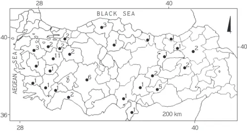
3 1 1 1 1 1 1 1 1 1 1 1 1 1 11 2 2 2 2 2 3 6 5 2 2 2 B L A C K S E A AEGEAN S E A 200 km 28 40 28 40 40 40 36
Figure 1. Collection localities of Martes foina in Turkey (figures indicate the number of specimens examined).
n = 1 (no. 3, 1 ¶, 7 September 1995); Karahüyük village, n = 1 (no. 9, 1 , 17 February 1996); May village, n = 1 (no. 2, 1 ¶, 28 August 1995); Akflehir, Çak›llar, n = 1 (no. 7, 1 , 22 January 1996); Üçhüyük village, n = 1 (no. 39, 1 , 15 July 1997); Konya, Çumra, Tumrafl village, n = 1 (no. 36, 1 , 22 April 1997); Kütahya, Merkez, n = 4 (no. 12, 1 ¶, 4 February 1996; no. 20, 1 ¶, 7 June 1996; no. 65, 1 ¶, 10 March 1998; no. 49, 1 , 6 October 1997); Bölcek village, n = 1 (no. 1, 1 , 25 June 1995); Aslanapa, Terziler village n = 1 (no. 43, 1 , 7 August 1997); Alt›ntafl, Zemme village, n = 1 (no. 30, 1 ¶, 13 February 1996); Il›ca, K›z›lcaören village n = 1 (no. 22, 1 , 17 July 1996); Tavflanl›, n = 1 (no. 37, 1 ¶, 28 April 1997); Bal›köy, n = 1 (no. 32, 1 , 20 February 1997); Yenimahalle, n = 1 (no. 5, 1 ¶, 18 November 1995); Malatya, Do¤anflehir, Erkene, n = 1 (no. 56, 1 , 4 December 1997); Han›mçiftli¤i village, n = 1 (no. 52, 1 ¶, 1 November 1997); Manisa, K›rka¤aç, Güvendik village, n = 1 (no. 6, 1 , 27 December 1995); Ordu, Tikence village, n = 1 (no. 44, 1 , 21 August 1997); Sakarya, Akyaz›, Dokurcul, n = 1 (no. 14, 1 ¶, 11 April 1996); Geyve, Do¤ançay, n = 1 (no. 15, 1 ¶, 17 April 1996); Trabzon, Sürmene, Çamburnu, n = 1 (no. 68, 1 ¶, 18 March 1998); Uflak, Ulubey, Omurca, n = 1 (no. 31, 1 ¶, 18 February 1997).
All the specimens examined in this study were deposited in the Department of Biology, Faculty of Sciences and Arts, University of Dumlup›nar.
Results
Young (n = 2: 1 , 1 ¶)
Pelage Color: In the 2 available young specimens (1 male, 1 female), both from western Turkey, the dorsal and ventral pelage are basically the same color as that of juveniles (see below), but the tips to the guard hairs, and the legs and tail are slightly paler.
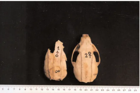
Skull: In the 2 available skulls the sagittal and lambdoidal crests were just visible at the posterior of the skull. When viewed dorsally, the temporal ridges ran parallel to each other along each side of the cranium, where the temporalis muscles originated. The temporal ridges started from behind the postorbital processes and extended through the lambdoidal crest (Figure 2). The interorbital and postorbital constrictions of 1 specimen were examined. In this specimen the postorbital constriction is wider than the interorbital constriction. The sutures on the anterior portion of the skull are obvious, because the pre-maxillae, maxillae, and nasal bones are not completely fused. The cusps of the upper incisors are pointed and the outer incisors are longer than the inner ones. The crown heights of the lower incisors are almost equal, the cusps being flat and running in the same axis as the tooth-row. The ridges of the crowns of pre-molars are sharp and the cusps of molars in both jaws are pointed. There are 13 tubercles on the chewing
surface of M1, which are clearly visible to the naked eye.
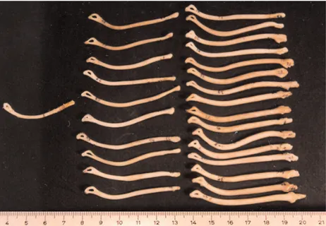
Baculum: The length of the only baculum is 42 mm and its weight is 0.10 g. The baculum is curved, starting from the middle and continuing distally. There is an ellipsoidal hole distally (Figure 3). There was no sign of testes or lactation in the young beech martens.
Juveniles (n = 22: 11 , 11 ¶¶)
Pelage Color: In total, 20 specimens were examined. On the dorsal and ventral sides of 11 male and 9 female specimens from Turkey, the underfur is generally dull grayish-brown, tinged with pale buff. The tips of the guard hairs, legs, and tail are the same color as those of adult specimens (see below). The pelage coloration of 1 female is indistinguishable from that of an adult.
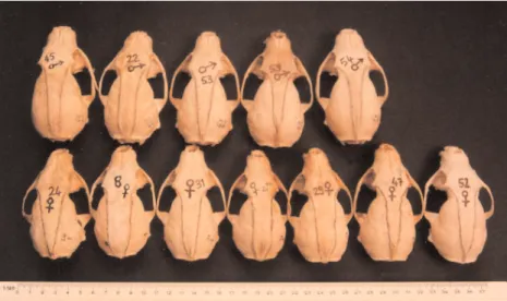
Skull: In the 22 skulls (11 , 11 ¶) that were examined the sagittal crest is clearly seen towards the posterior of the cranium. When viewed dorsally, the temporal ridges run from just behind the postorbital process and end at the lambdoidal crest. Thus, the temporal ridges form a V or U shape, narrowing posteriorly (Figure 4).
In all specimens the lambdoidal crest is clearly visible and the postorbital constriction is narrower than the interorbital constriction. The sutures on the anterior portion of the skull where the pre-maxillae, maxillae, and nasal bones join together are somewhat obvious. The cusps of the upper incisors are worn and not pointed. The outer incisors are longer than the inner ones. The lengths
of the lower incisors are equal and the cusps are flat, owing to wear. The lower incisors run along the same axis as the tooth-row. In most specimens the canines are pointed and the cusps are not worn (90.9%, n = 22). The ridges of the crowns of the pre-molars are sharp and the cusps of the molars in both jaws are pointed. Some small
tubercles were observed on the upper surface of M1,
which are slightly worn in almost half of the specimens (50.0%, n = 22).
Baculum: The lengths of 10 bacula range from 48.5 to 57.8 mm. Baculum weight ranges from 0.20-0.30 g. Bacula are thick and have a slight curve, and the proximal parts are somewhat swollen. There is a small projection proximally and an ellipsoidal hole distally (Figure 3). In 3 of the 11 male specimens the testes were clearly visible, but in the others they were less obvious. Mammary glands were not clearly visible in the 11 female specimens.
Adults (n = 33: 22 , 10 ¶¶, 1 unknown sex) Pelage Color: In total, 26 specimens were examined. On the dorsal and ventral sides of 16 male and 10 female specimens from throughout Turkey, the underfur is very light grayish-brown, tinged with pale buff. The guard hair tips are somewhat metallic brown and tinged with rust on both sides. The legs and tail are slightly blackish-brown. Two males and 1 female have darker tips on the guard hairs, and darker legs and tails than the other specimens.
There is no evidence of seasonal variation in pelage coloration.
Skull: In 33 skulls (22 , 10 ¶, and 1 unknown sex) the sagittal crest is clearly marked, especially towards the posterior of the skull. The temporal ridges arise from behind the postorbital processes and converge along the sagittal suture or sagittal crest.
Therefore, the temporal ridges form a Y shape. The sagittal crest varies in its depth from 1.0 to 4.4 mm (Figure 5).
In skulls with a deep sagittal crest, the sagittal and lambdoidal crests are well developed. In the 26 skulls belonging to this age class the postorbital constriction is narrower than the interorbital constriction. Upper incisors run along the same axis as the tooth-row and their cusps are flattened with wear. The canines are not pointed due to wear. The lower canines may be worn by up to half their crown heights in almost half of the specimens (45.45% n = 33). The ridges of the crowns of the premolars and the cusps of the molars in both jaws, as seen in younger age classes, are absent due to wear.
Tubercles on the upper surface of M1are also absent.
Figure 4. Dorsal views of skulls of juvenile M. foina, showing U- and V-shaped temporal ridges.
Figure 5. Dorsal views of skulls of adult M. foina, showing Y shape formed by the temporal ridges and sagittal crest.
Baculum: The lengths of 16 bacula belonging to this age class range from 56.5 to 66.0 mm and their weights range from 0.40 to 0.66 g (the baculum thickens gradually from the middle to the distal end, where it curves slightly (Figure 3).
At the proximal end of the baculum there are 3 small projections. From the field notes of 18 specimens it was recorded that the testes were well developed in all individuals. The distal scrotum in 2 of 16 summer specimens was reddish. Mammary glands were clearly visible in females, but only 2 females, which were collected in April and May, were lactating. There were only a few short hairs around the nipples in these lactating females. It was also observed that 1 female caught in March had an embryo.
Discussion
In the skulls of Turkish M. foina specimens examined herein, the sagittal crest is only just visible in young and juveniles, but it is well developed in adults. In young martens the temporal ridges are parallel, but in juveniles they form a U or V shape, whereas in adults the temporal ridges fuse to form a Y shape when viewed dorsally. In juveniles those skulls with U-shaped temporal ridges are from younger animals than those with V-shaped ridges. In the adult age class, the skulls with Y-shaped temporal ridges, which have a taller upright, are from individuals older than those with a shorter upright. According to the characteristics shown in Figures 4 and 5, there does not seem to be any effect of geographical location and sex on the observed variation in adults and juveniles. In the oldest individuals the height of the sagittal crest increases with age. The sagittal crest in juveniles and adult males is more pronounced than it is in females. The zygomatic breadth in adults is greater than it is in young and juveniles. These results are in agreement with observations by Ognev (1931), but contradict Harrison and Bates (1991), who stated that a sagittal crest did not occur in females.
Measurements of postorbital and interorbital constrictions were recorded for 1 young, 9 male and 11 female juveniles, and 17 male and 9 female adult beech martens. While the postorbital constriction is greater than the interorbital constriction in young beech marten
skulls, it is narrower in juvenile and adult age classes. The postorbital constriction decreases from young to adult. These findings are also in agreement with those of Ognev (1931). On the skull of one young individual, the suture where the pre-maxillae, maxillae, and nasal bones meet is clearly seen, but in most juveniles it is not.
The cusps of the upper incisors are much more pointed in young than in juveniles, and they are flattened in adults due to wear. In juveniles and youngs the outer incisors are taller than the inner ones. In adults the outer incisors are equal in length to the inner ones due to wear. In one young and most juveniles the canines are pointed, but not in adults. The lower canines may have been worn down by up to half their length in adults, which seems to be characteristic of adults.
The ridges of pre-molars and the cusps of molars in the upper and lower jaws of young and juveniles are sharp. The cusps are pointed in young and juveniles, while they are worn in adults. Small tubercles on the upper
surface of M1are clearly visible in juveniles, but are barely
developed in the young and are absent in adults.
The length of bacula range from 48.5 to 57.8 mm in juveniles and 56.5 to 66.0 mm in adults; baculum length is 42 mm long in the only young specimen that was available. The baculum is slender in the one young specimen examined and increases in thickness with age. The curvature of the baculum is greatest in the young animal, except for a few (3) adult specimens. The curvature generally begins in the middle of the baculum in young, close to the middle in juveniles, and occurs distally in adults. There is 1 bony projection on the proximal baculum of juveniles, 3 in those of adults, and none in those of young specimens. In all 3 age classes the distal baculum has an ellipsoidal hole in it. Bacular measurements of adults are significantly greater than those of juveniles (P < 0.05). These measurements and weights are very similar to those given by Heptner and Naumov (2002) for juvenile and adult (> 2 years old) age classes in the former Soviet Union.
According to the field notes, the testes in young individuals were not developed. However, in a few juvenile specimens (n = 2) and all adults the testes were clearly visible. Adult female beech martens have visible mammary glands, whereas juveniles do not.
References
Churcher, C.S. 1960. Cranial variations in the North American red fox. J. Mammal. 41:349-360.
Harrison, D.L. and Bates, P.J.J. 1991. The Mammals of Arabia. Second Edition, Sevenoaks, Kent, Harrison Zoological Museum, England. Helldin, J.O. 1997. Age determination of Eurasian pine martens by radiographs of teeth in situ. Wildlife Society Bulletin 25: 83-88. Heptner, V.G. and Naumov, N.P. 2002. Mammals of the Soviet Union
Volume II, Part 1b Carnivora (Weasels: Additional species). Enfield: Science Publishers Inc.
Kurtonur, C., Albayrak, ‹., K›vanç, E., Kefelio¤lu, H. and Özkan, B. 1996. Memeliler (Mammalia). 7-23, in: Türkiye Omurgal›lar Tür Listesi, (A. Kence, C. Bilgin, eds.), DPT/TBAG-Çev.Sek., Nurol Matbaas›, Ankara, 1-183.
Labrid, M. 1986. La Marte (Martes martes Linnaeus, 1758). Encyclopédie des carnivores de France, Société Française pour L’etude et la protection des Mammiferes 1-22.
Ognev, S.I. 1931. Mammals of Eastern Europe and Northern Asia, Vol. II, Carnivora (Fissipedia). Moskva-Leningrad (Translated from Russian, Israel Program for Scientific Translations. Jerusalem 1962).
Ridgway, R. 1886. A nomenclature of naturalists and compendium of useful knowledge for ornithologists, Boston.
Stubbe, M. 1993. Martes martes (Linné, 1758) –Baum-, Edelmarder, 374-426, in: Handbuch der Säugetiere Europas. Carnivora (Fissipedia) Teil I: Canidae, Ursidae, Procyonidae, Mustelidae 1 (J., Niethammer, F., Krapp, Eds.). Aula-Verlag Wiesbaden 1-526. Tumanov, I.L. 1972. Age changes in morphophysiological characters of
some species of martens. Zool. Zh. 51: 694-703.
Yurgenson, P.B. 1956. Determining the age of pine martens. Zool. zh. 35: 781-783.