Immunohistochemical evaluation of akabane virus
infection in aborted and new-born calves
M. Haligur
1, S. Hasircioglu
2,
O. Ozmen
1, M. Kale
2, A. Aydogan
1 1Faculty of Ceyhan Veterinary Medicine, University of Cukurova, Saricam, Adana, Turkey 2Faculty of Veterinary Medicine, University of Mehmet Akif Ersoy, Istiklal Yerleskesi,Burdur, Turkey
ABSTRACT: The present study was aimed at the detection and describing the lesions of akabane virus in foetal and new-born calves tissues using immunohistochemical and immunofluorescence techniques. Akabane virus lesions were evaluated in 12 foetuses and three new-born calves using serological and pathological methods and immunohistochemistry and immunofluorescence. Macroscopically, prominent arthrogryposis and hydranen-cephaly (A-H syndrome) were the main symptoms. At the histopathological examination, lesions were especially localised in the midbrain, pons, and medulla oblongata in the central nervous system (CNS) in calves naturally infected with akabane virus. In these areas, degenerative and necrotic neurons were observed. There was promi-nent mononuclear infiltration in perivascular areas. While akabane virus antigen was only detected in brain using immunofluorescence, immunohistochemistry against akabane virus yielded positive antigenic reactions in the cerebrum, cerebellum, liver, spleen and kidneys. In addition to these findings, there was a relationship between akabane virus infection and neurofilament (NF), glial fibrillary acidic protein (GFAP), and N-methyl-D-aspartate receptor (NMDAR) immunoreaction in astrocytes and neurons.
Keywords: akabane virus; foetus; calves; immunofluorescence; immunoperoxidase; pathology; virology
Akabane virus is classified in the family Bunya- viridae and is among the most potent viral tera-togens of domestic animals (Konno et al. 1982; Taylor and Mellor 1994; Maxie and Youssef 2007). Multiple congenital anomalies due to akabane virus infection occur between days 29–48 in pregnancy. Congenital disease leads to a range of predomi-nantly neural abnormalities in calves, lambs and kids, but akabane virus is best known for pro-ducing akabane virus outbreaks which manifest chiefly as arthrogryposis and hydranencephaly in calves(Konno et al. 1982; Maxie and Youssef 2007; Schlafer and Miller 2007).Arthrogryposis appears early in the outbreak following foetal infection at five-six months of pregnancy. With increasing age of the foetus at the time of infection, the cavitating cerebral changes are less severe and grade towards porencephaly (Maxie and Youssef 2007).
Glial fibrillary acidic protein is an intermediate filament protein and is expressed by numerous cell types, especially in astrocyte and ependymal cells in the CNS (Jacque et al. 1978; Roessmann et al.
1980). GFAP plays an important role in CNS pro-cesses, including astrocyte-neuron interactions as well as cell-cell communication and the function of the blood brain barrier and helps to maintain astrocyte mechanical strength (Weinstein et al. 1991; Liedtke et al. 1996; Cullen et al. 2007). It is suggested that GFAP plays many critical roles in the CNS. Nevertheless, its exact function remains poorly understood, despite the large number of studies on GFAP function. GFAP is a type III inter-mediate filament and it can polymerise with other type III proteins or with NF protein (Reeves et al. 1989; Bongcam-Rudloff et al. 1991).
Neurofilament is an intermediate filament, which is a major component of the cell’s cytoskeleton and provides support for normal axonal radial growth. It is found specifically in neurons (Gotow 2000; Sanchez et al. 2000). It plays an important role in ensuring that proteins reach their final destina-tion (down axons) and is synthesised within the cell body. NF directly controls axonal diameter, which in turn controls how fast electrical signals
travel down the axon (Alberts et al. 2002; Ackerley et al. 2003). Immunohistochemically, NF is an im-portant marker for diagnostic neuropathology in differentiating neurons from glia (Menzies et al. 2002; Trimmer et al. 2004).
The N-methyl-D-aspartate receptor (NMDAR) is an ionotropic glutamate receptor important in regulating synaptic plasticity and memory func-tion (Li and Tsien 2009). The NMDAR forms a heterotetramer of subunits which are termed glu-tamate-binding NMDAR subunits. The NMDAR is activated in two distinct ways. The first way is ligand-gated and voltage-dependent; the second way requires co-activation by two ligands: gluta-mate and glycine. Activation of NMDAR results in the opening of an ion channel (Dingledine et al. 1999; Paoletti and Neyton 2007).
Akabane virus has been detected using polymer-ase chain reaction and serological and pathological methods in various animals (Akashi et al. 1999; Noda et al. 2001; Stram et al. 2004). However, only a small number of studies have used the immu-nohistochemical method to detect akabane virus and to describe akabane virus lesions. Therefore, in the present study, akabane virus and lesions were evaluated in aborted foetuses of cattle serologically, pathologically, and using immunohistochemical and immunofluorescence techniques.
MATERIAL AND METHODS
In this study, 12 of 30 foetuses and three new-born calves that had been aborted due to akabane virus infection and originating from five different flocks were examined serologically and pathologically and using immunohistochemical and immunofluores-cence methods. The 12 aborted foetuses and three new-born calves ranged in age from seven months (gestation age) to one day (on the day of birth) old. Fifteen calves (nine Holstein, four mixed and two Montafon breed) of both sexes (ten female and five male) were used. The cases are summarised in Table 1. Necropsy was performed on foetuses and new-born calves. During necropsy, tissue samples were taken from all foetuses and all organs, and then fixed in 10% buffered formalin. Following routine procedures, tis-sue samples were blocked in paraffin and cut at 5µm thickness, then stained with Haematoxylin and Eosin (HE) and examined under a light microscope.
For immunohistochemistry, after histopatho-logical examination, tissue sections were immu-nostained using the streptavidin-biotin complex peroxidase technique, in accordance with the man-ufacturer’s instructions for akabane virus antigen (bovine akabane virus antiserum), GFAP, NF and NMDAR. For immunohistochemical observations, paraffin wax was sectioned at 4 µm and sections were
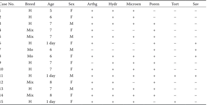
Table 1. Breed, age, sex and congenital malformations in calves naturally infected with akabane virus
Case No. Breed Age Sex Arthg Hydr Microen Poren Tort Sav
1 H 5 F + + + – – – 2 H 6 F + + + – – – 3 H 7 M + + + + – – 4 Mix 7 F + – – + – – 5 Mix 7 M + + + + – – 6 H 1 day F + – – + – + 7 Mo 6 M – – – + – + 8 Mo 6 F + + + + – + 9 H 7 F – + + + – – 10 H 7 F + + + + – – 11 H 1 day M + + + + + + 12 Mix 8 F + + – + – – 13 H 7 M + + + + – – 14 Mix 8 F + + + + – – 15 H 1 day F + + + – + –
H = Holstein, Mo = Montafon, Mix = mix breed; F = female, M = male, Age = gestation age (month) and birth days, Arthg = arthrogryposis, Hydr = hydranencephaly, Microen = microencephaly, Poren = porencephaly, Tort = torticollis, Sav = cerebro are avasculosa. Scoring of congenital anomalies: malformations, (+) present, (–) absent
attached to glass slides coated with poly-L-lysine. These were then deparaffinised and rehydrated. To reduce non-specific background staining due to en-dogenous peroxidase slides were incubated in 0.5% hydrogen peroxide/methanol for 10 min and boiled with 0.01% citrate buffer solution (pH 6.0) for 20 min, then incubated in diluted normal horse serum for 10 min. Subsequently the primary antibody for aka-bane virus antigen (commercial kit-Histofine SAB-PO kit, Nichirei, Tokyo, Japan, 1 : 4000 dilution) was ap-plied, and tissues were incubated overnight at +4 °C temperature in a refrigerator. Tissues were then in-cubated with biotinylated secondary antibody and streptavidin/peroxidase each for 10 min at room tem-perature. Tissues were further incubated for 10 min at room temperature in a solution of DAB (3, 3'-diami- nobenzidine) chromogen and counterstained with Mayer’s haematoxylin together with mounting me-dia. Normal rabbit serum (1 : 100) and mouse serum (1 : 100) were used as negative controls. Markers used for immunohistochemical examination are listed in Table 2.
For the indirect fluorescent antibody test (IFAT) tissue samples were taken from brain, spleen, liver and kidney post-mortem for viral antigen detec-tion. Tissue impressions were made from tissue samples by lightly touching tissues to a microscope slide. Following air-drying and acetone fixation the tissue impressions were incubated in a humidified chamber at 37 °Cfor 30 min with 50 microliters of a 1 : 100 dilution of a polyclonal antibody derived from anti-akabane virus serum from a cow naturally infected with akabane virus. The impressions were then gently rinsed with PBS, soaked for 10 min in PBS and blotted. Slides with 50 microliters of fluo-rescein isothiocyanate labeled caprine anti-bovine IgG1,2 (VMRD, Inc., Pulman, WA, USA) were incu-bated at 37 °C for 30 min and then washed as before. The slides were dried and a cover slip mounted with one drop of mounting fluid (VMRD). Slides were examined for fluorescence immediately using a Nikon fluorescent microscope at 400×.
Akabane virus antibodies in serum samples col-lected from aborted animals and new-born calves were detected using a commercial competitive ELISA kit (ID Vet Innovative Diagnostics, France). The test was performed according to the procedure described by the manufacturer.
RESULTS
Clinically, hyperaesthesia and recumbence were observed in three cases and torticollis was detected in two cases. Necropsy findings revealed promi-nent arthrogryposis and hydranencephaly (A-H syndrome). In three cases either arthrogryposis or hydranencephaly was observed. Arthrogryposis was seen in 13 of 15 cases and was prominent especially in the forelimb. Hydranencephaly, hydrocephalus, microencephaly and porencephaly were observed in the CNS. The cranial cavity was opened and the brain stem, pons, thalamus, medulla oblongata and a portion of cerebellum were generally observed. However, the gyrus of the brain was usually absent. The leptomemingeal membranes were filled with about 50–75 ml of cerebrospinal fluid (Figure 1A, B, C). Hydranencephaly was observed in 12 of 15 cases, porencephaly in 12 and microencephaly in 11 cases. In addition to these findings, meningocel and menin-goencephalocel was detected in two cases and area serebrovasculosa in the CNS in four calves (Figure 1A). All findings are summarised in Table 2. There was abdominal fluid and thoracic fluid in 12 aborted foetuses. Fibrin masses were detected within these fluids and the surface of visceral organs.
At the microscopical examination, lesions, espe-cially localised in the midbrain, pons, and medulla oblongata in foetuses and new-born calves natu-rally infected with akabane virus were observed in the CNS. In these areas, degenerative and necrotic neurons, perineuronal and perivascular oedema was observed. Encephalitis and meningitis were prominent in the CNS and generally localised in
Table 2. Antibodies used for assessment of akabane virus infection in aborted and new-born calves
Antibody Species Clon Dilutions Commercial company Antigen retrieval Bovine AKAV antisera bovine polyclonal 1 : 5 ID Vet Innov. Diag. (customer desing) yes
GFAP mouse monoclonal 1 : 2000 Abcam (ab4648-100) yes
NF mouse monoclonal 1 : 50 DAKO yes
NMDAR rabbit polyclonal 1 : 200 Abcam (ab28669) yes
Bovine AKAV antiserum = akabane virus anti serum, GFAP = glial fibrillary acidic protein, NF = neurofilament, NMDAR = N-methyl-D-aspartate receptor
perivascular inflammatory cell areas. These cells comprised lymphocytes, plasma cells, and mac-rophages (Figure 2). Infiltration of perivascular
mononuclear cells was observed in 13 cases and infiltration was severe in seven cases. Additionally, diffuse and focal proliferation of glial cells was seen
Table 3. Histopathological lesions in cattle naturally infected with akabane virus Case
No.
Central nervous system Visceral organs Muscular tissue myodegeneration
or polymyosistis
FG PMNCI ND-N NV NPV hepatitis nephritis
1 ++ +++ + ++ + + + ++ 2 + +++ ++ + ++ + + + 3 +++ +++ + + ++ – + ++ 4 + ++ + – + – + + 5 + +++ ++ – + – + + 6 + +++ +++ – + + – + 7 ++ + + – – – – ++ 8 +++ + + + – ++ – – 9 +++ + +++ + – ++ + + 10 + +++ + – + + – – 11 ++ – +++ ++ + – – + 12 ++ – ++ – + – + ++ 13 + ++ ++ + – – + ++ 14 + + + – ++ + – + 15 ++ +++ +++ ++ ++ – + +
FG = focal gliosis, PMNCI = perivascular mononuclear cells infiltration, ND-N = neuronal degeneration and necrosis, NV = neuronal vacuolisation, NPV = neuropil vacuolisation. Scoring of severity of lesions: (+++) severe, (++) moderate, (+) mild, (–) no lesion
Figures 1–4. 1. Macroscopic appearance of CNS anomalies (A, B, C) in calves infected with akabane virus. 2. Meningitis and perivascular cuffing (arrow), HE, bar = 200 µm. 3. Demonstration of viral antigen using immunofluorescence. 4. Demonstra-tion of viral agents (arrows) using immunohistochemistry, streptavidin method, with haematoxylin counter stain, bar = 50 µm
in the cerebrum and cerebellum. There was gliosis in all cases, but marked gliosis was observed only in three cases, moderate gliosis in five cases and mild gliosis was seen in seven cases. Vacuolisation of neuropil and demyelination were detected in the cerebrum. In addition, intracytoplasmic vacuolisa-tion was observed in neurons.
Moderate neuropil vacuolisation and demyelina-tion (four cases), mild neuropil vacuolisademyelina-tion (seven cases), moderate vacuolisation of neuron cytoplasm (three cases) and mild neuronal vacuolisation (five cases) were observed. Hepatitis and nephritis were observed in calves naturally infected with akabane virus. Kupffer cells were easily seen and bile pig-ments were localised in the cytoplasm of these cells. Neutrophil leukocytes had infiltrated both the liver and kidneys. Myodegeneration or polymyositis were marked in affected foetuses and new-born calves. All histopathological results are summarised in Table 3. Immunohistochemically, positive antigenic reac-tions were observed in the cytoplasm of neurons, Kupffer cells, mesangial cells of glomerulus and kid-ney tubular epithelial cells, and lymphocytes in the
spleen. Positive immunofluorescence reactions were only seen in the brain (Figure 3) while immunohis-tochemically akabane virus antigen was detected in the CNS, liver, kidney and spleen (Figure 4). Mild-to-severe positive immunoreactions were especially seen in the CNS. All immunoreactivity against akabane virus is summarised in Table 4. Serologically, anti-akabane virus antibodies were detected in aborted foetuses and new-born calf serum using competi-tive ELISA. An increased expression of GFAP was observed in the brain using immunohistochemical methods (Figure 5). A marked increase in NF expres-sion manifested in the axons of neurons and motor neurons as an accumulation of dark-brown colour (Figure 6). Marked positive immunoreaction of NMDAR was detected in neurons (Figure 7).
DISCUSSION
Akabane virus infection can cause many congeni-tal abnormalities, especially in the central nervous system. The initial manifestation of neuronal
ab-Figures 5–7. 5. Positive immunoreaction for GFAP in neurons (arrows) and perineuronal oedema (arrow heads), streptavidin-biotin complex peroxidase tech-nique, with haematoxylin counter stain, bar = 200 µm.
6. Immunopositive NF reaction in neuronal axons
(arrows), streptavidin-biotin complex peroxidase tech-nique, with haematoxylin counter stain, bar = 100 µm.
7. Immunoreaction for NMDAR in neurons (arrows),
streptavidin-biotin complex peroxidase technique, with haematoxylin counter stain, bar = 100 µm
normality in a field outbreak is the birth of incoor-dinate calves while microencephaly and cerebellar hypoplasia occur occasionally as manifestations of late infections. Akabane virus has been isolated from different species such as buffalo, camels, horses, sheep and goats. The virus passes through the placenta and infects the calf, specifically the calf’s central nervous system where it may cause congenital abnormalities of varying severity in ru-minants. Congenital abnormalities and prominent arthrogryposis and hydranencephaly are observed in both natural and experimental infection with akabane virus in foetuses. With respect to con-genital anomalies microcephaly, porencephaly, hydranencephaly, hydrocephalus, hydromyelia, arthrogryposis, kyphosis, lordosis, scoliosis, brach-ygnatia, muscular atrophy, spina bifida, cyclops, Arnold-Chiari syndrome and atresia ani have been reported in various animals. Diagnosis of the vi-rus is often made on the basis of clinical signs and can be confirmed by detection of antibody in the blood of the calf or in the blood of the dam (Konno et al. 1982; Uchida et al. 2000; Kamata et al. 2009). The pathogenesis of akabane virus in-fection in ruminant foetuses has been described previously (e.g. Narita and Kawashima 1993). In the present study, congenital anomalies such as arthrogryposis, hydranencephaly, hydrocephalus,
microencephaly, porencephaly and cerebellar cavi-tation and meningitis were commonly observed but vertebral deformities such as scoliosis, kyphosis and thoracolumbar lordosis were not seen in any of the aborted foetuses infected with akabane virus. In addition to these findings torticollis was seen in two cases. Diagnosis of akabane virus infection in aborted calves was made on the basis of serological, pathological, immunohistochemical and immuno-fluorescence techniques. Positive immunoreactions were observed in the brain, kidney, liver and spleen of aborted foetuses.
If foetal infection occurs between days 41 and 50 of gestation, the virus can cause thick perivascular cuffing of macrophages, lymphocytes and plasma cells, coupled with very prominent glial nodules. Most of the central nervous system malformations caused by akabane virus are related to differences in the gestational age of the foetus at the time of infection. The vulnerability of neurons is related to direct injury, and the nature of the tissue response to such injury. Lesions in the brain and muscles may be due to the direct cytopathic effect of the virus on developing neurons. Neuronal death in CNS is followed by neurogenic muscle atrophy and arthrogryposis (Schlafer and Miller 2007). In this study, histopathological findings were similar to those reported previously.
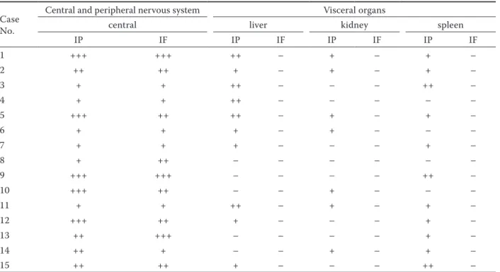
Table 4. Localisation of immunoreactions in organs naturally infected with akabane virus Case
No.
Central and peripheral nervous system Visceral organs
central liver kidney spleen
IP IF IP IF IP IF IP IF 1 +++ +++ ++ – + – + – 2 ++ ++ + – + – + – 3 + + ++ – – – ++ – 4 + + ++ – – – – – 5 +++ ++ ++ – + – + – 6 + + + – + – – – 7 + + + – – – + – 8 + ++ – – – – – – 9 +++ +++ – – – – ++ – 10 +++ ++ – – + – – – 11 + + ++ – + – + – 12 +++ ++ + – – – + – 13 ++ +++ – – – – + – 14 ++ + – – + – + – 15 ++ ++ + – – – ++ –
IP = immunoperoxidase, IF = Immunoflouresence. Scoring of the immunoreactivitiy: (+++) severe, (++) moderate, (+) mild, (–) no reaction
Serologically, akabane virus antigen was detect-ed using competitive ELISA in serum taken from aborted animals and new-born calves. Akabane virus antigen can be detected using immunohis-tochemical and serological methods in brain tissues (Noda et al. 2001). In the present study, akabane virus antigen detection in brain was performed us-ing immunofluorescence and immunohistochemi-cal techniques. Viral antigens were detected in the kidney, liver and spleen only using the immunohis-tochemical technique. Positive immunoreactions in lymphocytes of the spleen may be explained by the role of lymphocytes in viral dissemination in the foetus and their contribution to the pathogenesis of akabane virus infection.
GFAP has been shown to be important in repair after CNS injury, more specifically for its role in the formation of glial scars in a multitude of locations throughout the CNS including the eye (Tuccari et al. 1986) and brain (Paetau et al. 1985). GFAP regu-lation is disrupted in multiple disorders and injury can cause glial cells to react in detrimental ways (Bunge et al. 1961). The expression of some GFAP isoforms has been reported to decrease in response to acute infection or neurodegeneration (Johnston-Wilson et al. 2000). Increased expression of GFAP by astrocytes is well recognised in pathological conditions affecting the CNS (Eng 1980; Selkoe et al. 1982). Our results suggest that akabane virus infection may contribute to cell damage and the increase in GFAP expression may be attributed to akabane virus infection as a neurodegenerative dis-ease. It is probable that the regulation and expres-sion of GFAP is impaired in akabane virus infection leading to a failure to repair damage to the CNS. GFAP is the major constituent of the intermediate filaments of astrocytes in the CNS and has been used as a marker for these cells (Eng 1980; Eng and DeArmond 1983). Severe demyelisation both in the CNS and peripheral nervous system associ-ated with gliosis and infiltration of macrophages (globoid cells) are the main neuropathological fea-tures (Kobayashi et al. 1986). In the present study inflammation of mononuclear cells, vacuolisation of neurons and demyelinisation were observed. In addition, cell-to-cell communication or astrocyte differentiation can be disturbed and so akabane virus infection in the foetal CNS may inhibit cell migration.
Neurofilament (NF) proteins are exclusively ex-pressed in the axons of neurons and constitute key building blocks of the axonal cytoskeleton. This
protein plays an important role as an indicator of normal biological and pathogenic processes (Norgren et al. 2003; Petzold 2005). Generally, ac-cumulation and abnormal assembly of NF proteins are common pathological findings in several types of neurodegenerative disease in humans and ani-mals (Yu et al. 1993; Figlewicz et al. 1994). Motor neuron degeneration is related to NF protein ac-cumulation (Collard et al. 1995). Additionally, ax-onal degeneration owing to NF protein has been demonstrated. When neurons die or axonal degen-eration occurs, axonal membrane disintegration occurs inevitably (Petzold 2005). Neurofilaments are expressed in all neurons of the central and pe-ripheral nervous system (Liem et al. 1978). In the present study, NF accumulation was commonly seen in axonal neurons. This finding suggests that akabane virus infection may constitute a severe neurodegenerative disease.
NMDAR has four isoforms named NR1, NR2, NR3 and NR4. In invertebrates only NR2 is found; however, all four forms are expressed in verte-brates (Teng et al. 2010). NR2 subunits contain the binding site for the neurotransmitter gluta-mate and have been detected in various cell types. NR2B in particular, is mainly present in immature neurons and in extra synaptic locations (Liu et al. 2004). Many endogenous and exogenous com-pounds modulate NMDA receptors which play a crucial role in a wide range of physiological and pathological processes (Huggins and Grant 2005). Some ions such as Na+, K+ and Ca2+ pass through
the NMDA receptor channel and modulate NMDA receptor activity. NMDA receptor activity is regu-lated by a “redox modulatory site” (Aizenman et al. 1989). This chemical reduction and oxidation is very important for the function of the receptor. While reductants dramatically enhance NMDA channel activity, oxidants either reverse the effects of reductants or depress native responses. The NMDA receptors allow Ca2+, Na+, and K+ to pass
into the cell. The excitatory postsynaptic potential (EPSP) produced by activation of an NMDA recep-tor increases the concentration of Ca2+ in the cell
(Purves et al. 2008). In the present study, mainly the NMDA receptor was activated by akabane virus infection and thus neuronal degeneration and necrosis may occur in akabane virus cases. Activation of the NMDAR can lead to the open-ing of ion channels and can cause uncontrolled transfer of non-specific cations. This ion transfer, in turn, can lead to cause cell damage.
CONCLUSION
This study revealed that akabane virus infection is an important abortive agent in bovines. Viral an-tigen was detected in the kidney, spleen, liver and central nervous system. The presence of infective lymphocytes with akabane virus was determined im-munohistochemically. Diagnosis of akabane virus was made using immunohistochemical and immu-nofluorescence techniques in this study and these techniques may be considered important tools for antigenic determination. Our results show that im-munohistochemical techniques are more sensitive than immunofluorescence. Additionally, analysis of GFAP, NF and NAMDR expression suggests that ak-abane virus may lead to neurodegenerative disease.
REFERENCES
Ackerley S, Thornhill P, Grierson AJ, Brownlees J, An-derton BH, Leigh PN, Shaw CE, Miller CCJ (2003): Neurofilament heavy chain side arm phosphorylation regulates axonal transport of neurofilaments. Journal of Cell Biology 161, 489–495.
Akashi H, Onuma S, Nagano H, Ohta M, Fukutomi T (1999): Detection and differentiation of Aino and Aka-bane Simbu serogroup bunyaviruses by nested polymer-ase chain reaction. Archives of Virology 144, 2101–2109. Aizenman E, Lipton SA, Loring RH (1989): Selective
modulation of NMDA responses by reduction and oxidation. Neuron 2, 1257–1263.
Alberts B, Johnson A, Lewis J, Raff M, Roberts K, Wal-ter P (eds.) (2002): Molecular Biology of the Cell. 4th ed. Garland Science, New York. 924 pp.
Bongcam-Rudloff E, Nister M, Betsholtz C, Wang JL, Stenman G, Huebner K, Croce CM, Westermark B (1991): Human glial fibrillary acidic protein: comple-mentary DNA cloning, chromosome localization, and messenger RNA expression in human glioma cell lines of various phenotypes. Cancer Research 51, 1553–1560. Bunge MB, Bunge RP, Ris H (1961): Ultrastructural study
of remyelination in an experimental lesion in adult cat cord. Journal of Biophysical and Biochemical Cytology 10, 67–94.
Collard JF, Cote F, Julien JP (1995): Defective axonal transport in a transgenic mouse model of amyotrophic lateral sclerosis. Nature 375, 61–64.
Cullen DK, Simon CM, Laplaca MC (2007): Strain rate-dependent induction of reactive astrogliosis and cell death in three-dimensional neuronal-astrocyte co-cultures. Brain Research 1158, 103–115.
Dingledine R, Borges K, Bowie D, Traynelis SF (1999): The glutamate receptor ion channels. Pharmacologi-cal Reviews 51, 7–61.
Eng LF (1980): The glial fibrillary acidic (GFA) protein. In: Bradshaw RA, Schneider DM (eds.): Proteins of the Nerv-ous System. 2nd ed. Raven Press, New York. 85–117. Eng LF, DeArmond SJ (1983): Immunochemistry of the
glialfibrillary acidic protein, progress in neuropathol-ogy. In: Zimmerman HM (eds.): Neuroglia. Raven Press, New York. 19–39.
Figlewicz DA, Krizus A, Martinoli MG, Meininger M, Meininger V, Dib M, Rouleau GA, Julien JP (1994): Variants of the heavy neurofilament subunit are as-sociated with the development of amyotrophic lateral sclerosis. Human Molecular Genetics 3, 1757–1761. Gotow T (2000): Neurofilaments in health and disease.
Medical Electron Microscopy 33, 173–199.
Huggins DJ, Grant GH (2005): The function of the amino terminal domain in NMDA receptor modulation. Jour-nal of Molecular Graphics and Modelling 23, 381–388. Jacque CM, Vinner C, Kujas M, Raoul M, Racadot J, Bau-mann NA (1978): Determination of glial fibrillary acidic protein (GFAP) in human brain tumors. Journal of the Neurological Sciences 35, 147–155.
Johnston-Wilson NL, Sims CD, Hofmann JP, Anderson L, Shore AD, Torrey EF, Yolken RH (2000): Disease-specific alterations in frontal cortex brain proteins in schizophrenia, bipolar disorder, and major depressive disorder. Stanley Neuropathology Consortium, Mo-lecular Psychiatry 5, 142–149.
Kamata H, Inai K, Maeda K, Nishimura T, Arita S, Tsuda T, Sato M (2009): Encephalomyelitis of cattle caused by akabane virus in Southern Japan in 2006. Journal of Comparative Pathology 140, 187–193.
Kobayashi S, Chiu FC, Katayama M, Sacchi RS, Suzuki K, Suzuki K (1986): Expression of glial fibrillary acidic protein in the CNS and PNS of murine globoid cell leukodystrophy, the twitcher. American Journal of Pa-thology 125, 227–243.
Konno S, Moriwaki M, Nakagawa M (1982): Akabane disease in cattle: congenital abnormalities caused by viral infection. Spontaneous disease. Veterinary Pa-thology 19, 246–266.
Li F, Tsien JZ (2009): Clinical implications of basic re-search: memory and the NMDA receptors. New Eng-land Journal of Medicine 361, 302–303.
Liedtke W, Edelmann W, Bieri PL, Chiu FC, Cowan NJ, Kucherlapati R, Raine CS (1996): GFAP is necessary for the integrity of CNS white matter architecture and long-term maintenance of myelination. Neuron 17, 607–615. Liem RK, Yen SH, Salomon GD, Shelanski ML (1978):
Intermediate filaments in nervous tissues. Journal of Cell Biology 79, 637–645.
Liu XB, Murray KD, Jones EG (2004): Switching of NMDA receptor 2A and 2B subunits at thalamic and cortical synapses during early postnatal development. Journal of Neuroscience 24, 8885–8895.
Maxie MG, Youssef S (2007): Nervous system. In: Maxie MG (eds.), Jubb, Kennedy, and Palmer’s Pathology of Domestic Animals. 5th ed. Elsevier Saunders, Philadel-phia, PA. 304–318.
Menzies FM, Grierson AJ, Cookson MR, Heath PR, Tom-kins J, Figlewicz DA, Ince, PG, Shaw PJ (2002): Selective loss of neurofilament expression in Cu/Zn superoxide dismutase (SOD1) linked amyotrophic lateral sclerosis. Journal of Neurochemistry 82, 118–1128.
Narita M, Kawashima K (1993): Detection of Akabane vi-ral antigen and immunoglobulin-containing cells in ovine fetuses by use of immunoperoxidase staining. American Journal of Veterinary Research 54, 420–424.
Noda Y, Yokoyama H, Katsuki T, Kurashige S, Uchinuno Y, Navita M (2001): Demonstration of akabane virus an-tigen using immunohistochemistry in naturally infected newborn calves. Veterinary Pathology 38, 216–218. Norgren N, Rosengren L, Stigbrand T (2003): Elevated
neurofilament levels in neurological diseases. Brain Re-search 987, 25–31.
Paetau A, Elovaara I, Paasivuo R, Virtanen I, Palo J, Haltia M (1985): Glial filaments are a major brain fraction in infantile neuronal ceroid-lipofuscinosis. Acta Neuro-pathologica 65, 190–194.
Paoletti P, Neyton J (2007): NMDA receptor subunits: func-tion and pharmacology. Current Opinion in Pharmacol-ogy 7, 39–47.
Petzold A (2005): Neurofilament phosphoforms: surrogate markers for axonal injury, degeneration and loss. Journal Neurological Science 233, 183–198.
Purves D, Augustine GJ, Fitzpatrick D, Hall WC, Lamantia AS, McNamara JO, White LE (eds.) (2008): Neurosci-ence. 4th ed. Springer Verlag, Berlin-Heidelberg, New York. 129–131.
Reeves SA, Helman LJ, Allison A, Israel MA (1989): Mo-lecular cloning and primary structure of human glial fibrillary acidic protein. Proceedings of the National Academy of Science. USA 86, 5178–182.
Roessmann U, Velasco ME, Sindely SD, Gambetti P (1980): Glial fibrillary acidic protein (GFAP) in ependymal cells during development. An immunocytochemical study. Brain Research 200, 13–21.
Sanchez I, Hassinger L, Sihag RK, Cleveland DW, Mohan P, Nixon RA (2000): Local control of neurofilament
ac-cumulation during radial growth of myelinating axons in vivo. Selective role of site specific phosphorylation. Journal of Cell Biology 151, 1013–1024.
Schlafer DH, Miller RB (2007): Female genital system. In: MG Maxie MG (eds.): Jubb, Kennedy, and Palmer’s Pa-thology of Domestic Animals. 5th ed. Elsevier Saunders, Philadelphia, PA. 534–534.
Selkoe DJ, Salazar FJ, Abraham C, Kosik KS (1982): Hunt-ing-ton’s disease: Changes in striatal proteins reflect astrocytic gliosis. Brain Research 245, 117–125. Stram Y, Kuznetzova L, Guini M, Rogel A, Meirom R, Chai
D, Yadin H, Brenner J (2004): Detection and quantitation of Akabane and Aino viruses by multiplex real-time reverse-transcriptase PCR. Journal of Virological Meth-ods 116, 147–154.
Taylor WP, Mellor PS (1994): The distribution of Akabane virus in the Middle East. Epidemiology and Infection 113, 175–185.
Teng HJ, Cai WS, Zhou LL, J. Liu ZQ, Wang YQ, Sun ZS (2010): Evolutionary mode and functional divergence of vertebrate nmda receptor subunit 2 genes. PLoS One 5, DOI: ARTN e13342; DOI 10.1371/journal.
Trimmer PA, Borland MK, Keeney PM, Bennett JP, Parker WD (2004): Parkinson’s disease transgenic mitochondrial cybrids generate Lewy inclusion bodies. Journal of Neu-rochemistry 88, 800–812.
Tuccari G, Trombetta C, Giardinelli MM, Arena F, Barresi G (1986): Distribution of glial fibrillary acidic protein in normal and gliotic human retina. Basic and Applied His-tochemistry 30, 425–432.
Uchida K, Murakami T, Sueyoshi M, Tsuda T, Inai K, Acorda JA, Yamaguchi R, Tateyama S (2000): Detection of Akabane viral antigens in spontaneous lymphohistio-cytic encephalomyelitis in cattle. Journal of Veterinary Diagnostic Investigation 12, 518–524.
Weinstein DE, Shelanski ML, Liem RK (1991): Suppression by antisense mRNA demonstrates a requirement for the glial fibrillary acidic protein in the formation of stable astrocytic processes in response to neurons. Journal of Cell Biology 112, 1205–1213.
Yu ZS, Cork LC, Griffin JW (1993): Increased expression of neurofilament subunit NF-L produces morphopatho-logical alterations that resemble the pathology of human motor neurondisease. Cell 73, 23–33.
Received: 2012–06–26 Accepted after corrections: 2014–06–29 Corresponding Author:
Dr. Mehmet Haligur, PhD, University of Cukurova, Faculty of Ceyhan Veterinary Medicine, Departments of Pathology, Balcali, 01330, Saricam, Adana, Turkey