ORIGINAL ARTICLE
Polyoxy-Derivatized Perylenediimide as Selective
Fluorescent Ag (I) Chemosensor
Merve Zeyrek Ongun1&Kadriye Ertekin2&Said Nadeem3,4&Ozgül Birel3
Received: 19 July 2016 / Accepted: 26 August 2016 / Published online: 13 September 2016 # Springer Science+Business Media New York 2016
Abstract Recent investigations indicated that same concen-trations of the ionic silver have harmful effects on aquatic life, bacteria and human cells. Herein we report chemosensory
properties of N,N′-Bis(4-{2-[2-(2-methoxyethoxy)ethoxy]
eth-oxy}phenyl) -3,4:9,10-perylene tetracarboxydiimide (PERKAT) towards ionic silver. The dye doped sensing agents were prepared utilizing ethyl cellulose (EC) and poly (methylmethacrylate) (PMMA) and then forwarded to electrospinning to prepare sensing fibers or mats. The PERKAT exhibited bright emission in embedded forms in EC or in the solvents of N,N-Dimethylformamide (DMF), Dichloromethane (DCM), Tetrahydrofurane (THF) and in the mixture of DCM/ethanol. The PERKAT exhibited selec-tive and linear response for ionic silver in the concentration
range of 10−10– 10−5M Ag (I) at pH 5.5. Detection limits
were found to be 2.6 × 10⁻¹⁰and 4.3 × 10⁻¹¹M, in solution
phase studies and PERKAT doped sensing films, respectively. Cross sensitivity of the PERKAT towards pH and some metal
ions was also studied. There were no response for the Li+,
Na+, K+, Ca2+, Ba2+, Mg2+, NH4
+
, Ni2+, Co2+, Cu2+,Pb2+,
Al3+, Cr3+,Mn2+, Sn2+, Hg+, Hg2+, Fe2+and Fe3+in buffered
solutions. To the best of our knowledge, this is the first study investigating silver sensing abilities of the PERKAT. Keywords Polyoxy perylenediimide . Electrospinning . Ag (I) . Fluorescence . Sensor
Introduction
Contamination of the fresh waters with ionic silver has be-come a matter of concern due to the increasing use of silver in industry medicine and technology. Different forms of silver are found in functional products for water purification, bio-films, dental treatment water, wound healing bandages, pool water, integrated into textile for medicinal benefits, in washing machines and refrigerators and numerous others. This inten-sive interest towards silver arises from its antibacterial effi-ciency. Today exposure to silver compounds is widespread owing to the intensive use of soluble silver formulations. On the other hand the Environmental Protection Agency (EPA) limits concentrations of the Ag(I) lower than 1.6 nM for aquat-ic life and maquat-icroorganisms, and to 0.9 mM in drinking water
[1]. Consequently, analysis of trace amounts of ionic silver is
very important. Atomic absorption spectrometry, inductively coupled plasma-atomic emission spectrometry, voltammetry potentiometric applications and spectrofluorimetry have been utilized for detection of Ag(I) in a variety of moieties includ-ing biological and environmental samples. Among them fluorescence-based detection methods generally possess ad-vantages of selectivity, sensitivity, and simplicity as compared to the other analytical techniques. The fluorescent based sens-ing approaches utilize fluorescent dyes and ionophores or their integrated form, fluoroionophores. Up to now a number of fluorescent chemosensors for silver ion have been designed * Kadriye Ertekin
1 Chemistry Technology Program, Izmir Vocational School,
University of Dokuz Eylul, 35160 Izmir, Turkey
2
Faculty of Sciences, Department of Chemistry, University of Dokuz Eylul, 35160 Izmir, Turkey
3
Faculty of Sciences, Department of Chemistry, Mugla Sıtkı Koçman
University, Kötekli, 48121 Mugla, Turkey
4 Department of Medicinal and Aromatic Plants, Köyceyiz Vocational
School, Mugla Sıtkı Koçman University, Koyceyiz,
and tested successfully [2–9]. Most of these studies have been performed in the solution phase. Obviously, studies performed in liquid phase provided valuable information for researchers. Nevertheless, the integration of sensing ionophores with solid state components is necessary for better detection limits. Previous studies on the determination of Ag + ions are
com-pared in detail in Table1 in terms of the sensing material,
analysis media, working range, detection limit, and selectivity. According to the numbers, studies performed in solid state present better detection limits (7, 10–13).
Here we have successfully combined the solid state mate-rials with optical sensing technology for silver detection at sub-nanomolar levels utilizing the electrospun fiber materials. In this study, matrix materials of poly (methyl methacrylate) (PMMA) and ethyl cellulose (EC) were used to produce silver sensing mats. The fluorescent probe: N,N′-Bis(4-{2-[2-(2-methoxyethoxy)ethoxy]ethoxy}phenyl)-3,4:9,10-perylene tetracarboxydiimide (PERKAT) was chosen as the indicator due to the strong absorbance, bright luminescence, large Stoke’s shift and excellent photostability.
The electrospun fibers were characterized using scanning electron microscopy (SEM) and their average diameters were evaluated. To our knowledge this is the first attempt using the PERKAT as the fluoroionophore-along with electrospinning approach for silver sensing at sub-nanomolar levels.
Silver Sensing Ionophore and Used Chemicals
Synthesis of N,N′-Bis(4-{2-[2-(2-Methoxyethoxy)Ethoxy]
Ethoxy}Phenyl)-3,4:9,10-Perylene Tetracarboxydiimide (PERKAT)
PERKAT was synthesized in our labs according to the reported
procedure [13, 14]. Perylene diimide utilized here bears
polyoxyethylene substituent groups. Polyoxyethylene chains enhance solubility of the molecule and improve compatibility of the dye with non-ionic polymeric matrices. A short summary
of the followed synthetic procedure is given here [14]
Perylene-3,4:9,10-tetracarboxylic dianhydride (0.69 mmol), 1 3,4-di{2-[2-(2 methoxyethoxy) ethoxy] ethoxy}aniline (5 mmol) and imidazole (5 g) were heated at 140 °C for 4.5 h under Ar atmosphere. Then HCl (200 mL 2 N) was added into the reac-tion solureac-tion and the resulting mixture was stirred for 1 h at
room temperature. Then extracted with CHCl3. The organic
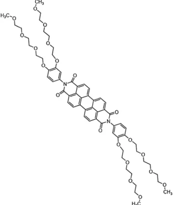
phase evaporated under vacuum and crude product was puri-fied by column chromatography. The schematic structure of the
employed fluoroionophore is shown in Fig.1.
(CH2Cl2: MeOH, 10:1). FT–IR (cm−1): 2922, 2867, 1704
and 1663 (imide group), 1595, 1512, 1455, 1404, 1361,
1299,1255, 1178, 1124. 1H NMR (CDCl3; δ, ppm): 8.68–
8.59 (q, 8H, ArH (perylene)), 7.20–7.02 (6H, ArH),4.15
(8H, ArOCH2–), 3.84 (8H, ArOCH2CH2–), 3.7 (8H,
−OCH2–CH2OCH3), 3.63–3.61 (16H, −OCH2CH2O–), 3.5
(8H,−CH2OCH3), 3.33 (12H,−OCH3), C64H74N2O20.
The polymers of ethyl cellulose (with an ethoxy content of 46 %) and poly (methyl methacrylate) were purchased from Acros and Aldrich companies, respectively. The plasticizer, dioctyl phthalate (DOP) was supplied from Aldrich. The ionic liquid, 1-butyl-3-methylimidazolium tetrafluoroborate
(BMIMBF4) and potassium tetrakis-(4-chlorophenyl) borate
were supplied from Fluka. All of the solvents and other
chemicals (AAS standards or nitrate salts of the Li+, Na+,
K+, Ca2+, Ba2+, Mg2+, NH4+, Ni2+, Co2+, Cu2+,Pb2+, Al3+,
Cr3+,Mn2+, Sn2+, Hg+, Hg2+, Fe2+and Fe3+) were of
analyt-ical grade and purchased from Merck, Fluka, and Riedel, re-spectively. Aqueous solutions were prepared with freshly de-ionized ultra pure water (specific resistance >18 MΏ cm, pH 5.5) from a Millipore reagent grade water system.
AgNO3was used for the calibration studies.
Preparation of Sensing Composites and Electrospun Nanofibers
In this study, electrospinning was chosen to fabricate the sens-ing materials. Conditions of the electrospinnsens-ing were opti-mized in order to form bead-free PMMA or EC based contin-uous fibers by varying the concentrations of plasticizer, PMMA or EC and Room Temperature Ionic Liquids (RTILs) in the composites. The sensing composites were pre-pared by mixing 240 mg of polymer (PMMA or EC), 192 mg of plasticizer (DOP), 48 mg of ionic liquid and 3 mg of PERKAT in 2.0 mL of DCM:EtOH (25:75). IL-free forms were also prepared for comparison. Then, the viscous solution was taken in a plastic syringe and an electric potential of 27 kV was applied between the needle of the syringe and the substrate coated with an aluminum foil. The distance be-tween the needle and the electrode was 10 cm while the diam-eter of the needle was 0.40 mm. Flow rate of the solution was maintained at 0.5 mL/h programming the syringe pump.
The concentration of RTIL was varied from 0.0 up to 50.0 % w/w (0.0, 5.0, 10.0, 20.0, 40.0, and 50.0 w/w), with respect to the content of PMMA or EC. It was found that the presence of the optimum amount of RTILs in the PMMA solutions facilitates the electrospinning of bead-free fibers from the lower polymer concentrations. This behavior can be attributed to the ionic conductivity and proper viscosity of the RTIL doped precursor polymer solutions. Schematic structure of the electrospinning apparatus has been Publisher
earlier [9] Fig.2reveals SEM images of EC and PMMA based
electrospun membranes under various magnifications. While, the EC based cocktails exhibiting a micro scale porous structure, the PMMA based ones were in fiber forms. In both cases, the empty spaces of the holes within the net-work structures allow diffusion of ionic silver into the plasti-cized matrix. It was observed that the electrospun membranes
Ta b le 1 F ormer st udies o n the determination of A g (I)ions in terms of the chemosen sor , an alys is me dia, w o rki n g ran ge, d etec tion li m it , and se lec tivi ty Che m ose n sing ag ent M at rix /Re sponse W o rking range LOD Sta bili ty/Re v ersibi lity se lec tivi ty Re f No Boradiazaindacenes, R at iometric chemose n sor w ith lar g e ps eudo-Stokes ’ shi ft (( λ ex ), 48 0 n m) TH F A g + (0 –10 μ M ) -N o int erfe re nce for P b 2+ ,Mn 2+ ,F e 2+,H g 2+,a n d C o 2+ [ 2 ] Peripherall y functi onali ze d zin c-p hthal ocyanine bearing b enzofu ra n d er ivative (λ ex 34 5 n m , 616 nm. TH F / Qu en ch ing in emis sio n intens ity 1.0 –50 μ MA g + --[ 3 ] Rosamine based fluo re sce n t probe ,λ ex = 520 nm. E th anol/ E nhancement in fl uorescence int en sit y 0 .0 –5.0 μ M A g+ 10 – 7M A g+ S el ec tivi ty over C d 2+,C u 2+ ,H g 2+,N a +,M g 2+,N i 2+ , Pb 2+ ,F e 3+,a n d Z n 2+) [ 4 ] Bis-py re n e deri vative bearin g two pyri dine group s (λ ex = 344 nm Ra ti ometri c res ponse in D MSO-HE P E S 0 .0 –20 μ MA g + -S el ec ti vi ty over A l 3+,C a 2+ ,C d 2+,C o 2+,C u 2+ ,F e 2+, Fe 3+,H g 2+ ,K +,L i +,M g 2+ ,M n 2+,N a +,N i 2+, Pb 2+ ,S r 2+ ,a n d Z n 2+ [ 6 ] T w o d if ferent Zn (II) phthalocyanine deriv at ives (( λ ex ) 640, (λ em ) 7 17 nm) Plasticized Ethyl cellulose (EC ) (Thin F ilm Form) 10 – 10 -1 0 − 4MA g + 7.6 × 1 0 − 12 and 2.3 × 1 0 − 11 M No int erfe re nce for Li +,Z n 2+ ,C u 2+,N i 2+ ,C o 2+ , Hg 2+,P d 2+ ,S b 3+ ,A l 3+,B i 3+ S tabel At le as t 180 days [ 7 ] Bis-triazolocou m arins o n sugar templates (( λ ex ) 345, (λ em ) 400 –550 n m S alty w at er Fluores ce nce quenchi ng Ag +(0 –10 μ M) -H igh se n sit ivi ty , n o cross sens iti vti y towards (Na +,K +,M g 2+ ,C a 2+,C u 2+ ,C o 2+ ,C d 2+,M n 2+ , Ni 2+,Zn 2+ ) [ 8 ] Azacrown[N,S,O] in to furoquinoline fluoro phore (λ ex ) 380 nm) λ em 519 nm, ra tiometi c resp onse Et hanol -High se n sit ivt iy for Ag +,N oc ro ss se n t.F o r (C r 3+ , Fe 3+,C u 2+,H g 2+,Z n 2+ ,C d 2+,a n d P b 2+, , sli ght sens it ivty for C a 2+,M g 2+ ,C o 2+,N i 2+,K +,a n d Mn 2+ [ 6 ] (1,2-bi s(4-methox ybenzyli dene) hydrazine) pol y(me thyl meth ac ryl ate) P M M A, EC and io nic li quid, 1-eth yl-3-methy limi daz o liu m tet ra fl uorobo ra te (Elec trospun n an o fiber) 10 – 12 -1 0 − 6MA g + No int erfe re nce for Li +,N a +,K +,C a 2+,B a 2+ , Mg 2+,N H4 +,N i 2+ ,C o 2+,C u 2+,P b 2+,A l 3+ , Cr 3+,M n 2+ ,S n 2+ ,H g +,H g 2+ ,F e 2+ and F e 3+ No selectivity for F −,C l −,B r −,NO3 −,N O2 −,SO 4 2− and P O4 3− [ 9 ] Fluores ce nt ly la b el ed sing le-strand ed DNA (s sDNA ) pro be that adsorb s on nano-C60, 10 mM 3-(N -m o rpholi no)propanes ulfoni c aci d (MOPS ) buf fe r cont aini ng 50 mM NaNO 3 (pH 7 .0). 0– 40 0 n M A g + 1n MA g + S ele cti v it y o v er C a 2+ ,C d 2+ ,C o 2+,C u 2+,F e 2+ ,F e 3+, Mg 2+,M n 2+ ,Ni 2+,P b 2+ ,a n d Z n 2+ , [ 10 ] Graphene oxi de ex onuclease III-assi sted si gnal amplificatio n. In the presence of A g(I), the self-h ybridization of cytos ine-rich ss -DNA la b el ed w it h the flu o re sc en t tag F A M − /Strong amplification o f fluo re sce n ce . 1– 25 0 n M 0.1 nM S el ec tivi ty over F e 3+,Z n 2+,C u 2+,H g 2+,C o 2+,C d 2+ [ 11 ] Ion-i m p rinted fl uorescent on –of f sensor/ m o nome r (E)-3_ ,6 _-bis (allyloxy)-2-((th iazol-2-yl m et hylene)amino)-4 a_ , 9a_-di hydros piro[i soin doli ne-1,9_-xant hen]-3-one Detect ion at 490 nm /p hosphat e b uf fer pH 7.4 10 to 60 μ M -S ele cti v it y o v er A l 3+,B a 2+,C a 2+ ,C o 2+,C u 2+,F e 2+ , Hg 2+,N i 2+ ,P b 2+ ,Z n 2+ [ 12 ] Polyo xy-derivat ized P eryl enedii m id e in D CM -E tOH and in the p olymers of ethyl ce ll ul ose and pol y(me thyl meth ac ryl ate) Fluores ce nce quenchi ng, Detecti on at 490 nm /i n D CM-Et O H Detect ion at 390 nm in so li d state S ensor calib ra te d w ith A g (I) con taining ace ta te buf fe r at p H 5 .5 10 − 9–10 − 3M [Ag+ ] in DCM -Et OH and 1 0 − 10–10 − 5MA g+ in so lid st ate 2.6 × 1 0 − 10 and 4.3 × 1 0 − 11 M [Ag+ ] for solu tion and so lid state, respectively S ele cti v it y o v er 1 0 − 3Mo f L i +,N a +,K +,C a 2+, Ba 2+,M g 2+ ,N H4 +,N i 2+,C o 2+ ,C u 2+ ,P b 2+, Al 3+,C r 3+,M n 2+,S n 2+,H g +,H g 2+,F e 2+ and F e 3+ ions Thi s work
made up of PMMA exhibited 3D network like structure with a random fiber orientation that was evenly distributed on the
substrate. The diameters of the fibers varied between 1.2μm
and 146 nm (See Fig.2). This type of fibrous-structure of the
electrospun membrane provided higher surface area than that of the conventional continuous thin films. Further increase of the surface area may be achieved by changing the conditions of the electrospinning process such as solvent composition, viscosity, concentration, temperature, humidity and working distance, which results in either smaller diameter fibers or increased porosity at the fiber surface.
Apparatus
vis spectra were recorded by using a Shimadzu 2101 UV-visible spectrophotometer. Emission spectra were recorded using a Varian Cary Eclipse or FLS920 instrument from Edinburg Instruments. Lifetime measurements were recorded by a Time Correlated Single Photon Counting TCSPC) sys-tem (Edinburgh Instruments (UK). The instrument was equipped with a Standard15W Xe lamp and laser or a micro-second flash lamp for steady- state and lifetime measurements, respectively. During measurements, the Instrument Response Function (IRF) was obtained from a non-fluorescing suspen-sion of a colloidal silica (LUDOX30%, Sigma Aldrich) in water, held in 10 mm path length quartz cell and was consid-ered to be wavelength independent. All lifetimes were fit to a χ2 value of less than1.1and with residuals trace symmetrically distributed around the zero axes. All of the measurements were performed at room temperature.
F i g . 1 S t r u c t u r e o f t h e N , N′ B i s ( 4 { 2 [ 2 ( 2
-methoxyethoxy)ethoxy]ethoxy}phenyl)-3,4:9,10-perylene tetracarboxydiimide (PERKAT)
Fig. 2 SEM images of EC and PMMA based electrospun membranes under various magnifications
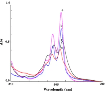
Spectral Characterization of the Fluoroionophore Absorption, excitation and emission spectra of the PERKAT were recorded in the solvents of DMF, DCM, THF, and in mixture of toluene/ethanol 80:20 (v/v), re-spectively. In all of the solvents the dye exhibited two distinct absorption maxima with high molar extinction coefficients, around 490 and 526 nm, respectively (See
Fig. 3). The dye exhibited highest absorption efficiency
in DMF. Absorption maxima observed in the visible side of the spectrum located between 487 and 530 nm
corre-sponds to π– π* singlet transitions reported for the
perylenediimide.
Spectral data of the dye were shown in Table2. Upon
excitation around 485 nm, the dye displayed emission maxima at 532, 532, 525, and 539 nm in the solvents of DMF, DCM, THF and DCM:EtOH, respectively. As can be seen from
Table2, the dye can be excited around 485 nm or further
wavelengths in the solvents. However when encapsulated in polymeric matrices the excitation maximum shifts to shorter wavelengths, 340 or 349 nm for EC and PMMA, respectively.
The highest and lowest Stoke’s shift values of 50 and 36 nm
were observed in EC and in PMMA, respectively. Due to the higher Stoke’s shift, the EC matrix was chosen for further sensing studies.
Cross Sensitivity towards Acidic or Alkaline Species In most cases the calibration standards are either in acidic form or should be acidified to prevent the precipitation of the ion under investigation. Therefore, cross sensitivity of the chromoionophore towards pH should be questioned.
We investigated effect of the acidic and alkaline species o n e m i s s i o n p e r f o r m a n c e o f P E R K AT . W e
spectrofluorimetrically titrated 10−5 M solution of the
PERKAT (in DCM) with non-aqueous HClO4 (10−3 M
in dioxane) and strong quaternary ammonium base;
tetrabutylammonium hydoxide (10−3 M in 2-propanol).
We recorded a high sensitivity both in the acidic and basic regions during titrations. However when encapsulated in
EC matrix along with ionic liquid BMIMBF4, the
acid-base sensitivity of the PERKAT diminished significantly.
Figure 4 I and II reveals acid-base response of the EC
encapsulated PERKAT towards strong acids and bases, respectively.
Even in encapsulated forms; the dye is still sensitive to strong acidity around pH 1.0. However at higher pH values of 3.0, this effect is not significant and can be overcome uti-lizing the calibration standards in the buffered solutions. The encapsulated forms of the PERKAT along with the ionic liquid exhibited both short and long term stability with respect to the IL-free forms. The tuned sensitivity and long term stability of the PERKAT in EC can be attributed to the intrinsic buffering
effect of the ionic liquid. The BMIMBF4acts like a buffer
system by neutralizing the acidic species due to the formation of weak Lewis acid–base complexes between proton and an-ionic side of the RTIL. Formation of such a buffer-like system tunes the sensitivity of the sensor and enhances the long-term stability of the indicator since it slowly acts as a sink for acidic species in the ambient air. Similar effects of the RTILs have
been observed in our previous studies [15,16].
For further metal ion sensing studies we employed buffered solutions keeping the acidity constant at pH 5.5. The effect of pH on the complexation of PERKAT with Ag (I) ions was investigated between pH 2.5 and 7.0 at fixed metal ion
con-centration of 10−5mol L−1. The relative signal change; (I0− I)/
I0produced by the Ag (I) ions was high and stable around
pH 5.5. Distribution of the chemical species in the working conditions was theoretically checked with chemical equilibri-um software programme (Visual MINTEQ). At pH 5.5
abun-d a n c e o f t h e a c e t at e ( C H3COO−( a q ) ) a c e t i c a ci d
CH3COOH(aq) and Ag
+
(aq) were 36.4 %, 66.6 % and 100.0 %, respectively. Due to the excellent solubility of silver, the acetic acid/acetate buffered solutions of pH 5.5 were cho-sen as working moiety further studies.
Silver Uptake into the Mats and Fluorescence Based Response
When doped into the EC matrices along with the anionic ad-ditive, potassium tetrakis-(4-chlorophenyl) borate, the PERKAT dye becomes a Ag (I) selective probe. In this sys-tem, silver ions are selectively extracted into the porous films (mats) by the anionic additive meanwhile potassium ions dif-fuse from the membrane into the aqueous phase due to the Fig. 3 Absorption spectra of the PERKAT in different solvents a) DMF
mechanism of ion-exchange. Physical aspect of the response mechanism of PERKAT dye can be explained by the
follow-ing ion-exchange pathway shown in Eq. (1).
PERKATð Þ pinkorg ð Þþ 2Kþð Þorg þ 2TpCIPB−ð Þorg
þ 2Agþð Þ↔PERKATAgaq 2þðorgÞ colorlessð Þ þ 2TpCIPB− org ð Þþ 2Kþð Þaq ð1Þ
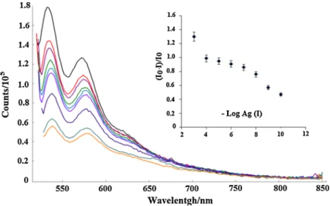
Figures5and6show the change in fluorescence spectra of
solution and electrospun materials as a function of different concentrations of silver ions. Upon exposure to Ag (I) ions, both the solution and EC based electrospun materials exhibited sim-ilar fluorescence signal change in direction of fluorescence quenching. Signal drops observed at the emission maxima of 534 nm or 572 nm can be followed as the analytical signal for
the concentration range of 10−9–10−3M in solution phase. In EC
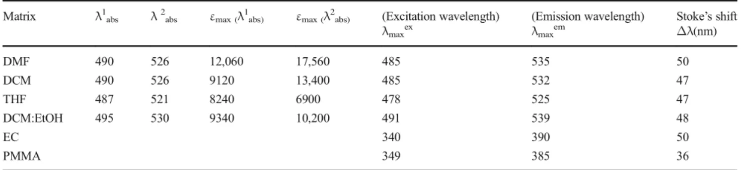
Table 2 Absorption and emission based spectral data of the dye acquired in different moieties
Matrix λ1abs λ2abs εmax (λ1abs) εmax (λ2abs) (Excitation wavelength)
λmaxex (Emission wavelength) λmaxem Stoke’s shift Δλ(nm) DMF 490 526 12,060 17,560 485 535 50 DCM 490 526 9120 13,400 485 532 47 THF 487 521 8240 6900 478 525 47 DCM:EtOH 495 530 9340 10,200 491 539 48 EC 340 390 50 PMMA 349 385 36
Fig. 4 I: Acid base sensitivity of PERKAT encapsulated in EC in
the pH range of 1.0–7.0. II: pH
range of 7.0–11.0. Buffer
solutions were prepared with
0.01 M CH3COOH, 0.01 NaOH,
0.01 M NaH2PO4, and 0.01 M
based forms the signal quenching at 390 nm is very stable and repeatable. The working range shifted to lower concentrations,
and exhibited a more liner response between10−10 -10−5M
Ag(I) when the dye was embedded in the EC (See Figs.5and
6). The calibration curves were plotted by taking the mean
values of four different solutions (n = 4) of the same medium. Detection limits in solution and PERKAT doped sensing
films were found to be 2.6 × 10⁻¹⁰and 4.3 × 10⁻¹¹M,
respec-tively (The LOD values have been calculated utilizing concen-tration of the metal ion giving a signal equal to average of the blank signal (for n = 20) plus three standard deviations). The
data given in Table1indicate that, the LOD associated with the
proposed sensing material for Ag(I) ion is undoubtedly superior with respect to that of most of other silver probes. The intensity decrease is known to be due to the quenching of the PERKAT by Ag (I) ions. In many instances the fluorophore can be quenched both by collisions and by complex formation. The
intensity based data (I0/I) or (I0− I)/I0exhibiting an
upward-concave curvature towards the y-axis is the evidence of
combined quenching both by collisions (dynamic quenching) and by complex formation (static quenching) with the same
quencher [17]. In case of dynamic quenching the collisions
between the quencher and the fluorophore affect only the excit-ed state of the fluorophore, no changes in the absorption or excitation spectrum are expected. On the contrary, the formation of ground-state complex in static quenching will perturb the absorption spectra of the fluorophore.
Thus, a careful examination of the absorption spectrum would be helpful to distinguish static and dynamic quenching.
Figure7depicts the absorption spectrum of PERKAT dye in
the absence and presence of Ag (I) ions (See spectrumBa^ and
Bb^). Dramatic changes observed in the absorption integral upon exposure to ionic silver reveal formation of a non-fluorescent complex with Ag (I) in the ground state. When the linear shape of the fluorescence intensity based response data were evaluated together, the quenching mechanism
be-tween PERKAT and Ag (I) can be concluded as Bstatic^
quenching. Measurement of fluorescence lifetimes also Fig. 5 Fluorescence response of
the (a) Ag-free solution in
DCM-EtOH, (b) 10−9M, (c) 10−8M, (d)
10−7M, (e) 10−6M, (f) 10−5M,
(g) 10−4M, (h) 10−3M Ag (I).
Inset: calibration plot for the
concentration range of 10−9to
10−3M Ag (I)
Fig. 6 Excitation–emission
based response of the dye-doped EC based electrospun mats to Ag (I) ions at pH 5.5. (a) Ag-free
buffer, (b) 10−10M, (c) 10−9M,
(d) 10−8M, (e) 10−7M, (f)
10−6M, (g) 10−5M Ag (I). Inset:
calibration plot for the
concentration range of 10–10 to
supported our findings. Figure8compares decay curves of the Ag (I)-free and Ag (I) containing solutions of the PERKAT. Bi-exponential decay times of 2.15 ± 0.1 (7.2 %) and 13.4 ± 0.1 (92.8 %) nanoseconds were measured for the Ag-free forms. The decay times of 2.14 ± 0.1 (7.3 %) and 13.6 ± 0.1 (92.7 %) nanoseconds reported for Ag containing forms can be concluded as the evidence of static quenching.
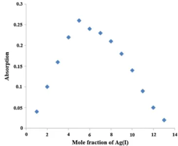
The method of continuous variation (Job’s method) was
used to determine thestoichiometryof the binding and a 1:2
complex formation of PERKAT: 2Ag+ was suggested,
consistent with Job’s plot analysis (See Fig.9).
Selectivity
Calcium, magnesium, potassium and sodium, which are the most abundant ions in natural water samples as well as phys-iologically relevant species and could be potential interferents for determination of the Ag (I). In chemosensing based ap-proaches, affinity of chromoionophores for the above Fig. 7 Absorption spectra of the PERKAT in DCM: EtOH (20:80) a: Ag(I) free b: Ag (I) containing moieties
Fig. 8 Decay curves of PERKAT in DCM: EtOH (20:80) in the absence and presence of the ionic silver
Fig. 9 Job’s plot based on the absorption intensity of PERKAT-Ag(I) at
4 8 6 n m i n D C M : Et O H so l u t i o n (2 0 : 8 0 v / v ) , [ P E R K AT +
Ag(I)] = 100μmol L−1
Fig. 10 Response of EC based fibers for the 10−3M concentrations of
metal ions at pH 5.5. II: Response of the same composition towards conventional anions and proton
mentioned metal ions is still a problem. In order to reveal the selectivity of the proposed method on ionic silver, the influ-ence of a number of cations were investigated.
Tests were performed in presence of 10−3M of Li+, Na+, K+,
Ca2+, Ba2+, Mg2+, NH4+, Ni2+, Co2+, Cu2+,Pb2+, Al3+,
Cr3+,Mn2+, Sn2+, Hg+, Hg2+, Fe2+ and Fe3+ ions in acetic
acid/acetate buffered separete solutions at pH 5.5. From
Fig.10-I, it can be concluded that, the sensing membranes are
capable of determining Ag (I) with a high selectivity over other ions. The fluorescence was dramatically quenched in the
pres-ence of Ag + at 390 nm exhibiting an I0/I ratio of 41. The
inter-ference effects of the anions of F-,Cl−, Br−, NO3−, NO2−,SO4
2−
PO43−and proton were also tested. The sensing agents exhibited
negligible signal changes when exposed to all of the conventional
anions at pH 5.5 (See Fig.10-II). Only very high concentrations
of proton caused a considerable signal change which can be overcome using buffered solutions under test conditions. Response Time, Regeneration and Stability
The sensor exhibited a very fast but non-reversible response
towards 10−4M of Ag (I) in solution phase studies. The response
time (τ90) was less than 30 s. However, in solid state the
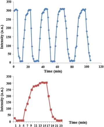
re-sponse was fully reversible and only a slight drift (1.89 %) on the upper signal level has been observed after 15 cycles. Response
and regeneration experiments were carried out utilizing 10−4M
Ag (I) containing CH3COOH/CH3COO−buffer (10−3M) and
slightly acidic 0.1 M EDTA solutions, alternatively. Approximately100% regeneration performance was succeeded with 0.1 M thiourea. However due to the toxicity considerations,
for further regeneration treatments 0.01 M CH3COOH/
CH3COO−buffered EDTA solutions (at pH: 4.5) were preferred.
The average response and regeneration times for EC based struc-tures were measured as 3.5 and 7.5 min (n = 15). Between the 1st and 15th cycles, the level of reproducibility achieved was quite good and exhibited a SD of 309.5 ± 6.7 and RSD% 2.2 for upper
signal level, respectively (see Fig.11). Figure11also reveals
short term stability of the encapsulated forms of PERKAT. We
have demonstrated that the BIMIMBF4doped sensor fibers and
mesoprorous slides exhibited a stable and reproducible response for silver measurements for a large concentration range. The sensing composites were left in the lab atmosphere in a dessicator and long term stabilities were tested within certain time intervals, during 8 months. There was no significant signal drift or instability in their response to oxygen even after 14 months. Our long term stability tests are still in progress.
Conclusion
Herein we report sensing properties of N,N′
-Bis(4-{2-[2-(2-methoxyethoxy)ethoxy] eth- oxy}phenyl) -3,4:9,10-perylene tetracarboxydiimide (PERKAT) towards ionic silver as well as other potential interferants. In this work, we performed coupling of polymeric electrospun materials with fluorescence-based measurement technique without scattering and other side ef-fects. We performed to measure the Ag (I) concentrations as
low as 10−11M exploiting the dye along with solid state
mate-rials. Our sensing approach resulted with large linear working
ranges extending to 10−11–10−5mol L−1Ag (I). Utilization of
the ionic liquid within the matrix enhanced the long term stabil-ity of the molecule considerably. Further efforts will focus on exploring new sensing materials and polymer compositions, controlling the size of the electrospun membranes, and optimiz-ing the sensitivities for the detection of a variety of analytes.
Acknowledgments Funding this research was provided by Scientific
Research Funds of Dokuz Eylul University and the Scientific and Technological Research Council of Turkey (TUBITAK).
References
1. EPA (Environmental Protection Agency) (1980) Ambient water
quality criteria for silver. EPA 4405–80-071. Office of Water Regulations, Washington DC
2. Coskun A, Akkaya EU (2005) Ion sensing coupled to resonance
energy transfer: a highly selective and sensitive ratiometric fluores-cent chemosensor for Ag (I) by a modular approach. J Am Chem
Soc 127:10464–10465
Fig. 11 The response and regeneration dynamics of the EC based
3. Kandaz M, Güney O, Senkal FB (2009) Fluorescent chemosensor for Ag (I) based on amplified fluorescence quenching of a new phthalocyanine bearing derivative of benzofuran. Polyhedron 28:
3110–3114
4. Iyoshi S, Taki M, Yamamoto Y (2008) Rosamine-based fluorescent
chemosensor for selective detection of silver (I) in an aqueous
so-lution. Inorg Chem 47:3946–3948
5. Shamsipur M, Alizadeh K, Hosseini M, Caltagirone C, Lippolis V
(2006) A selective optode membrane for silver ion based on fluo-rescence quenching of the dansylamidopropyl pendant arm deriva-tive of 1-aza-4,7,10-trithiacyclododecane([12]aneNS3. Sensors
Actuators B Chem 113:892–899
6. Wang H, Xue L, Qian Y, Jiang H (2010) Novel ratiometric
fluores-cent sensor for silver ions. Org Lett 12:292–295
7. Topal SZ, Gürek AG, Ertekin K, Yenigul B, Ahsen V (2010)
Fluorescent probes for silver detection employing phthalocyanines in polymer matrices. Sens Lett 8:1–8
8. He XP, Song Z, Wang ZZ, Shi XX, Chen K, Chen GR (2011)
Creation of 3,4-bis-triazolocoumarinesugar conjugates via flourogenic dual click chemistry and their quenching specificity with silver(I) in aqueous media. Tetrahedron 67:3343–3347
9. Kacmaz S, Ertekin K, Suslu A, Ozdemir M, Ergun Y, Celik E,
Cocen U (2011) Emission based sub-nanomolar silver sensing with electrospun nanofibers. Sensors Actuators B Chem 153:205–213
10. Li H, Zhai J, Sun X (2011) Highly sensitive and selective detection
of silver (I) ion using nano-C60 as an effective fluorescent sensing
platform. Analyst 136(10):2040–2043
11. Lang M, Li Q, Huang H, Yu F, Chen Q (2016) Highly sensitive
exonuclease III-assisted fluorometric determination of silver(I) based on graphene oxide and self-hybridization of cytosine-rich
ss-DNA. Microchim Acta 183:1659–1665
12. Sun H, Lai JP, Lin DS, Huang XX, Zuo Y, Lia YL (2016) A novel
fluorescent multi-functional monomer for preparation of silver
ion-imprinted fluorescent on–off chemosensor. Sensors Actuators B
Chem 224:485–491
13. Langhals H, Ismael R, Yuruk O (2000) Persistent fluorescence of
perylene dyes by steric inhibition of aggregation. Tetrahedron 56:
5435–5441
14. Birel OH, Zafer C, Dincalp H, Aydin B, Can M (2011) Highly
soluble polyoxyethylene-perylene diimide: optical, electrochemical and photovoltaic studies. J Chem Soc Pak 33(4):562–569
15. Oter O, Ertekin K, Derinkuyu S (2008) Ratiometric sensing of CO2
in ionic liquid modified ethyl cellulose matrix. Talanta 76:557–563
16. Oter O, Ertekin K, Topkaya D, Alp S (2006) Emission-based optical
carbon dioxide sensing with HPTS in green chemistry reagents: room-temperature ionic liquids. Anal Bioanal Chem 386:1225–1234
17. Lakowicz JR (2006) Principles of Fluorescence Spectroscopy, 3rd