Ankara Üniv Vet Fak Derg, 57, 259-261, 2010
Short Communication / Kısa Bilimsel Çalışma
Treatment of rostral mandibular body fractures in 3 cats
Mehmet SAĞLAM1, M.Alper ÇETİNKAYA2
1Ankara University, Faculty of Veterinary Medicine, Department of Surgery, Ankara; 2Hacettepe University, Faculty of Medicine,
Medical and Surgical Research Lab., Ankara, Turkey.
Summary: Three cats which had rostral mandibular body fracture just distal to canine teeth; formed the material of the study.
Partial mandibulectomy and acrylic external fixator techniques were used in cases for treatment. The purpose of this study is to present some techniques that are useful for treatment of these fractures in cats.
Key words: cat, fracture, mandible, oral and maxillofacial surgery
Üç kedide rostral corpus mandibulae kırığının sağaltımı
Özet: Canin dişlerin hemen gerisinde rostral corpus mandibula kırığı bulunan 3 kedi çalışmanın materyalini oluşturdu.
Olgularda sağaltım amacıyla rostral parsiyel mandibulektomi ve akrilik eksternal fiksatör teknikleri kullanıldı. Bu çalışmanın amacı, kedilerde bu tarz kırıkların sağaltımında kullanılan bazı yöntemleri sunmaktır.
Anahtar Sözcükler: kedi, kırık, mandibula, oral ve maksillofasiyal cerrahi.
An animal with a mandibular or maxillofacial fracture generally has been subjected to significant trauma. Treatment must not only include fracture fixation, but also include the soft tissue, the dentition and the maintenance of nutrition. Central nervous system should carefully be evaluated and observed (3 - 5, 8, 10). Principles of treatment of mandibular fractures are: to establish accurate anatomic reduction, rigid fixation and correct occlusion, re-established nutrition, to avoid excessive operative trauma to soft tissue, damage to teeth and neurovascular structures with implants, and indiscriminate extraction of teeth (7 – 11). Mandibular fracture repair techniques in cats have limited use, because cats do not tolerate many techniques. Location and shape of fracture and presence of teeth also effect the application of many techniques (4, 7, 8).
The purpose of the study reported here was, to present mandibulectomy and acrylic external fixator (AEF) techniques that can be useful for treatment of these fractures in cats.
Three cats which had rostral mandibular body fracture just distal to canine teeth formed the material of the study. After clinical examinations; V/D (ventrodorsal), D/V (dorsoventral) and 20° oblique L/L (laterolateral) radiographical images were assessed, and bilateral fracture in a cat and unilateral fracture in two cats were determined.
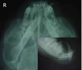
Figure 1- Preoperative radiographs of case no. 1. Şekil 1- Olgu no. 1’in preoperatif radyografileri.
Case no.1: The case had bilateral communitive fracture around canine teeth and it was treated by bilateral rostral partial mandibulectomy (Fig. 1 and 2). Communitive part of rostral mandibular body was resected bilaterally with canine teeth from mucosal gingival attachments. Closure of soft tissue over the resection stumps was achieved without tension.
Case no.2: The case had unilateral communitive and open fracture around left canine tooth and the fixation
Mehmet Sağlam - M. Alper Çetinkaya 260
was achieved by AEF technique. However the fixator was removed because of the infection at the fracture site and in pin tracts. Then case was treated by unilateral rostral partial mandibulectomy. Left rostral mandibular body including canine tooth and infected bone tissue was resected in 11th day (Fig. 3).
Figure 2- Postoperative 13th day (a) and 46th day (b)
radiographs of case no. 1.
Şekil 2- Olgu no. 1’in postoperatif 13. gün (a) ve 46. gün (b) radyografileri.
Figure 3- Postoperative radiographs of case no.2; AEF was removed because of developed infection (a) and unilateral rostral partial mandibulectomy was performed (b).
Şekil 3- Olgu no. 2’nin postoperatif radyografileri; enfeksiyon nedeni ile uygulanan AEF uzaklaştırıldı (a) ve unilateral rostral parsiyel mandibulektomi uygulandı (b).
Figure 4- Preoperative (a) and postoperative (b) radiographs of case no. 3.
Şekil 4- Olgu no. 3’ün preoperatif (a) ve postoperatif (b) radyografileri.
Figure 5- Postoperative 84th day radiographs of case no. 3 (a)
and (b), white arrow shows traumatized canine tooth root in intraoral dental film (b).
Şekil 5- Olgu no. 3’ün postoperatif 84. gün radyografileri (a) ve (b), intraoral diş filminde (b) beyaz ok canin diş kökündeki lezyonu göstermekte.
In the first (Fig. 2) and second cases a 0.8 mm kirschner wire was performed transversally to achieve hemimandibular stability until bony union occurred in rostral mandibular region; to maintain normal occlusion between upper and lower teeth; and to prevent abnormal temporomandibular joint laxity and reduce joint degeneration.
Case no.3: The case had unilateral open fracture just distal to canine tooth and the fracture was stabilized by AEF. Kirschner wires (0.8 mm) were applied, avoiding tooth roots and neurovascular structures. Acrylic was prepared and moulded with hand, and placed over the wires 0.8 cm away from skin, before toughened (in doughty stage). Fracture ends were held in reduction and normal occlusion about 10-15 minutes until the acrylic polymerized (Fig. 4 and 5).
Postoperative oral antibiotics (amoxicillin-clavulanic acid combination) and antiseptics (Chlorhexidine gluconate %0.2) were applied for a week. Antiseptics were also used following fixation removal for 3-4 days. Liquid diets for the first 4-5 days, then soft food feeding were recommended. Cases had been evaluated clinically and radiographically until clinical and functional healing was occurred.
Rostral partial mandibulectomies were well tolerated and functional recovery was obtained in both cases with in 2nd day followed by operation. Transversal
pins were removed in 46th day in 1st case and 24th day in
2nd case. Functional recovery was obtained in both cases.
In these cases, there was no easily visible cosmetic imperfection on the operation site after completely growth of clipped hairs.
Where there are multiple small fragments of fractured bone and broken teeth, an alternative approach for comminuted fractures is partial mandibulectomy. Mandibulectomy has been recommended for the management of fractures where primary repair is likely to
Ankara Üniv Vet Fak Derg, 57, 2010 261
fail because of the presence of extensive trauma, bone loss or infection, or in cases where primary repair has already failed and resulted in an inability to eat or drink (4, 6 - 8).
Acrylic external fixator (case no. 3) was well tolerated and the cat was able to use its mouth for eating soft foods within 24 hours following the fracture fixation. During the follow-up period, occlusion and fracture stability were excellent and no complications were identified. The fixator was removed 84th day after
fracture healing. In this case, there was only left canine tooth trauma (Fig. 5b) due to cranial applied pin which had been used as a full pin just distal to canine teeth.
Fractures of rostral mandibular body just distal to canine teeth can be treated with external fixation, interdental wire and intraoral splint applications (1, 2, 4, 8, 9). Many external fixation devices are heavy and bulky for cats. Use of AEF has many advantages; include, price, weight and flexibility; however, the technique is useless in bilateral fractures of this region because of regional anatomy (canine teeth roots, the fibrocartilaginous symphysis and insufficient bone surface around canine teeth).
In conclusion, numerous techniques can be used for the fixation of mandibular body fractures, but many of these techniques can not be useful for rostral fractures just distal to the canine teeth, because of the specific structures of regional anatomy and the small shape of head in cats. In our study we would like to present some techniques for the treatment of rostral mandibular body fractures, and their results for clinicians that are interested in this subject.
References
1. Aron DN (1998): Acrylic pin splint external skeletal
fixators for mandibular fractures. 980-984. In: M.J. Bojrab
(Ed), Current Techniques in Small Animal Surgery. Williams & Wilkins Co, Baltimore.
2. Bennett JW, Kapatkin AS, Manfra Maretta S (1994):
Dental composite for the fixation of mandibular fractures and luxations in 11 cats and 6 dogs. Vet Surg, 23, 190-194.
3. Brinker WO, Piermattei DL, Flo GL (1997): Fractures
and luxations of the mandible and maxilla. 659-675. In:
W.O. Brinker, D.L. Piermattei, G.L. Flo (Eds), Handbook of Small Animal Orthopedics and Fracture Repair. WB Saunders Co, Philadelphia.
4. Egger EL (1993): Skull and mandibular fractures. 1910-1921. In: D. Slatter (Ed), Textbook of Small Animal Surgery. WB Saunders Co, Philadelphia.
5. Fossum TW (1997): Maxillary and mandibular fractures. 767-778. In: T.W. Fossum (Ed), Small Animal Surgery. Mosby-Year Book Inc, St.Louis.
6. Lantz GC, Salisbury SK (1987): Partial mandibulectomy
for treatment of mandibular fractures in dogs: eight cases (1981-1984). J Am Vet Med Assoc, 191, 243-245.
7. Sağlam M, Çetinkaya MA (2003): Clinical studies of
orthopaedic treatments of maxillar and mandibular traumatic lesions in cats. JTVS, 9, 5-10.
8. Scott HW (1998): The skull and mandible. 115-132. In: A. Coughlan, A. Miller (Eds), Manual of Small Animal Fracture Repair and Management. British Small Animal Veterinary Association, Hampshire.
9. Smith MM, Kern DA (1995): Skull trauma and
mandibular fractures. Vet Clin North Am Small Anim
Pract, 25, 1127-1148.
10. Taylor RA (1998): Surgical repair of mandibular
fractures. 977-980. In: M.J. Bojrab (Ed), Current
Techniques in Small Animal Surgery. Williams & Wilkins Co, Baltimore.
11. Turner TM (1995): Fractures of the skull and mandible. 171-179. In: ML Olmstead (Ed), Small Animal Orthopedics. Mosby-Year Book Inc, St.Louis.
Geliş tarihi: 10.06.2008 / Kabul tarihi: 07.01.2010
Address for correspondance:
Mehmet Sağlam, DVM, PhD
Ankara Univesity, Faculty of Veterinary Medicine Department of Surgery 06110 Diskapi – Ankara TURKEY E-mail: [email protected]