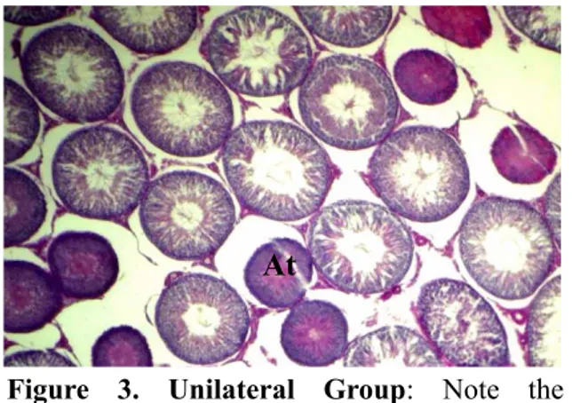
Effects of unilateral and bilateral epididymectomy on testes of rats
Tam metin
Şekil

Benzer Belgeler
In the present study, the average lethal concen- tration of Ni (96-hours LC 50 ) for Gambusia holbrooki was determined as 16.811 mg/l. Gambusia species can rapidly adapt
In this chapter we explore some of the applications of the definite integral by using it to compute areas between curves, volumes of solids, and the work done by a varying force....
In the construction industry, one of the risk areas is lack of planning and management on the project budget, project time, and the project scope in the briefing
In fact, even though I exaggerated, I mention “the Anatolian Journal of Cardiology owes its level to devoted efforts of serious referees and frequent trainings of authors provided
Objective: The objective of the study was to research the oxidant and antioxidant activity of Thymoquinone (TQ) in the testicular tissue of Reserpinized rats. Methods: Eighteen
have reported significantly lower plasma cholesterol level in rats fed protein deficient diet compared with rats fed control diets (13, 5, 19), others reported not
The group of paclitaxel + cisplatin combination therapy, was not found to exhibit a statistically significant difference regarding reduction in the number of primary follicles
“second” stages of labor, and age, gravida, parity, gestational week, caesarean indication, hospital stay, blood transfusion requirement, maternal outcomes such as intraoperative and
