Adıyaman Üniversitesi Sağlık Bilimleri Dergisi, 2020;6(3):304-310 doi:10.30569.adiyamansaglik.788181
Bu eser, Creative Commons Atıf-GayriTicari 4.0 Uluslararası Lisansı ile lisanslanmıştır. Telif Hakkı © 2020 Adıyaman Üniversitesi Rektörlüğü
Research Article/Özgün Araştırma
The evaluation of age and gender related changes of the choanae height and
width sizes with computed tomography
Bilgisayarlı tomografi ile choanae yükseklik ve genişlik ölçümlerinin yaş ve
cinsiyete bağlı değişimlerinin değerlendirilmesi
Mahmut ÖKSÜZLER1 , Sema ÖZANDAÇ POLAT2 , Ayşe Gül KABAKCI2
1Department of Diagnostic and Interventional Radiology, Private Medline Adana Hospital, 01360, Adana-Turkey 2Department of Anatomy, Faculty of Medicine, Çukurova University, 01380, Adana-Turkey
Atıf gösterme/Cite this article as: Öksüzler M, Özandaç Polat S, Kabakcı AG. The evaluation of age and gender
related changes of the choanae height and width sizes with computed tomography. ADYÜ Sağlık Bilimleri Derg. 2020;6(3):304-310. doi:10.30569.adiyamansaglik.788181
Abstract
Aim: It is the evaluation of height and width
morphometry of choanae according to age and gender in healthy Turkish population between 18-80 years old.
Materials and Methods: 281 subjects (140 males and
141 females) having brain Computed Tomography in the Radiology Department, participated in this study. ANOVA Test, Paired Samples T test and Pearson Correlation analysis were used to determine the relation and significance between measurements.
Results: The overall means and standard deviations of
the choanae height (right/left) and choanae width (right/left) were found as 24.29±3.35mm/24.20±3.23mm,
13.17±1.43mm/12.76±1.43mm in females, respectively whereas, the same measurements were 22.97±3.12mm/23.01±3.28mm;
13.14±1.67mm/12.60±1.38mm in males, respectively. When analyzed the values, there were an increase from 18 years to 80 years (from decade 2 to decade 8) in choanae heights of two sides (right/left).
Conclusion: The knowledge of the choanae ideal and
reference values may play a key role in success of the surgical procedure.
Keywords: Choanae height; Choanae width; Choanal
atresia.
Öz
Amaç: 18-80 yaş arası sağlıklı Türk popülasyonunda
yaşa ve cinsiyete göre kohana yükseklik ve genişlik morfometrisinin değerlendirilmesidir.
Gereç ve Yöntem: Çalışma, Radyoloji bölümünde
Bilgisayarlı Tomografi görüntüleri bulunan 281 (140 erkek ve 141 kadın) katılımcıdan oluşmaktadır. Ölçümler arasındaki ilişki ve anlamlılığı belirlemek için ANOVA Test, Paired Samples T test ve Pearson korelasyon analizi kullanılmıştır.
Bulgular: Kohana yüksekliği (sağ/sol), kohana
genişlik (sağ/sol) ölçümlerinin ortalama ve standart sapma değerleri kadınlarda sırasıyla; 24,29±3,35mm/24,20±3,23mm,
13,17±1,43mm/12,76±1,43mm iken erkeklerde aynı değerler sırasıyla, 22,97±3,12mm/23,01±3,28mm; 13,14±1,67mm/12,60±1,38mm bulunmuştur. Ölçümler analiz edildiğinde 18 yaşından 80 yaşına kadar (2. de kattan 8. de kata) her iki tarafta (sağ/sol) kohana yüksekliğinde artış bulunmuştur.
Sonuç: Kohana yükseklik ve genişlik ölçümlerinin
ideal ve referans değerlerinin bilinmesi cerrahi süreçlerin başarısında anahtar rol oynayabilir.
Anahtar Kelimeler: Kohana yüksekliği; Kohana
genişliği; Kohanal atrezi.
Yazışma Adresi/Address for Correspondence: Ayşe Gül KABAKCI, Department of Anatomy, Faculty of Medicine, Çukurova
University, 01380, Adana-Turkey, E-mail: [email protected]
Geliş Tarihi/Received:02.09.2020 Kabul Tarihi/Accepted:15.10.2020 Yayım Tarihi/Published online:03.12.2020
https://dergipark.org.tr/tr/pub/adiyamansaglik
Bu makale araştırma ve yayın etiğine uygun hazırlanmıştır. intihal incelemesinden geçirilmiştir.
305 Introduction
Nasal cavity which is the beginning section of respiratory system, opens anteriorly via the nostril. It connects posteriorly with nasopharynx via the choanae. It is divided with nasal septum into two sections as right and left. Nasal cavity lateral wall forms three structures named conchae and turbinates.1 Choanae is valued as posterior nasal aperture. Nasal airway keeps up with posterior nares. Air from nasal cavity continues into the lungs via the choanal apertures. Choanal atresia (CA) is defined as the anatomical closure of the posterior choanae in the nasal cavity. In childhood, choanae may be congenitally closed. Also, bilateral or unilateral CA nasal obstruction may develop. Bilateral CA is frequent during the first 6 weeks of life. Therefore, bilatateral CA is thought as an immediate in pediatric subjects.2 The development of choanae occurs between the 4th and 11th weeks of gestation.3 CA is a rarely congenital disease. Also, it ends up the developmental failure of the posterior nasal cavity to link up the nasopharynx. The incidence of CA is 1 per 5000–8000 birth. Posterior choanae anatomic borders involves the under of the body of the sphenoid bones from superior, the medial pterygoid lamina laterally, the vomer from medial, and the palatal bone horizontal part inferiorly. The main restriction may be caused by one of these bony components. It is important to determine exactly the point of choana blockage for surgical success.4-6 There are few studies about choanae morphometry.1,7-9 Additionally, these studies were about chonal atresia and ages of subjects ranged from 1 to 18 years. So, this is the first study about age of between 18-80 and gender related changes of the choanae morphometry in Turkish population.
This study was aimed to exhibit the normal values of choanae width and height dependent on age groups (18-80) and gender in healthy Turkish population.
Materials and Methods
This study was carried out 281 healthy subjects (140 Males; 141 females) aged between 18 and 80 years. This study was a
retrospective observational study. This study was approved by Clinical Researches Ethics Committee (Decision No:2019/100-86). All the test procedures were performed after ethics committee approval according to the Helsinki Declaration of Principles and and the measures were done in Department of Radiology at Medline Hospital in Turkey. This study was a retrospective observational study carried out in Department of Radiology at Medline Hospital in Turkey and subjects who were admitted to the hospital for various reasons between January 2014 and December 2019 were participated. Subjects who participated in the study signed a voluntary consent form. All CT scans were obtained using a 64x2-slice multidetector CT (Siemens
Somatom Definition AS, Siemens
Healthcare). The coronal image was used to obtain the morphometry (width and height) of the choanae. Exclusion criteria for this study; history of tumors or pathology of the nasal septum and choanae. Having surgery related to rhinoplasty or septoplasty and serious medical complications.
The data were divided into both two groups as healthy female and male subjects and six groups according to ages (Group I, 18-30 years; Group II, 31-40 years; Group III, 41-50 years; Group IV, 51-60 years; Group V, 61-70 years; and Group 6, 71-80 years). Estimations were expressed as millimeters. Measurements were performed with two sides as right and left.
These parameters were as follows:
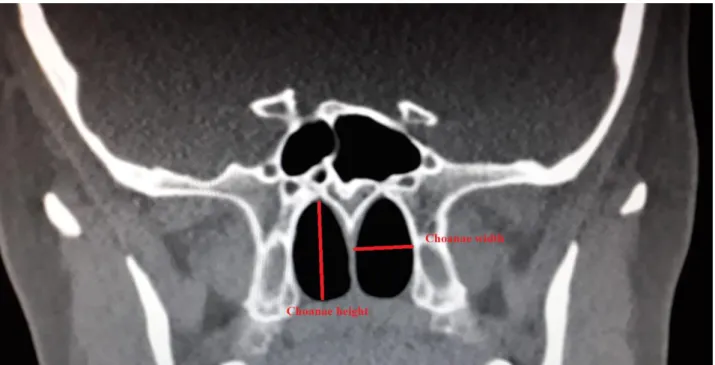
Choanae width: The distance between medial and lateral walls of the choanae in midline was measured (Figure 1).9
Choanae height: The distance between superior and inferior walls of the choanae in vertical plane was measured (Figure 1).9
Kolmogorov Smirnov Test was used to determine whether the data showed normal distribution or not and to decide which test to use (parametric or non-parametric tests). According to test result, ANOVA was chosen from parametric tests. Also, Independent's Sample T test were used for comparison of choanae heights and widths of two sides. Pearson Correlation analysis was performed
306
for relation between two sides of parameters. The SPSS 21.0 program was used for statistical analysis. From these measurements, means, standard deviations (SD), minimum
and maximum values were calculated. In all statistical analyses; p value under 0.05 was considered as statistically significant.
Figure 1. Choanae height and choanae width measurements. Results
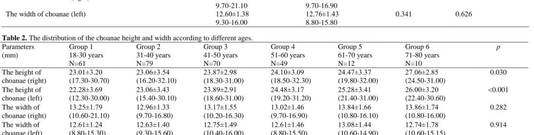
The values of minimum, maximum, mean and standard deviation of the choanae width and height calculated in 281 healthy subjects (141 males and 140 females) were shown in Table 1-2. The measurement comparisons of right and left sides were given in Table 1. All measurements were higher in females than males. Also, there were significant difference in choanae height (p=0.001, right; p=0.002, left), however there were no significant different in the choanae width measurements (p>0.05) between female and male subjects (Table 1). The significant difference was no found in the width of choanae (right) (p=0.282) and the width of choanae (left) (p=0.914) between Group 1 (18-30 years), Group 2 (31-40 years), Group 3 (41-50 years), Group 4 (51-60 years), Group 5 (61-70 years) and Group 6 (71-80 years) while there was a significant difference in the height of choanae (right) (p=0.030) and the height of choanae (left) (p<0.001) (Table 2). In evaluation of the comparison of measurements as right and left sides, there were a significant difference in choanae width of both right and left sides. In correlation analysis, a significant and very high correlation was found in the heights of
choanae (r=0.912) and a significant and moderate correlation in the widths of choanae (r=0.626) (Table 1). Additionally, in males and females, the lowest value of right choanae height was obtained in 38 years in males (16.20 mm) and 36 years in females (16.80 mm). The highest value of same measurement was found in 66 years in males (32.00 mm) and in 53 years in females (33.30 mm), respectively (Table 2).
Discussion
Choanae are holes positioned at postero-superior part of the hard palate. These holes open the nasal cavity. This is separated with vomer in the middle.10 The height of choanae is a signal for the the sphenoid bone bottom in a neonate and it shows the vertical boundary.6,11 The knowledge of regional anatomy is important for the safe implementation of bilateral choanal atresia repair and although skull base injuries are uncommon, they correspond to one of the most serious potential intraoperative complications of choanal atresia repair. This risk increases by unusual anatomy or bony defects.6,11,12 It was stated that the reason for Blacks’ achievement in many sports branches more than Whites may be related to the size
307
of choanae diameters in relation to apertura piriformis in a study conducted by Aksu et al.9 For this reason, it can be thought that subjects having high choanae width may have more physical capacity in direct proportion. But, a study investigated relation between physiological
respiration and choanae diameter was not found.9,13 The decrease in physical capacity as she/he gets older may induce the decrease in choanae width. However, such a relationship was no found between age and choanae width in this study.9,14
Table 1. The choanae measurements according to gender.
Parameters Males (140) Females (141) p Correlation (r)
The height of choanae (right) 22.97±3.12 16.20-32.00
24.29±3.35 16.80-32.30
0.001 The height of choanae (left) 23.01±3.28
15.40-31.20
24.20±3.23 16.50-31.00
0.002 0.912 The width of choanae (right) 13.14±1.67
9.70-21.10
13.17±1.43 9.70-16.90
0.861 The width of choanae (left) 12.60±1.38
9.30-16.00
12.76±1.43 8.80-15.80
0.341 0.626
Table 2. The distribution of the choanae height and width according to different ages.
Parameters (mm) Group 1 18-30 years N=61 Group 2 31-40 years N=79 Group 3 41-50 years N=70 Group 4 51-60 years N=49 Group 5 61-70 years N=12 Group 6 71-80 years N=10 p The height of choanae (right) 23.01±3.20 (17.30-30.70) 23.06±3.54 (16.20-32.10) 23.87±2.98 (18.30-31.00) 24.10±3.09 (18.50-32.30) 24.47±3.37 (19.80-32.00) 27.06±2.85 (24.50-31.00) 0.030 The height of choanae (left) 22.28±3.69 (12.30-30.00) 23.06±3.43 (15.40-30.10) 23.89±2.91 (18.60-31.00) 24.48±3.17 (19.20-31.20) 25.28±3.41 (21.40-31.00) 26.00±3.20 (22.40-30.60) <0.001 The width of choanae (right) 13.25±1.79 (10.60-21.10) 12.96±1.33 (9.70-16.80) 13.17±1.55 (10.20-16.30) 13.02±1.46 (9.70-16.90) 13.84±1.66 (10.80-16.10) 13.86±1.74 (10.80-16.00) 0.282 The width of choanae (left) 12.61±1.24 (8.80-15.30) 12.63±1.40 (9.30-15.60) 12.75±1.49 (10.40-16.00) 12.61±1.46 (8.80-15.50) 13.08±1.44 (10.60-14.90) 12.74±1.78 (10.60-15.15) 0.914
Choanal atresia is described as a defect in the development of communication between the nasal cavity and nasopharynx. An atresia is determined in every 5-8 thousand live births. It is a life -threatening condition if double sided. A unilateral choanal atresia may not be recognized until an advanced age. Therefore, the relationship between the right and the left side was especially evaluated in our study. Furthermore, incomplete atresia are defined as choanal stenosis.16,17 Therefore, cohanal diameter (cohonal height and width) measurements using imaging methods are important in the diagnosis of cohanal
atresia. Few studies about choanae size with choanal atresia such as study performed with 9 subjects having choanal atresia aged ranged from newborn to 1 year the choanae width and healthy subjects aged ranged from newborn to 1 year by Aslan et al.4 Another study compared the choanae height and choanae width parameters of two sides (right and left) in the Western Anatolian population by Aksu et al.9 A study measured the choanae height and width of two sides in subjects aged between 1 and 18 years by Ertekin et al.1, a different study of Fitzpatrick et al.6 evaluated choanae width and height in
308
Australian patients having bilateral choanal atresia. Another study done with United Kingdom population aged between 18 and 73 years by Violaris et al.8 and the study performed with 72 children by Violaris et al.7 Moreover, a different study performed with dried skull of Polat et al’s13 were evaluated (Table 3). The lowest values of righ and left sides were obtained in the age of 1 in both females and males, whereas the highest values were measured in the age of 18 in females; in the age of 16 in males in both right and left sides.1 The highest value was found in the age of 17 in males and females, whereas, the lowest value was obtained in the age of 1 years in males an females.1 In this study, in males and females, the lowest value of right choanae height was obtained in 38 years in males (16.20 mm); and 36 years in females (16.80 mm). The highest value of same measurement was found in 66 years in males (32.00 mm); and in 53 years in females (33.30 mm), respectively. In males and females, the lowest value of left choanae height was obtained in 31 years in males (15.40 mm); and 36 years in females (16.50 mm). The highest value of same measurement was found in 59 years in males (31.20 mm); and in 50 years in females (31.00 mm) respectively. In choanae width measurements of right side, the lowest value was obtained in 35 years for males (9.70 mm) in 51 years for females (9.70 mm), whereas the highest value was measured in 29 years of males, in 53 years for females (16.90 mm). In left choanae width the lowest value was measured as 9.30 mm in males (in 31 years), 8.80 mm in females (in 51 years). The highest value of left choanae widths were 16.00 mm in males (in 48 years); 15.80 mm in females (in 43 years). Due to these data, we found differences in the mean values of choanae height and width of United Kingdom, Australian and Turkish population, United Kingdom population with our population (Table 3). Interestingly, when dried skull choanae findings of Turkish population were compared with this study findings, our choanae height and width results were similar to West Anatolian population. Additionally, choanae width findings of Anatolian
population in Polat et al’s13 study with dry skulls were lower than ours, whereas choanae heights were greater than our study. These discrepancies can a result of such factors like materials (dried bone), and demographic variables including age, gender and race. When the other studies performed with subjects in the range from newborn to 1 years were evaluated, we observed that our choanae width and height findings were greater.
Conclusion
As a result, according to this study data, important differences between the Turkish population and other nations were found. Also, the differences may be originated from age, gender and race. A detailed knowledge about choanae size of normal healthy subjects is great importance in evaluating the structures of choanae in clinical and pathological situations. Clinically, it is known that highly sensitive anatomical knowledge is required in corrective rhinoplasty operations performed by breaking nasal passage bone structures, especially after traffic and sports accidents In this respect, knowing the average values in adult individuals may contribute to aesthetic surgery. Moreover, Cohanal diameters (cohanal height and width) are important in the diagnosis of cohanal atresia caused by cohanal stenosis. For this reason, we think that knowing the normal cohanal diameters (cohanal height and width) in healthy individuals in our study will guide the diagnosis of cohanal atresia. Also, we think that this study will also help to perform endoscopic nasal surgeries safely.
Limitations of the study; Subjects with cohanal atresia were not taken in this study. The limited number of literature on subjects with cohanal atresia was compared with the healthy population measurements in our study. Therefore, it is recommended to make measurements with subjects with choanal atresia compare them with the healthy population, and to conduct studies with more people.
309 Table 3. The distribution of the choanae measurements according to literature
Literature studies
Width (mm) Height (mm)
Male (Right)
Female (Right) Male (Left)
Female (Left)
Male (Right)
Female (Right) Male (Left)
Female (Left) Aslan et al. 4
(age of 1)
0.94±0.16 (with choanal atresia) 1.32±0.14 (healthy subjects) Aksu et al.9 13.09 13.33 24.45 23.77 Ertekin et al.1(age of 1) 7.45 7.29 7.46 7.31 9.73 8.64 9.85 8.61 Ertekin et al.1(age of 18) 12.64 12.17 12.17 12.17 21.78 20.73 21.72 20.67 Fitzpatrick et al.6 1.67±3.14 3.68±2.93 Violaris et al.8 1.5 cm 1.5 cm 2.6 cm 2.5 cm
(range from 0.9 cm to 2.2 cm) (range from 1.5 cm to 4.0 cm) Violaris et al.7 3 (under 1 year old)
6.1 (above 1 years of age)
5.5 (under 1 year old) 9.3 (above 1 years of age)
Polat et al.13 12.10 11.90 26.40 26.80
Present study 13.14±1.67 13.17±1.43 12.60±1.38 12.76±1.43 22.97±3.12 24.29±3.35 23.01±3.28 24.20±3.23
Ethics Committee Approval
This study was approved by Clinical Researches Ethics Committee (Decision No:2019/100-86).
Informed Consent
Subjects who participated in the study signed a voluntary consent form.
Author Contributions
Idea, design, collection of resources, analysis and interpretation of results and literature, written and critical: MÖ, SP and AGK.
Acknowledgments
We would like to thank for our gone but not forgotten Prof. Dr. Ahmet Hilmi Yücel supervising on us.
Conflict of Interest
There is no conflict of interest among the authors.
Financial Disclosure
There is no financial disclosure.
Statements
These research results have not previously been presented.
Peer-review
Externally peer-reviewed.
References
1. Ertekin T, Değirmenci M, Nisari M, Unu E, Coşkun A. Age-related changes of nasal cavity and conchae volumes and volume fractions in children: a stereological study. Folia Morphologica. 2016;75(1):38-47.
2. Thiagarajan B, Kothandaraman S. Choanal atresia a literature review. Webmed Central: ENT Scholar. 2012;3(11):1-8.
3. Dunham ME, Miller RP. Bilateral choanal atresia associated with malformation of the anterior skull base: embryogenesis and clinical implications. Annals of Otology, Rhinology & Laryngology. 1992;101(11):916-9.
310
4. Aslan S, Yilmazer C, Yildirim T, Akkuzu B, Yilmaz İ. Comparison of nasal region dimensions in bilateral choanal atresia patients and normal controls: A computed tomographic analysis with clinical implications. International Journal of
Pediatric Otorhinolaryngology. 2009;73:329-35.
5. Kwoong KM. Current updates on choanal atresia. Frontiers in
Pediatrics. 2015;3:52-8.
6. Fitzpatrick NS, Bartley AC, Bekhit E, Berkowitz RG. Skull base anatomy and surgical safety in isolated and charge-associated bilateral choanal atresia. International Journal of
Pediatric Otorhinolaryngology. 2018;115:61-4.
7. Violaris NS, Pahor AL, Chavda S. Objective assessment of posterior choanae and subglottis. Rhinology. 1994;32:148–50. 8. Violaris NS, Patel K, Chavda S, Pahor AL. Does nasal septal
deviation influence adult posterior choanal size. Rhinology. 1994;32:84-6.
9. Aksu F, Mas NG, Kahveci O, Çırpan S, Karabekir S. Apertura Piriformis ve Choana Çapları: Anatomik Bir Çalışma. Dokuz
Eylül Üniversitesi Tıp Fakültesi Dergisi. 2013; 27(1):1-6.
10. Yücel AH. Dere Anatomi Atlası ve Ders Kitabı. 7th Ed. Adana, Akademisyen Yayınevi, 2018.
11. Hughes DC, Kaduthodil MJ, Connolly DJA, Griffiths PD. Dimensions and ossification of the normal anterior cranial fossa in children. American Journal of Neuroradiology. 2010;31 (7): 1268-72.
12. LaCour JB, Patel MR, Zdanski C. Image-guided endoscopic and microdebrider assisted repair of choanal atresia in a neonate. International Journal of Pediatric Otorhinolaryngology. 2009;4(1):21-4
13. Polat S, Kabakcı AG, Yücel AH. The investigation of anatomy of the choana and airway in dry bone skull. 4th International Multidisiplinary Studies Congress. Proceeding Book Kyrenia. 2018:83-9.
14. Hommerich CP, Riegel A. Measuring of the piriform aperture in humans with 3D-SSD-CT-reconstructions. Annals of
Anatomy. 2002;184:455-9.
15. Sweeney KD, Deskin RW, Hokanson JA, Thompson CP, Yoo JK. Establishment of normal values of nasal choanal size in children: comparison of nasal choanal size in children with and without symptoms of nasal obstruction. International Journal
of Pediatric Otorhinolaryngology. 1997;39:51-7.
16. Sarıca S, Altınışık M, Bilal N, Orhan İ. Choanal Atresia: Is a Stent Necessary? Turkish Journal of Pediatric Disease. 2017; 2: 108-11.
17. Bakır S, Özbay M, Kınış V, Gün R, Yorgancılar E. Bilateral choanal atresia in adults. Kulak Burun Bogaz Ihtisas Dergisi. 2014;24(2):114-7.