and kidney damage in an experimental
model of obstructive jaundice
Ann Ital Chir, 2020 91, 1: 122-130pii: S0003469X20030511 Epub Ahead of Print 15 October 2019
free reading: www.annitalchir.com
Pervenuto in Redazione Febbraio 2019. Accettato per la pubblicazione April 2019
Correspodnce to: Tolga Dinc,Ankara Numune Eğitim ve Araştırma Hastanesi, Genel Cerrahi Servisi, Anafartalar Mh, Tolga Dinc, Talatpaşa
Omer Vefik Ozozan*, Tolga Dinc**, Veli Vural***, Candan Ozogul°, Mehmet Mahir Ozmen°°
Faruk Coskun**
*Istinye Univesity, Department of General Surgery, Istanbul, Turkey.
**Ankara Numune Training and Reserach Hospital, Department of General Surgery, Ankara, Turkey. ***Akdeniz University, Department of General Surgery, Antalya, Turkey.
°Gazi University, Department of Histology and Embryology, Ankara, Turkey. °°Private Liv Hospital, Department of General Surgery, Ankara, Turkey.
An electron microscopy study of liver and kidney damage in an experimental model of obstructive jaundice.
With this experimental study we investigated the consequences of ligation of the common bile duct (CBD) on hepatic cells and on the renal ultrastructure by electron microscopy and also determine the effects after liberation of the ductus joint in order to clarify the mechanisms of renal failure commonly observed in cholestatic liver disease.
The study was conducted on 53 Wistar albino rats divided into 4 subgroups. In the comparison group (sham) we pro-ceeded to the simple laparotomy. After preparation of the common bile duct of all the rats of the four groups, and liga-tion of the duct at the level of the distal third, eight rats in each group were sacrificed on the 3rd, 7th, 10th and 14th day after surgery, taking blood samples to measure the serum levels of ALP and bilirubin, and liver and renal tissue samples for histological evaluation. In four rats of each group the common bile duct was unligated at the same deadlines to obtain free drainage of the bile for a week. At the end of this week, the rats were sacrificed by collecting blood and liver and kidney tissue samples.
RESULTS: after CBD ligation in both groups, the ALP value, total and direct bilurubin levels were proportionally
increa-sed. After duct release, bilurubin levels decreased significantly.
In group II, while large lipid granules were observed to indicate oxidative damage, mitochondrial swelling and crystals were observed after duct liberation. Areas of glycogen and normal mitochondria were observed in group IV. After duct release in this group, increases in Ito granules, lipid granules and normal mitochondria were observed, which may reflect the evolution of hepatic regeneration.
When renal tissue was examined in group II, fusion processes in the feet, thickening of the basement membrane and mesengium were observed, and mitochondrial crystals were observed in renal tissue as well as in the liver after duct release. Damage in group III and group IV was increased parallel to prolongation of jaundice and after loosening per-sistent damage with mitochondrial crystals.
CONCLUSION: Ultrastructural changes in rat liver tissue in conditions of obstructive jaundice may be reversible after
restoration of drainage. On the other hand, ultrastructural changes in renal tissue in cases of prolonged jaundice are irreversible even if the internal drainage is restored.
KEY WORDS: Bile Duct, Liver, Kidney, Obstructive Jaundice
pulmonary, renal disfunction and sepsis. The high mor-tality rates show correlation with the rise of bilirubin levels related to the duration of the obstruction 1.
Patients are especially under the risk of acute renal fai-lure during major abdominal surgery. The mechanism lying under the development of acute renal failure has not been yet established. But it has been suggested that as a result of bile acids accumulation in plasma being excreted only by the kidneys make it nephrotoxic. Introduction
Obstructive jaundice, progresses with high mortality and morbidity rates due to many serious complications like
READ-ONLY
COPY
PRINTING
of obstructive jaundice in various times and levels on the liver and kidney by electron microscopy and by inve-stigating the changes after bile duct unligation to enli-ghten the mechanism of renal failure.
Materials and Methods
EXPERIMENT GROUPS AND STUDY PLAN
After the approval of ethical committee, 53 Wistar albi-no rats with average weight of 266 (250-280) gr divi-ded into 4 subgroups. All of the rats were kept on a standard rat diet of chow and drinking water till the day of the study and were left starved the night before the study.
The rats’ anesthesias were given by intraperitoneal injec-tions of 5mg/kg Ketamine HCl (50 mg/ml ketamin hydrochloride E. Werner Lambert). The abdomen was opened by a median laparotomy under sterile conditions. Sham group underwent laparotomy. After exposing the common bile duct of all rats in group I, group II, group III and group IV(n=12), at a level one third distal of the duct ligated with 6/0 PG. Eight rats in each group are consecutively sacrificed at day 3. 7. 10 and 14th days
after ligation and blood samples are collected for serum ALP and bilirubine levels.
The groups and the procedures followed are shown below.
GROUPS
Control group (n=5): only median line laparotomy. Group I, II, III, IV (n=8): After being ligated on 3rd, 7th, 10th and 14th days the common bile duct, the rats were sacrificed and blood and tissue samples were taken from the liver and kidney.
Group Iu, IIu, IIIu, IVu (n =4): After being ligated the common bile duct was unligated and the rats were sacri-ficed on the 10th, 14th, 17th and 21st days, tissue and
blood samples were taken from the liver and kidney.
BIOCHEMICAL ANALYSIS
Blood samples were obtained by cardiac puncture. 6ml of blood was taken. 3mls of the blood was put in glass tubes for ALP, total bilirubin, direct bilirubin and indi-rect bilirubin levels. Biochemical analyses were carried out immediately. 3mls of blood was centrifuged at 3000 rpm, 4°C for 10 minutes and serum and plasma was separated. Serum total bilirubin, direct bilirubin and indirect bilirubin were studied by spectrophoto-metric methods (Hitachi Mod. 912, Boehrinder – Mannheim)
ELECTRON MICROSCOPY METHODS
Half thin sections obtained from tissue blocks were stai-ned with toluidine blue and observed by BH2 olympus photomicroscope and captured photos were evaluated. The thin cross section slices taken from the marked sam-ples were stained with lead citrate and uranyl acetate. The samples were studied by Carl-Zeis EM 900 and photos were evaluated.
Statistical Analysis
All data collected was evaluated using SPSS 21.0 for windows (SPSS Inc., Chicago, Illinois, USA). All values are shown as mean (SD). The evaluation was carried out using One-way ANOVA and Tukey posthoc tests. p<0.05 was accepted as statistically significant.
Results
During the experiment no experiment related mortality occurred. The results of biochemical and histopathologic research are given below.
EVALUATION OF BIOCHEMICAL PARAMETERS
The mean serum total bilirubin, direct bilirubin and ALP (Alkaline Phosphatase) values of the groups are given in Table I.
The differences between the total bilirubin values of the control group and group II and group III were found statistically significant (p<0.001, p<0.003). The ces between Group II and group IIu, and the differen-ces between group III and group IIIu were also found significant. After the unligation of the bile duct in both the 10th, 14th day and 17th and 21st day unligation
groups, a significant decrease in the bilirubin levels (p<0.05) was observed although the decrease was not to normal levels.
TABLEI - Total bilirubin, direct bilirubin and ALP levels of the groups.
Total Bilirubin Direct Bilirubin ALP(U/l)
(mg/dl) (mg/dl) Control 1.66(1.15) 1.0(0.0) 307.5(77.2) Group I 4.92(1.05) 4.5(0.88) 652.1(33.8) Group Iu 2.12(2.12) 1.69(1.92) 440.3(34.1) Group II 8.97(2.03) 8.45(2.18) 647.6(51.3) Group IIu 2.64(1.3) 2.09(1.38) 354.0(14.7) Group III 7.66(1.83) 6.1(1.79) 495.0(166.3) Group IIIu 2.11(3.02) 1.81(2.7) 364.0(19.46) Group IV 9.3(7.7) 1.23(2.8) 719.0(28.88) Group IVu 0.73(0.54) 0.55(0.44) 297.5(10.6)
READ-ONLY
COPY
PRINTING
PROHIBITED
The difference between the direct bilirubin levels of the control group and the groups of which the bile duct was ligated (Group II, Group III) was also found signi-ficant (p<0.001, p<0.014). The differences between Group II, group IIu, group III and the control group were also considered statistically significant (p<0.023, p<0,014).
The difference of ALP levels between the control group and group IV, and the difference between group IV and group IVu were also found significant (p<0.014, p<0.023). The ALP levels increased paralel to the serum bilirubin levels in groups ligated. After unligated it was observed that ALP levels decreased towards normal but no statistically significant difference was observed.
THE ELECTRON MICROSCOPIC ANALYSIS OF THE LIVER
The hepatocytes in group l are totally normal. The nuclear structure, the structure of the granulated endo-plasmic reticulum is normal and does not contain dila-tations. Normally dark cytoplasm indicates an active cell or apoptosis. But if the cell structure is normal, hetero-chrome and the cytosol is electron dense this is not a sign of apoptosis (Fig. 1). In Group Iu the findings are close to the control group findings. Its’ difference from Group I is that there is a few number of lipid granu-les in the cytoplasm and this can be accepted as nor-mal (Fig. 2).
In group II the mitochondria are dense due to matrix concentration, the cristae are normal. The number of
Fig. 1: GrupI: GER=granulated endoplasmic reticulum, m= mito-chondria, Uranyl acetate-lead citrate x9000.
Fig. 2: GrupIu: m=mitochondria, SER=smooth endoplazmic reticu-lum, L=Lipid granules, Uranyl acetate-lead citrate x9000.
Fig. 3: GrupII: m=mitochondria, L=Lipid granules, Uranyl acetate-Lead citrate x9000.
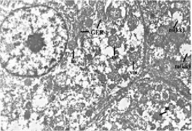
Fig. 4: Grup IIu: m=mitochondria, GER=granulated endoplasmic reti-culum, m(kk)= mitochondrial crystalysis, vm= vesicular mitochondria,
READ-ONLY
COPY
PRINTING
lipid granules is increased and the lipid granules are enlarged (Fig. 3). This indicates to increased steroid synthesis by the endoplasmic reticulum and may be a sign of oxidative injury. The dense mitochondria and the observation of lipid granules may indicate that the cel-ls are overworking.
In group IIu, the hepatocytes are normal, and no cry-stalizes was observed. Because oxidative phosphorylation cannot be performed, the tubular mitochondria have tur-ned into vesicular mitochondria (Fig. 4). This situation may indicate that antioxidant mechanisms are malfunc-tioning. In Group IIu, lipids are observed, besides nor-mal mitochondria, mitochondria with cristae loss and swallowed degenerated mitochondria are observed. At the same time the liver sinusoids are totally obstructed and collapsed, even the sinusoid endothelium is nearly
unob-servable. The lumen which is normally totally open and wide is narrowed, only to allow a single erythrocyte pass through.
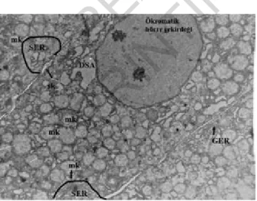
In group III, all mitochondria are swollen and there is significant mitochondrial cristae loss. The bile canaliculi are dilated and there are significant microvilli in the lumen. Also the endoplasmic reticulum cistern number is reduced, and due to mitochondrial swelling the cyto-plasm appears full with mitochondria. The rest of the organelles can not be observed due to the same reason. Also heterogeneous secondary lysosomes that phagocytes the damaged cell elements are present. This indicates to the start of phagocytosis. On the right in the light micro-scopy cross section many bile ducts are visible and pro-liferation is seen in these ducts (Fig. 5). In Group lllu the rough endoplasmic reticulum cisternae are totally
Fig. 5: Grup III: Left side ; SL= secondary lysosome, dm= degene-rated mitochondria, L= Lipid, sk= bile canaliculi , Uranyl acetate-lead citrate x13200 Right side light mikroscope , Toluidin blue.
Fig. 6: Grup IIIu: SER=smooth endoplazmic reticulum, GER= Granulated Endoplasmic Retikuculum, mk= mitochondrial crystaly-sis , sk= bile canaliculi, DSA= degenerative cytoplasmic area Uranyl acetate-lead citrate x9000.
Fig. 7: Grup IV: m=mitochondria, GER= Granulated Endoplasmic Reticulum, Uranyl acetate-lead citrate x9000.
Fig. 8: Grup IVu: left side light microscope, Toluidin blue, SK= bile canaliculi, right side EM, Uranyl acetate-lead citrate x9000.
READ-ONLY
COPY
PRINTING
dilated and all mitochondria have lost their cristae. Smooth endoplasmic reticulum is also dilated and dama-ge has increased. At the same time there are dedama-genera- degenera-tive cytoplasmic areas, the nucleus is totally euchroma-tic, healthy nuclei are not seen, and the nucleus of the cell going to necrosis and the nucleus inner and outer membrane can be totally distinguished (Fig. 6). In this group despite unligation the damage is irreversible. In group IV, the sinusoids are totally collapsed and an acinar appearance can be observed. The mitochondria are dense; the matrix is dense although the cristae are healthy (Fig. 7). The rough endoplasmic reticulum is slightly dilated in comparison to the control group and group I and there are significant areas of glycogen accumula-tion. This indicates that regeneration has started (NO effect?). In group IVu, under light microscopy neutrophil infiltration, connective tissue increase, fibroblast increa-se, collagen increase (toluidine blue), bile ducts and the portal area can be seen on the left side. The sinusoids with acinar appearance that function as secretory glands can be observed. In Ito cells lipid granules are
increa-sed by number and there are large lipid granules (Fig. 8). This shows that active cell synthesis and glycogen synthesis has begun. The cell nucleus in normal hetero-chromia, smooth endoplasmic reticulum is partially dila-ted but it is near to normal. The lipid granules are increased by number, the inner nucleus structure is nor-mal and the sinusoids are collapsed.
THE ELECTRON MICROSCOPIC EVALUATION OF THE KIDNEY
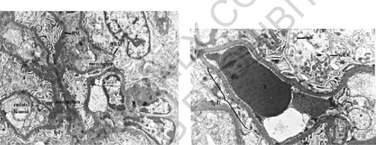
In group I the podocytes, the filtration splits foot pro-cesses (pedicules), the basement membrane, the mesan-gial cells and mesengium are totally normal (Fig. 9). The criteria for the kidneys are irregularity in the basement membrane (thickness, thinness), fibrillary appearance in the basement membrane and fusion of the foot proces-ses. The obstruction of filtration splits causes derange-ment in filtration. The endothelium, basederange-ment membra-ne, the blood-urine barrier, open filtration splits and foot
Fig. 9: Grup I: p= podosit, BM= Basement membrane, m= mezen-gial cell, Uranyl acetate-lead citrate x9000.
Fig. 10: Grup Iu: E= Eritrocyte, BM= basement membrane, fp= foot
Fig. 11: Grup II: KBM= dens basement membrane, fpf= foot pro-cess fusion, E= Eritrocyte, mk= mitochondrial crystalysis, m=mito-chondria, dm= dens mesengium.
Fig. 12: Grup IIu: BM= Basement Membrane, mk= Mitochondrial
READ-ONLY
COPY
PRINTING
processes without fusion are easily visible. The relation-ship between the endothelia cells and the tubular epithe-lia composes the basement membrane.
In group lu, partial increase of mesengium, healthy foot processes, healthy filtration splits in most areas and besi-des these fusion between filtration splits and erythrocy-tes are observed (Fig. 10). They are very similiar to the control group.
In group II, in some areas of membrane thickening is observed and in most areas there is fusion in the foot processes. When fusion occurs the filtration splits close and the ultra-filtrate cannot be filtrated. The mesengium is dense. This can be accepted as the initiation of fibro-sis. 2 erythrocytes can be seen in the field (Fig. 11). Crystalysis is observed in the podocyte’s mitochondria. In group llu, basement membrane irregularity and disin-tegration of the blood – urine barrier can be seen. Partial mitochondrial crystalysis in the podocyte cytoplasm and
degenerative cell particles in the capillary lumen are seen (Fig. 12). The damage is significant.
The basement membrane in group III is irregular just like as in thin basement membrane nephropathy. The membrane is thin in some areas and thick in other areas. The mesangium appears dense and increased (Fig. 13). The areas where the endotelium lumen and capillaries should be are totally full with cell waste. Significant fusion is present in the foot processes. Also the filtra-tion splits are obstructed. In group IIIu, crystalysis is significant in the mitochondria and the cell nucleus is euchromatic. Mesangial accumulation and protein accu-mulation are present just as in nephritis. The protein accumulation is due to the disfunction of the blood-uri-ne barrier. Also fusion is present in the foot processes. And there is significant crystalysis in the podocyte mito-chondria (Fig. 14).
In group IV, the foot process fusion is significant and
Fig. 13: Grup III: BM= Basement membrane, fpf= foot process fusion, Uranyl acetate-lead citrate x9000.
Fig. 14: Grup IIIu: mb= mesengial storage, fpf= foot process fusion, mk= mitochondrial crystalizis, m=mitochondria, ö= , euchromatic cell nucleus Uranyl acetate-lead citrate x9000.
Fig. 15: Grup IV: fpf=foot process fusion, E= Erytrocyte, GER= Granulated Endoplasmic retıculum, mk= mitochondrial crystalysis, Uranyl acetate-lead citrate x21000.
Fig. 16: Grup IVu: dv= degenerative vacuole, fpf= foot process fusion, Ö= euchromatic cell nucleus, BM= Basement membrane, Uranyl ace-tate-lead citrate x9000.
READ-ONLY
COPY
PRINTING
the rough endoplasmic reticulum cisternae are dilated, also significant mitochondrial cristae loss is present. The filtration splits are almost unobservable. The foot pro-cesses are lost and there is no barrier present. Mitochondrial crystalysis is seen (Fig. 15). In group IVu, many degenerative vacuoles and significant nucleus inva-sion is present. The fuinva-sion of the foot processes is marked. Significant protein accumulation is present in the basement membrane and the epithelium cells appear degenerated (Fig. 16). In comparison to group IV the filtration splits are more regular but in some areas no filtration splits are present.
Discussion
Obstructive jaundice is characterized by the accumula-tion of hepatotoxic agents, mitochondrial dysfuncaccumula-tion and the disintegration on the livers’ antioxidant defense system 1,2. In cholestatic liver lesions, bile salts and
bili-rubin accumulated in the intracellular compartment act like mediators causing systemic complications and lead to hepatocyte damage3. It has been proven that tissue
injury and endotoxemia cause an increase in the forma-tion of free oxygen radicals and reactive oxygen metabo-lites which leads to increased lipid peroxydation 4,5. The
accumulation of hydrophobic bile acids and the insuffi-ciency of energy production of the mitochondria are shown as the main reason of hepatotoxicity 6. Both the
accumu-lation of bile acids and the disintegration of the mito-chondrial metabolism lead to increased production of free oxygen radicals which causes oxydative injury 7,8.
Free radicals cause a series of events leading to cellular injury either directly themselves or by causing cellular antioxidan systems to malfunction 9-12.
Experimental studies have shown that obstructive jaun-dice increases lipid peroxydation, causes mitochondrial disfunction and decrease of glutatyon levels 13,14.
Many studies drawing attention to the role of free radi-cals in cholestatic liver injury have shown that the antioxidant capacity of the liver is highly affected by the duration of cholestasis. It is for this reason that the timing of surgical intervention to prevent irreversible liver injury is critical.
In this study on an experimental model of obstructive jaundice the ultrastructural changes in the liver and kid-neys have been observed under electron microscopy in short and long term (3rd, 7th, 10th and 14th days) and
after unligation (or internal drainage).
In this study the increse of bilirubin and alkaline pho-sphatase levels after ligation and the decrease after unli-gation shows that an obsrtuctive jaundice model has been created.
In studies it has been mentioned that the cause of hepa-tic injury may be the reactive oxygen radicals formed as a result of oxydative stress. Because the mitochondria is the organelle where reactive oxygen molecules are
for-med most the mitochondria is the organel where chan-ges at the cellular level occur the most. The mechani-sms that favor linking to oxidative phosphorylation in electron transport dysfunction causing autoxidation for-ming superoxides. 2-5% of the oxygen used in hepa-tocyte mitochondria is turned into reactive oxygen meta-bolites and the rest is turned into H2O. TNF-α, bile acids, ischemic-reperfusion injury, ceramides increase the formation of reactive oxygen metabolites and this increa-se blocks the electron transport system 15-18.
Although alkaline phosphatase (ALP) levels rise under many other pathologies it is actually an ectoenzyme of the plasma membrane and the rise of serum ALP levels is related to the injury of liver cell membrane injury. The reasonable rise of ALP levels may also be an indi-cation of cholestasis and cellular injury.
In early ligation groups I and Iu, histologic findings were all close to those of the control group, only in group Iu a low number of lipid granules were observed. In group II which was ligated for 7 days oxidative injury initiates. In group II, vesicular mitochondria are seen which may be due to cessation of oxidative phosphory-lation. In group II mitochondrial swelling and cristae loss is also present. In previous studies, the liver tissue damage in rats with ligated bile ducts is shown to be necrosis, mitochondrial swelling, crystalysis and apopto-sis 15,19.
The most marked liver tissue damage occurred in group III (ligated on the 10th day). In this group due to dege-nerated mitochondria other organelles cannot be distin-guished. Also in group III lysosomes that phagocyte other cellular elements are present. In group IIIu the injury can be accepted as irreversible despite unligation. In this group the mitochondria have lost their cristae.
In the final group IV mitochondria with intact cristae and glycogen accumulation areas are seen. In this group after unligation an increase of Ito granules occurs and lipid granules and normal mitochondria are observed. This may indicate to initiation of regeneration. Hepatic endothelial cells and Ito cells can synthesize nitric oxi-de by iNOS expression 20.
Any hemodynamic stimulus that can cause tension stress in the liver leads to nitric oxide release and thus initia-tes the liver regeneration cascade. Obstructive jaundice causes functional disorder in hepatocytes. In our study due to the induced regeneration on the 14th day injury has been reduced.
Patients with intra or extrahepatic biliary obstruction are under the risk of acute renal failure during major abdo-minal surgery. The mechanism lying under the renal injury during obstructive jaundice has not been enligh-tened yet. But it has been suggested that because they can only be excreted by kidneys, the bile acids accu-mulated in the plasma act as nephrotoxins 21. But
the-re is not enough evidence to support this hypothesis. In a study of Kaler et al. it has been shown that 3-4 days after the bile ducts were ligated non-spesific changes
READ-ONLY
COPY
PRINTING
occured in the proximal tubules, and that these changes were most significant when the the plasma levels of bili-rubin and bile acids were highest 21.
The importance of this study is that concurrently for-med ultrastructural changes of the kidney and liver have been investigated both in the presence of elongated jaun-dice and after unligation (or internal drenage). When the kidney tissue of 3 day ligation rats were evaluated the findings were similiar to the control group and even though fusion is present in filtration splits in some areas after unligation, the kidney histology is close to the con-trol groups. These findings show that kidney damage does not occur in the early stage of obstructive jaundi-ce and this is parallel with the study of Kaler et Al. In the 2nd group that is after 7 day ligation basement
membrane thickening in some places, fusion of the foot processes and dense mesengium is observed. The forma-tion of fusion means the ultra-filtrate is not being excre-ted and the clinical reflection can be oliguria. After unli-gation in this group disintegration of the blood-urine barrier and crystalysis of the podocyte mitochondria has been observed. The decreased excretion of acumulated bile acids in plasma due to oliguria and their effects as nephrotoxins in a longer time span despite unligation may explain this situation22-24. Also because of the
con-current liver injury the decrease of antioxidan enzyme levels will potentially increase the injury 25.
In the rats of group III the basement membranes are fairly irregular, and the findings are similiar to those of the thin basement membrane nephropathy. This shows that injury significantly increases as jaundice continues. Even after unligation injury has continued to increase. Mitochondrial crystalysis and euchromatic cell nucleus are present. In this group it has been observed that the rough endoplasmic reticuli are dilated and secondary lysosomes phagocytating intracellular waste has appeared. This shows that oxydative stress has increased and waste formed after tissue damage is trying to be digested. In group IV, the 14 day ligation group, foot process fusion is marked, the rough endoplasmic reticulum cister-nae are dilated and mitochondrial crystalysis is very signi-ficant. In this group even after unligation many dege-nerative vacuoles have been observed and endothelium injury and mesangial accumulation is significant. Interestingly in this group despite the regenerative capa-city of the liver the kidney injury can be said to be irre-versible. In one out of ten patients developing obstruc-tive jaundice acute renal failure develops after major sur-gical intervention 26. Although there are many factors in
the etiology of renal failure, there are no studies in lite-rature exploring the long term effects and electron micro-scobic changes.
Conclusion
Finally, we can say that changes observed in rat models
with obstructive jaundice can be reversible after draina-ge related to the duration of ligation. The ultrastructu-ral changes observed in the kidney are especially irre-versible in elongated jaundice despite internal drainage and this is associated with higher morbidity and morta-lity is the most defining factor.
In patients with elongated obstructive jaundice precau-tions like hydration and hemodialysis accelerating bile acid excretion could prevent the damage to reach irre-versible levels. Because all these results and interpreta-tions depend on an experimental study, it has to be sup-ported by clinical trials to explain the changes in humans.
Riassunto
Con questo studio sperimentale abbiamo indagato le con-seguenze della legatura del dotto biliare comune (CBD) sulle cellule epatiche e sull’ultrastruttura renale median-te microscopia elettronica e demedian-terminare anche gli effet-ti dopo liberazione del dotto per slegatura, al fine di chiarire i meccanismi di insufficienza renale comune-mente osservati nella malattia epatica colestatica . Lo studio è stato condotto su 53 ratti albini Wistar divi-si in 4 sottogruppi. Nel gruppo di confronto (sham) divi-si è proceduto alla semplice laparotomia. Dopo prepara-zione del dotto biliare comune di tutti i ratti dei quat-tro gruppi, e legatura del dotto a livello del terzo dista-le, otto ratti in ciascun gruppo sono stati sacrificati in 3^, 7^, 10^ e 14^ giornata dall’intervento, prelevando campioni di sangue per dosare i ivelli sierici di ALP e bilirubina, e campioni di tessuto epatico e renale per la valutazione istologica.
In quattro ratti di ciascun gruppo il dotto biliare comu-ne è stato slacciato alle stesse scadenze per ottecomu-nere il libero drenaggio della bile per una settimana. Alla fine di questa settimana i ratti sono stati sacrificati racco-gliendo campioni di sangue e di tessuti epatici e renali. Risultati: dopo la legatura del CBD in entrambi i grup-pi, il valore di ALP, i livelli di bilurubina totale e diret-ta sono risuldiret-tati proporzionalmente aumendiret-tati. Dopo la liberazione del dotto i livelli della bilurubina sono dimi-nuiti in modo significativo.
Nel II gruppo, mentre si erano osservati grossi granuli lipidici ad indicare il danno ossidativo, dopo liberazio-ne del dotto si è osservato rigonfiamento mitocondriale e presenza di cristalli. Nel gruppo IV sono state osser-vate aree di glicogeno e mitocondri normali. Dopo la liberazione del dotto in questo gruppo sono stati osser-vati aumento dei granuli di Ito, granuli lipidici e mito-condri normali, che possono riflettere l’evoluzione di una rigenerazione epatica.
Quando il tessuto renale è stato esaminato nel gruppo II, sono stati osservati processi di fusione nei piedi, ispes-simento della membrana basale e del mesengio, e dopo la liberazione del dotto sono stati osservati cristalli mito-condriali nel tessuto renale come nel fegato.
READ-ONLY
COPY
PRINTING
Il danno nel gruppo III e nel gruppo IV è risultato aumentato parallelamente al prolungamento dell’ittero e dopo la slegatura un danno persistente con cristalli mito-condriali.
İn conclusione i cambiamenti ultrastrutturali del tessuto epatico del ratto in condizione di ittero ostruttivo pos-sono essere reversibili dopo ripristino del drenaggiobilia-re. D’altra parte i cambiamenti ultrastrutturali del tes-suto renale nei casi di ittero prolungato sono irreversi-bili anche se il drenaggio interno è restaurato.
References
1. Shachleton G, SinghE Chackraborty J, Barley M: Anti-oxidant
defences in the bile duct ligated rat. Gastroenterolog, 1992. 103:
1625-629.
2. Krahembuhi S, Talos C, Fisher S, Reichen J: Toxicity of bile
acids on the electron transport chain of isolated rat liver mitochondria.
Hepatology, 1994; 19:471-79.
3. Fiori E, Macchiarelli G, Schillaci A, Lamazza A, Burza A, Paparelli C, Cavallaro A, Cangemi V: Hepatocyte Ultrastructural
aspects after preoperative biliary drainage in pancreatic cancer patients with cholestatic jaundice. Anticancer Res, 2003; 23:4859-864.
4. Sherlock S, James Dooley: Diseases of the Liver And Biliary
System. Eleventh Edition. In: Syndrome of cholestasis
Wiley-Blackwell, 2008; 223-32.
5. Zimmer MJ, Schwartz SI, Ellis H: Maingot’s Abdominal
Operations. Connecticut: Appleton and Lange, 1997; 315-13.
6. Krahembuhl S, Talos C, Lauterburg BH, Reichen J: Reduced
antioxidative capacity in liver mitochondria from bile duct ligated rats.
Hepatology, 1995; 22:607-12.
7. Sokoj RJ, Devereaux M, Khandwala RA, O’Brien K: Evidence
for involvement of oxygen free radicals in bile acid toxixity to isolated rat hepatocytes. Hepatology, 1993; 17:869-81.
8. Halliwell B, Grootveld M: The measurement of free radical
reac-tions in humans. FEBS Lett, 1987; 213:9-14.
9. William C, Meyers R, Jones S: Disorders of the Biliary System. Philadelphia: Lippincot, 1990; 303-9.
10. Gennari R, Alexander W: Effects of hiperoxia on bacteriel
tran-slocation and mortality during gut-derived sepsis. Arch Surg, 1996;
131:57-62.
11. Benjamin IS: Biliary tract obstruction. In: Blumgart LH (ed). Surgery of the liver and biliary tract. London: Longman Group Limited, 1994; 135-45.
12. Dawson JL, Stirling CA: Protective effect of mannitol on anoxic
jaundiced kidney. Arc Pathol, 1964; 78:254-59.
13. Cağlikülekci M, Pata C, Apa DD, Dirlik M, Tamer L, Yaylak F, Kanik A, Aydin S: The effect of N-acetylcysteine (NAC) on liver
and renal tissue inducible nitric oxide synthase (iNOS) and tissue lipid peroxidation in obstructive jaundice stimulated by lipopolysaccharide (LPS). Pharmacol Res, 2004; 49: 227-38.
14. Kawamura K, Kobayashi F, Kageyama F: Enhanced hepatic lipid
peroxidation in patients with primary biliary cirrhosis. Am J
Gastroenterol, 2000; 95; 3596-601.
15. Kaplowitz N: Mechanisms of liver cell injury. J Hepatol, 2000. 32: 39-47.
16. Cai J, Jones DP: Superoxide in apopitosis. J Biol Chem, 1998. 273: 11401-1404.
17. Li W, Chan AC, Lau JY, Lee DW, Ng EK, Sung JJ, Chung SC: Superoxide and nitric oxide production by Kupffer cells in rats
with obstructive jaundice: Effect of internaland external drainage.
Hepatology, 2004; 19:160-5.
18. Schoen JM, Wang HH, Minuk GY, Lautt WW: Shear
stress-induced nitric oxide release triggers the liver regeneration cascade. Nitric
Oxide, 2001; 5:453-64.
19. Liu TZ, Lee KT, Chern CL, Cheng JT, Stern A, Tsai LY: Free
radical-triggered hepatic injury of experimental obstructive jaundice of rats involves overproduction of proinflammatory cytokines and enhan-ced activation of nuclear factor kappaB. Ann Clin Lab Sci, 2001;
31: 383-90.
20. Li J, Billiar TR: Nitric Oxide. IV. Determinants of nitric oxide
protection and toxicity in liver. Am J Physiol, 1999; 276: 1069-73.
21. Kaler B, Karram T, Morgan WA, Bach PH, Yousef IM, Bomzon A: Are bile acids involved in the renal dysfunction of obstructive
jaun-dice? An experimental study in bile duct ligated rats. Ren Fail, 2004;
26: 507-16.
22. Lee SF, Huang YT, Wu WS, Lin JK: Induction of
c-junpro-tooncogene expression by hydrogen peroxide through hydroxyl radical generation and p60SRC tyrosine kinase activation. Free Radic Biol
Med, 1996; 21:437-48.
23. Bingöl F, Aydın S, Açıkgöz Ş: Free Radicals. Medic Jour of Ank Hosp, 1993; 28:1-23.
24. Hallivell B: Oxygen radicals as key mediators in neurological
disea-se: Fact or fiction? Ann Neurol, 1992; 32:10-5.
25. Laskowska-Klita T, Szumiło M: Lipid peroxidation in
hyper-trophic rat kidney. Biochim Biophys Acta, 1987; 922: 386-89.
26. Kramer HJ: Impaired renal function in obstructive jauindice: Roles
of the tromboxane and endothelin systems. Nephron, 1997; 77:1-12.