• Sonuç bulunamadı

Docking study of resveratrol like molecules on histone deacetylase

N/A
N/A
Protected

Academic year: 2021

Share "Docking study of resveratrol like molecules on histone deacetylase"

Copied!
73
0
0

Yükleniyor.... (view fulltext now)

Tam metin

(1)
(2)
(3)

2

The modulation of histone acetylation plays a pivotal role in the regulation of gene expression by governing the state of lysine residues located on the amino – terminal tails of histone proteins. A dynamic balance of histone acetylation /deacetylation is maintained by histone acetyl transferases(HAT) and histone deacetylases(HDACs) . Due to their fundamental role in gene expression, HDAC family have been associated with basic cellular events and disease states such as cell growth, differentiation and cancer information. In particular, distinct class I and II are overexpressed in some cancer disease. HDAC inhibitors structurally can be grouped into hydroxamates, cyclic peptides, aliphatic acids, benzamine’s. HDAC inhibitors (HDACi) and histone acetyl transferase activators increase histone acetylation. HDAC8 is a class I HDAC implicated as a therapeutic target for a various disease including disorder cell growth. The structure of this enzyme reaveled unique features as its conformational flexibility. The architecture of the catalytic site and the channel rim of HDAC8 adapt to accommodate various ligands according to their size, shape and chemical properties. İn this study, the native TSA ligand of HDAC8 (code 1T64) re docked, to observe the inhibition constant (Ki) with different parameter such as rotatable bonds and X, Y, Z coordinates. From this approach, starting de nova design from resveratrol compound, that is suggested to play a role in the preventation of some disease such as inhibition of tumour initiation. The resveratrol scaffold was used to create 100 hundred analogues by substituting chemical groups to observe the changes in the binding energy and inhibition constant by molecular docking using Autodock4.2. These 100 analogues were evaluated in terms of the inhibition constant (Ki) and 20 of them selected due to their lowest inhibition constant (Ki) and binding energy. The fact that HDAC8 is a promising target for cancer therapy, these molecules can be potential anticancer agents if additional laboratory assays are carried out to ascertain their inhibitory effects.

(4)
(5)
(6)
(7)
(8)

7 Figure1 .Structure of the nucleosome .

Figure2. Overview of histone modification pathway. Figure3 . Reversible actions of HAT and HDAC enzymes Figure 4 .Histone modification regulates gene expression.

Figure 5. X-ray crystal structure of bacterial histone deacetylase-like protein and TSA. Figure 6. Trichostatin A.

Figure 7 .Common pharmacophore for HDAC inhibitors.

Figure 8 . The interactions of inhibitor interface with the HDAC enzyme. Figure 9 .The proposed mechanism of HDAC family class I and II .

Figure 10 . Over all HDAC8 structure.

Figure 12. Zinc and K ions in the active site group. Figure 11. The proposed mechanism of HDAC8. Figure 13. Enlarged view of the active site.

Figure 14 Top view of a surface representation of SAHA . Figure 15 . Flowchart of distributed docking methods. Figure 16 . The structure of resveratrol.

Figure 17. HDAC8 complexed with TSA. Figure18 .The shape of grid box .

Figure19 . the resveratrol scaffold . Figure 20 . The design with the lowest Ki.

(9)

8

“I, Fatma Alnemsi, confirm that the work presented in this thesis is my own .where information has been derived from other sources, I confirm that this has been indicated in the thesis “

(10)

9

I would like to express my deepest gratitude for my thesis supervisors Prof Kemal yeleçi and Asst .Prof: şebnam Essiz Ğokhan for the continuous support of my master study and my research. This would not have been possible without the help and her guidance.

Besides my advisor, I would like to express my acknowledge to Asst. prof Hatiç bahar for her support.

I would like to thank my husband Aseed algamodi and my family and all my classmates and my friend for their endless encouragements.

(11)
(12)

11

Histone deacetylase HDAC8 is a class 1 histone deacetylase implicated as a therapeutic target in various diseases including cancer, X- linked intellectual disability, and parasitic infections. It is a multi-target for therapeutic interactions. In cancer, HDAC8 is a major (epigenetic player) that is liked to deregulated expression or interactions with transcription factors critical to tumorigenesis. In the parasite Schistosoma Manzoni and in viral infections, HDAC8 is the novel target that used to subdue the infections (1).

Based on sequence homology, HDAC8 is considered to be a class I enzyme. Although the phylogenetic analysis has shown it to depose near the boundary of the class I and class II enzymes. The enzymes of class I are mostly nuclear. Among them HDAC8 is the most documented in terms of crystallographic data(2).

Structurally, it is a well – characterized enzyme that also deacetylases non- histone proteins. The three-dimensional structure of Human HDAC8 was the first to discovered. In addition, 14 human structures co – crystallized with different inhibitors are presently attainable PDB codes (1T64, 1T67, 1T69, 1VKG, 1W22, 2W22, 2V5W, 2V5X, 3EW8, 3EWF, 3EZF, 3EZT, 3FO6, 3FO7, 3FOR). These structures have assisted to understand how catalytic activity occurs within the HDAC family of enzymes, revealing unique features of HDAC8, such as its conformational flexibility proximal to the binding site pocket .The architecture of the solved HDACs is similar . However; the organization of the loop and the distal helicases is different in HDAC8, it shows more open and accessible structures binding site than other HDAC enzymes (3).

The active site topology of HDAC8 showed large structural differences depending on which inhibitor is bound (4). It’s significance has been revealed by knockdown experiments of selective HDAC isoforms showing it as integral for cell survival. Significant differences are observed in the length and structure of the loops surrounding the active site, including the presence of two potassium ions in HDAC8 structure (5) .

To date, a number of potential HDAC inhibitors are in clinical trials. Suberoylanilide hydroxamic acid (SAHA) was the first HDAC inhibitor approved by FDA. In terms of diverse

(13)

12

chemical structures, HDAC inhibitors include a wide range of scaffolds and can be classified into structural classes such as aliphatic acids, hydroxamic acids, cyclic peptides and benzamides(6).

In this study a new technique and or algorithm will not be performed. What makes this study different is the approach. Namely to investigate a potent inhibitors of HDAC8 (code 1T64), one hundred de nova designs will be docked to 1T64 structure by using discovery studio (54) and will scanned using Autodock 4.2 against the structure HDAC8 (PDB ID:1T64) with virtual screening methodology and the new analogues will be evaluated according to the binding energy and the inhibition constant.

The goal of this study is reveal anew ligands or inhibitors against HDAC8 that could be more effective and potent in silico. This discovery of a new analogues or inhibitors will be beneficial to better understanding of cancer therapy and infection disease.

(14)

13

Chromatin is the state in which DNA is packaged within the cell. The DNA wrapped around an octamer of core histones, which is composed of two molecules each of the histones H2A, H2B, H3, and H4 called a nucleosome Fig 1 .The nucleosomal DNA is dynamically packaged to varying degrees, resulting in different levels of chromatin compaction, ranging from the 10-nm fibre to higher order structures such as the condensed mitotic chromosomes (1). These chromatin structures not only serve as essential structural elements to preserve genetic information but also play active roles in governing the accessibility of DNA sequences to transcription factors and general transcription machinery .

Fig. 1 Structure of the nucleosome (7)

The regulation of chromatin structure is one of the most intensively studied issues in the field of eukaryotic gene regulation. The most known mechanism that regulates chromatin structure is called a covalent chromatin modification on DNA or histones (8) or epigenetic modifications that are considered as inheritable changes and intimately link environmental

(15)

14

factors to our genetic code (9). Histones are modified at many sites. There are over 60 different residues on histones where modifications have been detected either by specific antibodies or by mass spectrometry (10) Fig (2). This represents a huge underestimate of the number of covalent modifications that can take place on histones such as phosphorylation, methylation and acetylation of histones and other modifications processes (11). The significant of these processes refers to their impact on the structure regulation by not merely being there , but they also recruit remodelling enzymes to reposition nucleosome . In this way, the influence of such modifications can be on transcription (12), and much fundamental biological process (9). Specific tail modifications of histones, or combinations thereof, constitute a code actual or potential transcriptional states, this called histone code (13).

Fig 2 . Overview of histone modification pathway.

Phosphorylation of histones takes a place on serines, threonines and thyrosines that controlled by kinases and phosphatases. Histone kinase transfer a phosphate group from ATP to the hydroxyl group of the target amino acid .This action is adding a negative charge to the histones (14). For instance, phosphorylation involving Ser-10 of histone H3 has emerged as an important modification, both in transcriptional activation and in chromosome condensation during mitosis (15).

Histone methylation is unique among post-translational histone modifications by virtue of its stability. It is thought to be a relatively stable and heritable epigenetic mark. That tells our genes to switch on or off, our cells whether and how to read the genes (16) for gene-specific

(16)

15

regulation (17). DNA methylation refers to the methylation of deoxy cytosine (DC) bases at the 5 position to form deoxymethylcytosine (18). The charge of histone proteins will not be affected by this modification .However, increasing methyl addition (mono, di or tri) does increase its basicity and hydrophobicity (19), Leading to a change in the chromatin structure from an open, transcriptionally active form to a more compact, inactive form, inaccessible to the transcription machinery (18) .The various methyl marks act as binding sites for other proteins that compact nucleosomes together or bring additional regulatory proteins to chromatin sites marked by methylation.(14).

The acetylation is highly dynamic and regulated by opposing actions of histone acetyl transferase (HATs) and histone de acetyltransferase (HDACs) (16) which add or remove acetyl groups to and from target lysine residues within histones. Relatively short unstructured tail of core histones consist of the targeted lysine (11) Fig (3). These tails has been conserved during evolution; therefore, the lysine positions, which are accessible to this enzymatic acetylation and identical in a wide variety of organisms ranging from, yeast to human (18).

The presence of such an acetyl-group by HAT Enzymes neutralizes a positive charge within the N-terminal part of the histone molecule (18). Thus changes the electrostatic state of lysine from positive to neutral and it also increases the size of amino acid chain (19), which could result in the destabilization of both the inter- nucleosomal interaction and the negatively charged phosphate backbone of DNA (20).This reduces the affinity of the histone tail that protrudes from the nucleosome core of DNA. As a result, chromatin adopts a more relaxed structure, unwinding of the nucleosomal arrays, and enabling the recruitment of the transcriptional machinery .This changes plays an important role in modulating gene transcription (21). In the case of acetylation marks; certain modified lysines represent specific binding surfaces for bromodomain-containing proteins, which are part of large complexes controlling chromatin architecture. These proteins represent a large class of chromatin-associated factors with at least 75 members expressed in humans (19).

(17)

16

Fig 3 . Reversible actions of HAT and HDAC enzymes

In particular acetylation, may affect the capacity of the histones to inhibit ribonucleic acid synthesis in vivo (22). This findings introduce the possibility that histone effects on nuclear RNA metabolism may involve more than a simple inhibition of RNA synthesis, and that more subtle mechanisms may exist which permit both inhibition and reactivation of RNA production at different loci “ multiple positions” along the chromosome (22) . Furthermore, the function of many transcription factors, chaperones and structural proteins depends on their acetylation state, which in turn affects a multitude of physiological pathways. Acetylation can equally affect enzymatic activities, as acetylated lysines exhibit slightly different preferences for secondary structures than un- acetylated lysines. Additionally, the acetylation of lysines can create new docking sites for protein–protein interactions, for example via recognition by the bromodomain proteins .Different covalent modifications can furthermore compete for the same lysines important for signalling or the subcellular localisation of a protein (23)

On the other hand, deacetylation by HDAC enzymes can be thought as the opposite process of acetylation, removing the targeted acetyl group. This process is associated with a more condensed chromatin state and transcriptional gene silencing. Thus increases ionic interactions between the positively charged histones and negatively charged DNA, which yields a more compact chromatin structure and represses gene transcription by limiting the accessibility of the transcription machinery (24).

(18)

17

Emerging evidence suggests that a family of histone deacetylases may exist to regulate diverse cellular functions , including chromatin structure , gene expression , cell cycle progression , and oncogenesis (25) Fig (4) . Additionally, numerous properties of proteins including DNA-protein interactions, subcellular localization, and transcriptional activity can be affected (19).) In the past few years, increasing experimental evidence has accumulated to support the view that the role of histone is to inhibit chromosomal activity (21).

(19)

18 :

Histone deacetylases (HDACs) reverse the regulatory acetylation of histone proteins, influencing nucleosome structure and altering gene transcription. Although histone deacetylases suggested to act to silence genes, but genetic experiments in Drosophila and the yeast Saccharomyces cerevisiae have indicated that deacetylase activity can contribute to gene activation as well (27).

This reversible acetylation modification (deacetylation) of lysine residues in histone proteins, and their putative role in RNA synthesis was first described in 1964 by Allfrey et al. Since that, the natural antifungal antibiotic Trichostatin A (TSA) was found to act by inhibition of mammalian histone deacetylases (HDAC) .Subsequently, the first human HDAC named HD1, a homolog of yeast transcriptional regulator Rpd3 was isolated (28).

Depending on sequence similarity and co factors dependency, HDACs are grouped into two families NAD+ dependent family and Zn⁺² dependent family. The ZN⁺² family classified into four classes based on a phylogenetic analysis. Class I enzymes comprise HDACs 1, 2, 3 and 8 (homologous to yeast Rpd3) and class II HDACs include 4–7, 9 and 10 (homologous to yeast Hda1) ;which are divided into two subclasses: IIa (HDACs 4, 5, 7, 9) with one catalytic domain and IIb (HDACs 6, 10) with two HDAC domains. HDAC11 is distinct from those in classes I, and II. It has been placed in class IV, and class III refers to the unrelated, NAD-dependent sirtuin deacetylase and insensitive to inhibition by HDAC class I/II inhibitors like TSA (3). Among zinc-dependent HDACs human HDAC8 lies on the evolutionary boundary between class I and class II HDACs and has become the best characterized experimentally (21).

From a structural point of view all HDACs enzymes share an approximately 11-Å tube –like catalytic channel with accommodates a zinc ion that is responsible for their deacetylase activity. With a few exceptions, the residues lining on this active pocket in the catalytic domain are widely conserved among different isoforms making the design of selective inhibitors. The 11- Å channel rim has been suggested to be the best place to find exploitable structural differences for the rational design of selective inhibitors or modulators (2).

In 1999, the crystal structure of a bacterial HDAC homology (histone deacetylase –like protein) (HDLP), bound to an HDAC inhibitor, trichostatin A (TSA) was reported (20) Fig 5.

(20)

19

Originally this inhibitor developed as an antifungal drug and considered as a member of a large class of HDAC inhibitors that has a broad spectrum of epigenetic activities. TSA selectively inhibits class I and II mammalian HDAC, and alters gene expression by interfering with the removal of acetyl groups from histones by HDAC and therefore alters the ability of DNA transcription factors to access the DNA within chromatin (29) Fig 6.

Fig 5. X-ray crystal structure of bacterial histone deacetylase-like protein and Trichostatin A (TSA)(30)

(21)

20

The catalytic domain of HDLP was closely related to those of both classes of HDACs (I, II). Thus the mechanism of deacetylation is presumably conserved as well, which require Zn⁺² for activity. The most common HDAC inhibitors use hydroxamic acids (19), which consist organic compounds bearing the functional group RC (O) N (OH) R’, with R and R' as organic residues and CO as a carbonyl group (30) with zinc binding groups despite unfavourable pharmacokinetic properties (27) due to their rapid clearance and severe toxicity (31) . In general, inhibitors can be grouped into hydroxamates, cyclic peptide, aliphatic acids, benzamides boronic acid -based compounds, benzofuranone and sulfanomide. Superoylanilide hydroxamic acid (SAHA) was the first HDAC inhibitors approved by FDA (32) (Food and Drug Administration) granted regular approval to vorinostat, a histone deacetylase inhibitor, for the treatment of manifestations of cutaneous T-cell lymphoma (CTCL) in patients with progressive, persistent, or recurrent disease (33). Vorinostat is structurally related to (TSA) which is indicated for the treatment of replaced and refractory (CTCL) (34).

Most of HDAC inhibitors share three common features such a common pharmacophore, consisting of a zinc-binding group (ZBG) or warhead to chelate the catalytic zinc ion, a capping group binding on the surface of the active site pocket, and a linker between the ZBG and the cap fig 7. It is generally thought that the ZBG is to a significant extent responsible for the potency and sometimes the isoform selectivity of HDAC inhibitors (31) .The zinc-binding group and polar group connected by a straight chain alkyl, or aryl linker (35). The capping group is solvent exposed and interacts with amino acids near the entrance of the active site. Linker helps for high affinity interactions with proteins Fig 8. The cyclic peptides are the most structurally complex group of HDAC inhibitors of which depsipeptide (an oligo- or polypeptide containing one or more ester bonds as well as peptide bonds) is one of the most important members (7).

(22)

21

Fig 7 ,Common pharmacophore for HDAC inhibitors(31)

Fig 8 . The interactions of inhibitor interface with the HDAC enzyme (36)

Most HDAC inhibitors such as TSA and SAHA closely resemble the aliphatic acetyl-lysine substrate, and deliver a hydroxamic acid or other zinc-binding group to the catalytic zinc ion at the bottom of a narrow active site pocket. Then hydroxamic acid coordinates zinc cation and hydrogen bound with some of the active site residues. The aliphatic chain makes

(23)

22

several van der Waals contacts with the channels residues. The cap group contacts residues on the rim of the pocket, and possibly mimics the amino acids adjacent to the acetylated lysine residues in the histone. The carbonyl oxygen of the N- acetyl amide bond is thought to coordinate to the zinc cation, and to thereby position it closely to a bound water molecule and activate it for a nucleophilic attack by the water (20) as showed in Fig 9. The majority of available HDACi drugs in and out of clinical trials inhibit all HDAC isoforms non-specifically so called pan-inhibitors (37), or non- selective. Thus, the effect of these inhibitors is often studied by examining changes in bulk histone acetylation or the therapeutic effects observed in a given experimental model or in clinical trails (34).

(24)

23 :

HDAC8 was initially discovered in 2000 by Ilse Van den Wyngaert and his team when they identified the characterization of human HDAC8 by cloning, using database searches. Such as several overlapping expressed sequence tags ESTs (39). This technique is utilizing cDNA library that is constructed from a tissue or cell line of interest. Individual clones are picked from the library, and one sequence is generated from each end of the cDNA insert. Thus, each clone normally has a 5 and 3 EST associated with it (40). As a result generate recombinant cell lines to catalyse deacetylation of a number of acetylated histone variants in vitro (39). Among the different histone deacetylase (HDAC) isozymes, HDAC8 is the most highly malleable enzyme, and it exhibits the potential to accommodate structurally diverse ligands (albeit with moderate binding affinities) in its active site pocket (41).

HDAC8 is critically involved in regulating chromatin structure and gene expression. It is either localized a sex linked gene in the nucleus as (primary site), or in the cytoplasm (1). In human tissues, it has been showed to be expressed 1.7 Kb while in tumour cell plot revealed a transcript of 2.4 Kb but in several lines (25). The detailed knowledge of its catalytic mechanism is of high importance since it has been established as a most promising target for the development of new anti-tumour drugs (21)

HDAC8 is expressed in a variety of adult caner tissue, such as colon, breast, lung, and pancreas as well as in childhood cancer tissue such as neuroblastoma and has not been identified in any major corepressor complexes, and very few (HDACi) developed to date target HDAC8 with any potency. Besides cancer, lately HDAC8 have been discovered as an important target to fight the neglected parasitic disease schistosomiasis caused by Schistosoma flatworms that affects over 200 million people. HDAC8 was shown to catalyse in vitro deacetylation of a number of acetylated histone variants. These substrates included full-length H2A/ H2B, H3, and H4 histones, acetylated at non-specific lysines. On the other hand there are some unique non-histone substrates such as cohesion (transcriptional activator proteins). And the Estragon-Related Receptor α (ERRα) .This receptor is expressed in a number of organs (42), and considered as a key regulator of growth and differentiation in a broad range of target tissues (43).

(25)

24

When structural features of HDAC8 are considered, the structure comprises a single α/β domain that includes an eight – stranded parallel β sheet sandwiched between 13 α helices. Each domain binds one zinc ion and two K⁺ ions, forming a dimer interface that buries ˷ 1,300Å of molecular surface area (44). The substrate binding surface, composed of nine loops and an 11 Å tunnel leading to the active site, is proposed to be conformationally flexible based on the poor occupancy and varying positions of the loop residues in crystal structures (45). The inhibitor mediates the dimeric arrangement in the crystal structure with two capping groups (pyridine and thiopene moieties) of each inhibitor molecule stacking against each other (44) Fig 10. The C terminal (aa50 – 112) protein-binding domain of other HDAC is not present in HDAC8 (1).

While approximately half of the amino are contained in canonical secondary structure elements, the other half of residues in HDAC8 from loops that link the various elements of regular secondary structure. These long loops emanating from the C - terminal ends of the strands of the core β sheet form a substantial portion of the volume of the entire enzyme and encompass the residues from the enzyme, active site, and catalytic machinery. These loops create a number of different conformations that bind ligands. The varied conformations provide a palette of binding sites to accommodate a multiplicity of substrates. Furthermore, long range allosteric movements propagated through the loops may affect the active site and surrounding areas, potentially altering substrate preferences (45).

(26)

25

Fig 10 . Over all HDAC8 structure

Some studies revealed that HDAC8 has a core architecture similar to that found in two enzymes; arginase, which catalyses the conversion of arginine, and (HDLP) from the hyper thermophilic bacterium (Aquifex aeolicus) with approximately 31% sequence identify with HDAC8 (32). In addition the catalytic domain discovered from HDLP which was closely related to those of both classes of HDACs (I, II). Thus the mechanism of deacetylation is presumably conserved as well, which require Zn⁺² for activity (22).

HDAC8 shows that the surface in the vicinity of the entrance to the catalytic site is very flexible and in particular the L1 and L2 loops of HDAC8 interconvert between different states. The catalytic cycle of HDAC8 involves at least three steps, including binding of substrate, cleaving of the acetylated lysine, and release of the products (46). The substrate binds to HDAC8 with the catalytic metal coordinating both the carbonyl oxygen of the acetyl

(27)

26

Lysine substrate and a water molecule. In the first step of the mechanism, His142 functions as a general base to abstract a proton from the metal-bound water that is reacting with the carbonyl carbon to form a high energy tetrahedral intermediate. The oxyanion intermediate is proposed to be stabilized by coordination with the metal ion, hydrogen bonding with Tyr306, and electrostatic interactions with positively charged groups in the active site. Proton donation from an active site general acid to the amine leaving group accompanies breakdown of the tetrahedral intermediate to form the acetate and the deacetylated lysine products (45) .These changes derive from the presence of two deep pockets one leading to the active site and a second one close to the active site and lined by Tyr306 and Phe152. The opening of the second pocket is mediated by a movement of loop L1 (46). The flexibility of zinc coordination, which allows different coordination modes and fast ligand exchange has been suggested to be one key catalytic feature of the zinc ion .This makes it an invaluable metal in biological catalysis (47) Fig 11.

Fig 11. The proposed mechanism of HDAC8.

The catalytic mechanism originally proposed for HDAC8 is based on crystal structure of the HDLP (48). In spite of their highly conserved catalytic domain, one key structural difference

(28)

27

between HDAC8 and HDLP is that the former has two K+ ion binding sites while HDLP lack (31) Fig 12.

Fig 12. Zinc and K ions in the active site group. (21)

When the ligand binding site is considered, it consist of a metal – binding domain, a linker domain, and a hydrophobic group. Recent studies have explored flexibility of the HDAC8 binding site using molecular dynamics simulations and confirmed that, indeed, the binding site of HDAC8 is rather malleable and, if required, can be subjected to induced fit technology that accounts for receptor flexibility in ligand-receptor docking by iteratively combining rigid receptor docking with protein structure prediction and refinement (49).

The inhibition of HDAC8 is strongly regulated by the surface malleability. The most promising scaffold seems to be associated with bulky linkers, which can fit into the HDAC8 groove (46). In addition, the interpretation of the binding poses may be biased due to the fact that the conformation of the HDAC ligands co-crystallized with HDAC8 is influenced by the interactions between the ligand and neighbouring copies of the protein and/ or ligand in the crystal lattice (50). Because of The topology of the unbound state very likely offers a wide pocket that accommodates to the ligand after its coordination to ZN. The inhibitor

(29)

28

coordinates the zinc in a bidentate fashion, and simultaneously contacts residues likely to be involved in catalysis (HiS-142, HİS -143, and Tyr -306 by its hydroximate moiety. The cyclic linker region of the inhibitor fits in the hydrophobic channel, with its aromatic aryl moiety stacked between side chain of Phe -152 and 208(46) Fig 13. Additionally Van Der Waals interactions occur between the linker and side chain (44).

Fig 13. Enlarged view of the active site, polar interactions are shown as dashed yellow lines (51).

In the case of small hydroxamates such as SAHA. Hydroxamates are very potent bidentate zinc chelators. The hydroxamate moiety is small enough to fit in the narrow pocket of HDAC8 leads to weaker inhibition of HDAC8 because the malleability of the protein will remove several of the hydrophobic contacts in the linker region that contribute to the binding. (52) Fig 14.

(30)

29

Fig 14 .left: Top view of a surface representation of the most favourable orientations of the cap of SAHA . Right: surface representation of the surface channel opened to allow the interaction of SAHA with Tyr306.

Selective inhibitors (HDAC8i) with characterization in advanced preclinical trails still scare. HDAC8 has high flexibility and hence is able to accommodate a range of diverse inhibitors (1).

Docking is the process by which two molecules fit together in 3D space. , which predicts the preferred orientation of one molecule to a second when bound to each other to form a stable complex .It is frequently used to predict the binding orientation of small molecule drug candidates to their protein targets in order to in turn predict the affinity and activity of the small molecule. Hence docking plays an important role in the rational design of drugs. Molecular docking may be defined as an optimization problem, which would describe the “best-fit” orientation of a ligand that binds to a particular protein of interest. The focus of molecular docking is to computationally stimulate the molecular recognition process, aiming to achieve an optimized conformation for both the protein and ligand, and relative orientation between protein and ligand such that the free energy of the overall system is minimized(53), and increasingly used for lead discover. Typically by screening a large database of organic molecules for putative ligands that fit a binding site(54).

Earlier docking methods that treat both ligands and proteins as rigid objects are called rigid docking whlie methods that consider a ligand as an articulated object, and a protein as a rigid object are named flexible docking (55).

(31)

30

Molecular docking can be seen as a problem of lock –and – key in which protein can be considered as the lock and ligand can be seen as the key. Docking program fits molecules in the site, often in hundreds or even thousands of orientations and conformations, evaluates their complementarity, and ranks each molecule relative to the rest of the database of possible ligands. Serious challenges remain, whereas the technique has had some important successes in recent years. These challenges may be divided into two categories: problems with calculating binding energies, and problems with sampling the degrees of freedom available to interacting molecules (54).

:

Two essential parts of a docking approach are a scoring function and an efficient algorithm for searching conformation space. A good scoring function should be able to distinguish a correct binding mode from other putative modes. Different ligands can be bound to a receptor in enormous conformations with varying energies (55). Docking methods typically use an energy-based scoring function to identify the most favourable ligand conformation when bound to the target. The general hypothesis is that lower energy scores represent better protein-ligand bindings compared to higher energy values (56). An accurate scoring function should reflect this observation. However, due to the approximation to the free energy, the native structure may not always have the lowest energy using the existing scoring functions(55) .A scoring function is used to rank the bindings correctly. A variety of scoring methods have been used in molecular docking. The algorithms for automated docking sampling can be divided into two broad categories: matching methods, and conformational search methods. In matching approach, the enzyme and ligand are taken as rigid objects, and the search is reduced to finding energetically or geometrically favourable configurations of the ligand within the active site of the enzyme. A conformational search method, which is considered in this study, is often to dock conformationally flexible ligands by employing a simulation or optimization method to search through the space of ligand– receptor configurations where the ligand is flexible and the receptor is fixed (55).

More recently, evolutionary algorithms have become a popular choice in molecular docking applications and performed better than simulated annealing (a general probabilistic local

(32)

31

search algorithm, proposed 20 years ago by Cerny and Kirkpatrick to solve difficult optimization problems. Many large instances of practical difficult problems were successfully solved by simulated annealing to use a simulated annealing algorithm to define a set of solution of an optimization problem. Then a neighbourhood structure is defined) (58) in some applications. An evolutionary algorithm is a generally adaptable concept for problem solving; especially well-suited for solving difficult optimization problems .It is based on ideas borrowed from genetics and natural selection. Evolutionary algorithms have been used to solve problems involving large search spaces, where traditional optimization methods are less efficient. They can be applied in varying ways to the confirmation search, structure-based design problems, and protein prediction.

In recent years, virtual high throughput screening (VHTS) has become an essential technique for the discovery of new lead compounds (the pro compounds that have biological properties to be a drug), and it has served as an alternative to experimental high throughput screening HTS (that provides a practical method to investigate large numbers of synthetic compounds in miniaturized in vitro assays to identify those capable of modulating the biological target of interest (59) in drug discovery. Virtual screening is a technique that uses computers to predict a binding mode and affinity of a given compound for a target receptor. A critical issue in receptor-centric virtual screening is to incorporate a dynamic nature of receptor structures. Commonly in molecular docking algorithms, the target protein is kept rigid in a single low-energy conformation, and only conformational and positional flexibility of a ligand is considered Fig 15. The importance of VHTS in drug discovery is increasing simultaneously with numbers of small molecules that are being available and rapidly growing in corporate and public libraries (60).

(33)

32

Fig 15. Flowchart of distributed docking methods

In this project, we aimed to design new ligands (inhibitors) of HDAC8 by exploiting resveratrol as a De nova design of our docking. Resveratrol Fig (16) (3, 5,4P-trihydroxystilbene) was the first detected in grapevines in 1976, and then in wine in 1992 .It is synthesized in the leaf epidermis and the skin of grape berries. The time in contact with grape skins is the determining factor in resveratrol extraction during the fermentation process (60). Its physiological function is not well defined but is thought to reside in the protection of the plant from environmental stress and pathogenic attack. On the other hand, it has been suggested to play a role in the prevention of heart disease, associated with red wine consumption, as it inhibits platelet aggregation, alters eicosanoid synthesis and modulates lipid and lipoprotein metabolism. Resveratrol was also recently shown to inhibit cellular events associated with tumour initiation, promotion and progression. In the light of its very weak toxicity, at least in mouse tumour models, and suggested that resveratrol should be investigated as a cancer chemo preventive agent in humans. As a polyphenol molecule, it is a radical scavenger and has been shown to inhibit cyclooxygenase activity (61).

(34)

33

Fig 16 . The structure of resveratrol

A number of clinical investigations have assessed the pharmacokinetics and metabolism of resveratrol in humans following oral ingestion either of the single synthetic agent or as a constituent of a particular food or drink. An understanding of the clinical pharmacokinetics and metabolism of resveratrol is essential to enable appropriate doses to be chosen for larger efficacy trials, for defining relevant concentrations that should be used in preclinical mechanistic studies, and for identifying metabolites that may contribute to or diminish the activity of the parent compound (63).

(35)

34

In this study, our aim is to design a new ligands for the enzyme HDAC8 (code 1T64.pdb )(64), that was complexed with TSA trichostatin A, by observing the inhibition constant of the new candidates.

We carried out following several steps to test inhibitors:

 Preparation of the protein HDAC8 (1T64) structure for docking.  Preparation of known ligand to dock.

 Re docking the native ligand and some known ligand to 1T64 as a test case.  Re Docking Native ligands From Other Structures Code of the Enzyme HDAC8  Creating a library of De Nova ligands and docking them to the structure.

As previously mentioned in the Introduction section 2.5.2, two essential parts of a docking approach are a scoring function and an efficient algorithm for searching conformation space. AutoDock uses a semi empirical force field scoring function based on the AMBER force field. It uses a molecular mechanics model for enthalpic contributions, such as VDW and hydrogen bonding, and an empirical model for entropic changes upon binding. Each component is multiplied by empirical weights obtained from the calibration against a set of known binding constants.

AutoDock uses a Lamarckian genetic algorithm for the conformational search part. For each molecule, 50 independent runs were performed. 300 distinct ligand conformers were initially generated and positioned randomly in the binding pocket. They had randomly assigned torsion angles to rotatable bonds and a randomly assigned overall rotation. A maximum of 100 million energy evaluations was allowed for each docking (65).

(36)

35

The crystal structure of human histone deacetylase, HDAC8 enzyme (PDB entry code: 1T64, complexed with the inhibitor TSA), was extracted from the Protein Data Bank (64) Fig 17. The enzyme structure was cleaned of all water molecules and the inhibitor TSA, as well as all interacting ions (except zinc) before being used in the docking studies. To relieve the crystal structure tension and to make the protein available to use in the AutoDock( docking simulation program), the protein’s geometry was first optimized and then submitted to the “Clean Geometry” toolkit of Discovery Studio(65) for a more complete check using a fast Deriding-like force field. Missing hydrogen atoms were added based on the protonation state of the titratable residues at a pH of 7.4. Ionic strength was set to 0.145 and the dielectric constant was set to 10(66).

(37)

36

3.3 Preparation of Ligand to Dock:

The 3D ligand molecule TSA were downloaded from on European bioinformatics institute (68) with experimental inhibition constant. And used to dock in our optimized structure .The AutoDock Tools (ADT) graphical user interface programs were also employed here to generate the docking input files of ligand. It assigns the Gustier partial charges to each atom and prepares two parameter files, namely a grid parameter file GPF(The grid parameter file specifies the 3D search space by setting the number of points in each dimension, the center of the grids and the spacing between points . It also specifies the types of probe atoms to use, the filename of the receptor and the names of each the output grid maps, and a docking parameter file (DPF) (67).

3.4 Re Docking of Native Ligand to HDAC8:

HDAC8 inhibitor (TSA) was re-docked in order to compare the docking accuracy (63) between the inhibition constant of (TSA) that we have found on European bioinformatics institute (70) and the Ki after relocking it in side our optimized structure. For this procedure we have prepared the gpf file with a grid box Fig (18) of X50, X50, and X50 and considered X, Y, Z coordinates as 44.186, 35.183, and 52.406 respectively. The result described in Table 1.

(38)

37

Table 1, docking results of TSA (native ligand to HDAC8)

Ligand TSA Rotatable bonds İnhibition constant µM

R - TSA

6 25 expr

E- TSA

(39)

38

Further steps, we have applied to increase the opportunity of getting candidates that are targeted to be more potent ,effective and specific than other docking studied have applied before in silico .In this step, we have docked six ligands, which have native properties of TSA, and have been considered as natural ligands of other structures of HDAC8. The process was performed by downloading the ligand PDB files from protein data bank (64). ADT used as a next step of preparing files for docking (gpf, dpf), by using the same parameters that have exploited to (TSA) ligand and the result showed in Table (2).

Note: all the results changed to Micro molar unit.

Table 2, re docking results of native ligands from other structures code of the enzyme

code Ligand structure Chemical name Binding

Energy Kcal/mole Inhibition constant Ki µM CRİ 5-(4-METHYL-BENZOYLAMINO)- BIPHENYL-3,4'-DICARBOXYLICACID 3- DIMETHYLAMIDE-4'-HYDROXYAMIDE -9.16 0.219

(40)

39

SAHA SUBEROYLANİLİDE HYDROXAMİC

ACİD -7.65 2.49 TSN TRICHOSTATIN A -7.48 3.26 NHB N-HYDROXY-4-(METHYL{[5-(2- PYRIDINYL)-2-THIENYL]SULFONYL}AMINO)BEN ZAE -8.68 0.436 SHH OCTANEDIOICACID HYDROXYAMIDE PHENYLAMIDE -6.12 32.86 B3N 4- (DİMETHYLAMİNO)-N-[7- (HYDROXYAMİNO)-7-OXOHEPTYL] BEZAMİDE -7.46 3.43

(41)

40

From making use of resveratrol as scaffold of our library Fig 19 .we have designed one hundred hits for our original ligand. For this procedure, we were using some chemical group that have showed an effect of the structure of the docks and its binding energy and the inhibition effect as well and these groups are:

Fig 19 .the resveratrol scaffold with R positions coloured with red. Oh Ch3 Och3 Cl F OC-NH3

(42)

41

As a test system, we have docked resveratrol code STL on protein data bank) (64) as the first step in order to observe the inhibition constant that we can build on .Then we built on it by adding a different chemical group to resveratrol scaffold. All these process were applied in Discovery Studio where we can govern the structure, and make the changes that we need (65). Moreover, following previous step by using the new docks on Autodock tool, where we can prepare the (gpf, dpf) files for the ligand to be ready for docking process. The last step is utilizing the new docks by docking them to 1T64 structure with the same parameters that we used for the previous docking with 10 runs. The resulting poses are observed for the binding energy value and the inhibition constant.

(43)

42

Table 3 shown the result of docking 100 ligands (STL De nova) to 1T64:

STL ligands Run Binding energy values

Kcal/mol İnhibition constant µM exp scaffold Run 10 -6.39 20.8 1 Run 6 -7.69 2.31 2 Run 1 -7.98 1.41 3 Run 2 -8.05 1.26 4 Run 2 -7.72 2.18

(44)

43 5 Run 4 -7.84 1.79 6 Run 9 -6.87 9.25 7 Run 10 -8.27 0.873 8 Run 1 -7.15 5.76 9 Run 5 -8.59 0.508 10 Run 9 -6.74 11.42

(45)

44 11 Run 1 -7.33 4.23 12 Run 4 -8.04 1.28 13 Run 5 -7.41 3.72 14 Run 10 -8.78 0.369 15 Run 5 -7.39 3.81 16 Run 5 -6.41 19.93

(46)

45 17 Run 2 -6.60 14.65 18 Run 7 -5.91 46.22 19 Run 7 -5.79 57.13 20 Run 8 -7.36 4.05 21 Run 9 -8.52 0.569 22 Run 9 -7.27 4.67

(47)

46 23 Run 5 -6.75 11.37 24 Run 9 -7.78 1.97 25 Run 9 -7.73 2.15 26 Run 3 -8.26 0.880 27 Run 4 -6.64 13.55 28 Run 9 -7.16 5.67

(48)

47 29 Run 1 -6.67 12.97 30 Run 9 -7.06 6.63 31 Run 9 -7.23 5.06 32 Run 9 -7.35 4.09 33 Run 1 -7.16 5.65 34 Run 2 -6.80 10.37

(49)

48 35 Run 8 -8.43 0.662 36 Run 9 -6.41 20.01 37 Run 5 -7.94 1.51 38 Run 5 -7.48 3.30 39 Run 7 -8.29 0.838 40 Run 5 -8.59 0.505

(50)

49 41 Run 8 -8.24 0.901 42 Run 9 -8.20 0.968 43 Run 4 -9.21 0.176 44 Run 7 -8.82 0.430 45 Run 10 -7.34 4.13 46 Run 6 -7.38 3.89

(51)

50 47 Run 10 -6.87 9.22 48 Run 4 -6.34 22.54 49 Run 8 -7.27 4.69 50 Run 10 -6.99 7.75 51 Run 7 -6.95 8.04 52 Run 6 -9.09 0.217

(52)

51 53 Run 10 -6.76 11.15 54 Run 4 -8.91 0.295 55 Run 8 -6.58 15.05 56 Run 3 -6.32 23.41 57 Run 5 -7.09 6.31 58 Run 9 -6.78 10.67

(53)

52 59 Run 7 -8.75 0.388 60 Run 2 -7.16 5.68 61 Run 7 -8.68 0.434 62 Run 8 -8.05 1.26 63 Run 2 -6.97 7.74 64 Run 6 -6.85 9.48

(54)

53 65 Run 4 -7.37 3.99 66 Run 6 -7.72 2.21 67 Run 10 -6.87 9.20 68 Run 10 -5.50 92.78 69 Run 10 -7.29 4.54 70 Run 4 -10.19 0.033

(55)

54 71 Run 8 -6.77 10.87 72 Run 3 -7.48 3.30 73 Run2 -7.37 3.95 74 Run 7 -6.23 27.05 75 Run 9 -6.34 22.46 76 Run 10 -7.59 2.74

(56)

55 77 Run 9 -7.69 2.29 78 Run 6 -7.22 5.13 79 Run 6 -7.18 5.12 80 Run 3 -5.90 47.58 81 Run 2 -7.63 2.45 82 Run 4 -6.51 16.87

(57)

56 83 Run 8 -5.56 83.9 84 Run 2 -6.82 10.05 85 Run 4 -6.46 18.52 86 Run 1 -6.25 26.16 87 Run 8 -6.88 9.08 88 Run 9 -6.96 7.93

(58)

57 89 Run 2 -7.90 1.61 90 Run 5 -7.90 1.61 91 Run 4 -7.48 3.31 92 Run 7 -7.54 2.96 93 Run 2 -7.94 1.50 94 Run 4 -7.17 5.56

(59)

58 95 Run 3 -8.88 0.307 96 Run 4 -8.31 0.816 97 Run 5 -9.95 0.092 98 Run 6 -7.90 1.62 99 Run 8 -6.92 8.45 100 Run 6 -7.70 2.25

(60)

59

First, efforts to create selective HDAC inhibitors have received increased attention over the past several years, leading to several classes – including isoform selective inhibitors (40). In this study, as have shown in Table 1, we compared the potency of our novel molecule with the native ligand of HDAC8 (PDB ID; 1T64), TSA to evaluate the inhibitory potentials. We observed deviation of the docking results from the known experimental inhibition values of HDAC8-TSA complex, with that of docking being lower. Furthermore, we perform docking with some isoform ligands of TSA that considered as native docks for other isoform structures of 1T64 as showed in Table 2. Hence we attended to compare the experimental inhibition constant of those ligands, and consider the lowest Ki and of which structure belongs for. From that, we were able to choose the best scaffold for our search.

The lowest Ki constant out of known ligands was for the ligand CRİ with (Ki= 0.21µM, with -9.16 kcal/mole), Depending on some chemical features of the structure that we considered in last step. This ligand has three phenolic groups in its structure, from which we inspired our de nova structure (resveratrol).

Lastly, resveratrol has been shown to possess inhibition (HDACs) enzyme activity. Depending on the previous step, we considered the resveratrol (code STL on PDB) as scaffold for our analogues to design selective inhibitors, which configuring of 100 analogs by using different chemical groups to compare the Ki among all. As shown in table 3, results for inhibition constant within the ligands are ranging from 0.033µM up 1.26µM to which we have twenty analogs with lowest Ki at Nano molar level (table 4) with chemical groups that accompanied at different position on STL structure.

Notably, we can see various impacts of R groups that we considered to be as a variable component in our docking process. Groups such as CH3 and Cl have shown a good effect on the Ki of the ligands by decreasing it. In contrast, the effect of NH3, F was obvious in increasing the Ki of our scaffold.

As a conclusion, we can consider this candidate as ligand design of our structure (1T64), and can apply more procedure on this analogue to ensure its capacity and efficacy, such as simulation and drug design to expertise its activity.

(61)

60

The lowest Ki constant Fig 20 that have recorded was (Ki = 0.03, - 10.19 kcal/mole) (fig 20) for the analogues connected with (Cl, F, OCH3).

Fig. 20. The design with the lowest Ki The binding interaction of the best pose is showed in Fig 21.

(62)

61

Table 4 .The best and lowest Ki ligand structures.

Ligand Run ligand Run

70 Run 4 95 Run 3 97 Run 5 44 Run 7 43 Run 4 59 Run 7 52 Run 6 61 Run 7 54 Run 4 40 Run 5

(63)

62 95 Run 3 9 Run 5 44 Run 7 40 Run 5 59 Run 7 9 Run 5 61 Run 7 21 Run 9 40 Run 5 35 Run 8

(64)

63 9 Run 5 96 Run 4 21 Run 9 39 Run 7 35 Run 8 39 Run 7 96 Run 4 7 Run 10 39 Run 7 26 Run 3

(65)

64 7 Run 10 41 Run 8 26 Run 3 42 Run 9 41 Run 8 62 Run 8 42 Run 9

(66)

65

1. Chakrabarti, A., Oehme, I., Witt, O., Oliveira, G., Sippl, W., Romier, C., Pierce, R. and Jung, M. (2015). HDAC8: a multifaceted target for therapeutic interventions. Trends in Pharmacological Sciences, 36(7), pp.481-492.

2. Deschamps, N., Simões-Pires, C., Carrupt, P. and Nurisso, A. (2015). How the flexibility of human histone deacetylases influences ligand binding: an overview. Drug Discovery Today, 20(6), pp.736-742.

3. Estiu, G., West, N., Mazitschek, R., Greenberg, E., Bradner, J. and Wiest, O. (2010). On the inhibition of histone deacetylase 8. Bioorganic & Medicinal Chemistry, 18(11), pp.4103-4110.

4. Vannini, A., Volpari, C., Filocamo, G., Casavola, E., Brunetti, M., Renzoni, D., Chakravarty, P., Paolini, C., De Francesco, R., Gallinari, P., Steinkuhler, C. and Di Marco, S. (2004). Crystal structure of a eukaryotic zinc-dependent histone deacetylase, human HDAC8, complexed with a hydroxamic acid inhibitor. Proceedings of the National Academy of Sciences, 101(42), pp.15064-15069

5. Heinke, R., Carlino, L., Kannan, S., Jung, M. and Sippl, W. (2011). Computer- and structure-based lead design for epigenetic targets. Bioorganic & Medicinal Chemistry, 19(12), pp.3605-3615.

6. Thangapandian, S., John, S., Lee, Y., Kim, S. and Lee, K. (2011). Dynamic Structure-Based Pharmacophore Model Development: A New and Effective Addition in the Histone Deacetylase 8 (HDAC8) Inhibitor Discovery. IJMS, 12(12), pp.9440-9462. 7. Gantt, S.L., Gattis, S.G. and Fierke, C.A. (2006) ‘Catalytic activity and inhibition of

human Histone Deacetylase 8 is dependent on the identity of the active site metal ion †’, Biochemistry, 45(19), pp. 6170–6178. Doi: 10.1021/bi060212u.

8. Yang, X. and Seto, E. (2003). Collaborative spirit of histone deacetylases in regulating chromatin structure and gene expression. Current Opinion in Genetics & Development, 13(2), pp.143-153.

(67)

66

9. Bohacek, J. and Mansuy, I. (2012). Epigenetic Inheritance of Disease and Disease Risk. Neuro psychopharmacology, 38(1), pp.220-236.

10. Kouzarides, T. (2007). Chromatin Modifications and Their Function. Cell, 128(4), pp.693-705.

11. Khochbin, S. and Wolffe, A. (1997). The origin and utility of histone deacetylases. FEBS Letters, 419(2-3), pp.157-160.

12. Bannister, A. and Kouzarides, T. (2011). Regulation of chromatin by histone modifications. Cell Res, 21(3), pp.381-395.

13. Turner, B. (2002). Cellular Memory and the Histone Code. Cell, 111(3), pp.285-291. 14. Arrow smith, C., Bountra, C., Fish, P., Lee, K. and Schapira, M. (2012). Epigenetic

protein families: a new frontier for drug discovery. Nature Reviews Drug Discovery, 11(5), pp.384-400.

15. Berger, S. (2002). Histone modifications in transcriptional regulation. Current Opinion in Genetics & Development, 12(2), pp.142-148.

16. Bentley-Lewis, R. (2010) ‘You are what she ate’, Science Translational Medicine, 2(28), pp. 28ec63–28ec63. doi: 10.1126/scitranslmed.3001164.

17. McManus, K., Biron, V., Heit, R., Underhill, D. and Hendzel, M. (2005). Dynamic Changes in Histone H3 Lysine 9 Methylations: IDENTIFICATION OF A MITOSIS-SPECIFIC FUNCTION FOR DYNAMIC METHYLATION IN CHROMOSOME CONGRESSION AND SEGREGATION. Journal of Biological Chemistry, 281(13), pp.8888-8897.

18. Richardson, B. (2003). Impact of aging on DNA methylation. Ageing Research Reviews, 2(3), pp.245-261.

19. McManus, K.J., Biron, V.L., Heit, R., Underhill, D.A. and Hendzel, M.J. (2005) ‘Dynamic changes in Histone H3 Lysine 9 Methylations: IDENTIFICATION OF a MITOSIS-SPECIFIC FUNCTION FOR DYNAMIC METHYLATION IN CHROMOSOME CONGRESSION AND SEGREGATION’, Journal of Biological Chemistry, 281(13), pp. 8888–8897. Doi: 10.1074/jbc.m505323200.

(68)

67

20. Rice, J. and Allis, C. (2001). Histone methylation versus histone acetylation: new insights into epigenetic regulation. Current Opinion in Cell Biology, 13(3), pp.263-273.

21. Allfrey, V., Faulkner, R. and Mirsky, A. (1964). ACETYLATION AND METHYLATION OF HISTONES AND THEIR POSSIBLE ROLE IN THE REGULATION OF RNA SYNTHESIS .Proceedings of the National Academy of Sciences, 51(5), pp.786-794.

22. López-Rodas, G., Brosch, G., Georgieva, E., Sendra, R., Franco, L. and Loidl, P. (1993). Histone deacetylase. FEBS Letters, 317(3), pp.175-180.

23. Peserico, A. and Simone, C. (2011). Physical and Functional HAT/HDAC Interplay Regulates Protein Acetylation Balance. Journal of Biomedicine and Biotechnology, 2011, pp.1-10.

24. Grozinger, C.M. and Schreiber, S.L. (2002) ‘Deacetylase enzymes’, Chemistry & Biology, 9(1), pp. 3–16. doi: 10.1016/s1074-5521(02)00092-3.

25. Wu, R., Wang, S., Zhou, N., Cao, Z. and Zhang, Y. (2010). A Proton-Shuttle Reaction Mechanism for Histone Deacetylase 8 and the Catalytic Role of Metal Ions. J. Am. Chem. Soc., 132(27), pp.9471-9479.

26. Verdin, E. and Ott, M. (2014) ‘50 years of protein acetylation: From gene regulation to epigenetics, metabolism and beyond’, Nature Reviews Molecular Cell Biology, 16(4), pp. 258–264. doi: 10.1038/nrm3931.

27. Spange, S., Wagner, T., Heinzel, T. and Krämer, O.H. (2009) ‘Acetylation of non-histone proteins modulates cellular signalling at multiple levels’, The International Journal of Biochemistry & Cell Biology, 41(1), pp. 185–198. Doi: 10.1016/j.biocel.2008.08.027.

28. Ropero, S. and Esteller, M. (2007) ‘The role of histone deacetylases (HDACs) in human cancer’, Molecular Oncology, 1(1), pp. 19–25. Doi: 10.1016/j.molonc.2007.01.001

(69)

68

29. Hu, E. (2000) ‘Cloning and characterization of a novel human class I Histone Deacetylase that functions as a transcription Repressor’, Journal of Biological Chemistry, 275(20), pp. 15254–15264. Doi: 10.1074/jbc.m908988199.

30. Fischer, A., Sananbenesi, F., Mungenast, A. and Tsai, L.-H. (2010) ‘Targeting the correct HDAC(s) to treat cognitive disorders’, Trends in Pharmacological Sciences, 31(12), pp. 605–617. Doi: 10.1016/j.tips.2010.09.003.

31. Gregoretti, I., Lee, Y.-M. And Goodson, H.V. (2004) ‘Molecular evolution of the Histone Deacetylase family: Functional implications of Phylogenetic analyses, Journal of Molecular Biology, 338(1), pp. 17–31. Doi: 10.1016/j.jmb.2004.02.006.

32. Mann, B.S., Johnson, J.R., Cohen, M.H., Justice, R. and Pazdur, R. (2007) ‘FDA approval summary: Vorinostat for treatment of advanced primary Cutaneous t-cell lymphoma’, The Oncologist, 12(10), pp. 1247–1252. Doi: 10.1634/theoncologist.12-10-1247.

33. Beckers, T., Burkhardt, C., Wieland, H., Gimmnich, P., Ciossek, T., Maier, T. and Sanders, K. (2007) ‘Distinct pharmacological properties of second generation HDAC inhibitors with the benzamide or hydroxamate head group’, International Journal of Cancer, 121(5), pp. 1138–1148. doi: 10.1002/ijc.22751.

34. Mohseni, J., Zabidi-Hussin, Z.A.M.H. and Sasongko, T.H. (2013) ‘Histone deacetylase inhibitors as potential treatment for spinal muscular atrophy’, Genetics and Molecular Biology, 36(3), pp. 299–307. Doi: 10.1590/s1415-47572013000300001. 35. Small molecule inhibitors of histone deacetylase. (2002). Expert Opinion on

Therapeutic Patents, 12(6), pp.943-947.

36. https://en.wikipedia.org/wiki/Hydroxamic_acid

37. Chen, K., Xu, L. and Wiest, O. (2013) ‘Computational exploration of zinc binding groups for HDAC inhibition’, The Journal of Organic Chemistry, 78(10), pp. 5051– 5055. Doi: 10.1021/jo400406g.

38. Somoza, J.R., Skene, R.J., Katz, B.A., Mol, C., Ho, J.D., Jennings, A.J., Luong, C., Arvai, A., Buggy, J.J., Chi, E., Tang, J., Sang, B.-C., Verner, E., Wynands, R., Leahy, E.M.,

(70)

69

Dougan, D.R., Snell, G., Navre, M., Knuth, M.W., Swanson, R.V., McRee, D.E. and Tari, L.W. (2004) ‘Structural snapshots of human HDAC8 provide insights into the class I Histone Deacetylases’, Structure, 12(7), pp. 1325–1334. doi: 10.1016/j.str.2004.04.012.

39. Wang, X., Wei, X., Pang, Q. and Yi, F. (2012) ‘Histone deacetylases and their inhibitors: Molecular mechanisms and therapeutic implications in diabetes mellitus’, Acta Pharmaceutica Sinica B, 2(4), pp. 387–395. Doi: 10.1016/j.apsb.2012.06.005. 40. Wolfsberg, T.G. and Landsman, D. (1997) ‘A comparison of expressed sequence tags

(ESTs) to human Genomic sequences’, Nucleic Acids Research, 25(8), pp. 1626–1632. Doi: 10.1093/nar/25.8.1626.

41. Thangapandian, S., John, S., Lee, Y., Arulalapperumal, V. and Lee, K.W. (2012) ‘Molecular modelling study on tunnel behavior in different Histone Deacetylase Isoforms’, PLoS ONE, 7(11), p. e49327. Doi: 10.1371/journal.pone.0049327.

42. Zhao, C., Toresson, G., Xu, L., Koehler, K.F., Gustafsson, J.-Å. And Dahlman-Wright, K. (2005) ‘Mouse estrogen receptor β Isoforms exhibit differences in ligand selectivity and Coactivator recruitment’, Biochemistry, 44(22), pp. 7936–7944. doi: 10.1021/bi047691m.

43. Bieliauskas, A.V. and Pflum, M.K.H. (2008) ‘ChemInform abstract: Isoform-Selective Histone Deacetylase inhibitors’, ChemInform, 39(40). Doi: 10.1002/chin.200840275. 44. Hodawadekar, S.C. and Marmorstein, R. (2007) ‘Chemistry of acetyl transfer by

histone modifying enzymes: Structure, mechanism and implications for effector design’, Oncogene, 26(37), pp. 5528–5540. Doi: 10.1038/sj.onc.1210619.

45. Gantt, S., Gattis, S. and Fierke, C. (2006). Catalytic Activity and Inhibition of Human Histone Deacetylase 8 Is Dependent on the Identity of the Active Site Metal Ion. Biochemistry, 45(19), pp.6170-6178.

46. Singh, R.K., Suzuki, T., Mandal, T., Balsubramanian, N., Haldar, M., Mueller, D.J., Strode, J.A., Cook, G., Mallik, S. and Srivastava, D.K. (2014) ‘Thermodynamics of binding of structurally similar ligands to Histone Deacetylase 8 sheds light on

(71)

70

challenges in the rational design of potent and Isozyme-Selective inhibitors of the enzyme’, Biochemistry, 53(48), pp. 7445–7458. Doi: 10.1021/bi500711x.

47. Wolfson, N.A., Pitcairn, C.A. and Fierke, C.A. (2012) ‘HDAC8 substrates: Histones and beyond’, Biopolymers, 99(2), pp. 112–126. Doi: 10.1002/bip.22135.

48. Vannini, A., Volpari, C., Filocamo, G., Casavola, E.C., Brunetti, M., Renzoni, D., Chakravarty, P., Paolini, C., De Francesco, R., Gallinari, P., Steinkuhler, C. and Di Marco, S. (2004) ‘Crystal structure of a eukaryotic zinc-dependent histone deacetylase, human HDAC8, complexed with a hydroxamic acid inhibitor’, Proceedings of the National Academy of Sciences, 101(42), pp. 15064–15069. Doi: 10.1073/pnas.0404603101.

49. Kunze, M.B.A., Wright, D.W., Werbeck, N.D., Kirkpatrick, J., Coveney, P.V. and Hansen, D.F. (2013) ‘Loop interactions and dynamics tune the enzymatic activity of the human Histone Deacetylase 8’, Journal of the American Chemical Society, 135(47), pp. 17862–17868. Doi: 10.1021/ja408184x.

50. Wu, R., Hu, P., Wang, S., Cao, Z. and Zhang, Y. (2010) ‘Flexibility of catalytic zinc coordination in Thermolysin and HDAC8: A Born−Oppenheimer ab Initio QM/MM molecular dynamics study’, Journal of Chemical Theory and Computation, 6(1), pp. 337–343. Doi: 10.1021/ct9005322.

51. Gantt, S.L., Joseph, C.G. and Fierke, C.A. (2009) ‘Activation and inhibition of Histone Deacetylase 8 by Monovalent Cations’, Journal of Biological Chemistry, 285(9), pp. 6036–6043. Doi: 10.1074/jbc.m109.033399.

52. Sherman, W., Beard, H.S. and Farid, R. (2006) ‘Use of an induced fit receptor structure in virtual screening’, Chemical Biology <html_ent glyph=‘@amp;’ ASCII=‘&’/> Drug Design, 67(1), pp. 83–84. Doi: 10.1111/j.1747-0285.2005.00327.x. 53. Brunsteiner, M. and Petukhov, P.A. (2012) ‘Insights from comprehensive multiple

receptor docking to HDAC8’, Journal of Molecular Modelling, 18(8), pp. 3927–3939. Doi: 10.1007/s00894-011-1297-8.

(72)

71

54. Wei, B.Q., Weaver, L.H., Ferrari, A.M., Matthews, B.W. and Shoichet, B.K. (2004) ‘Testing a flexible-receptor Docking algorithm in a model binding site’, Journal of Molecular Biology, 337(5), pp. 1161–1182. Doi: 10.1016/j.jmb.2004.02.015.

55. Chen, H.-M., Liu, B.-F., Huang, H.-L., Hwang, S.-F. And Ho, S.-Y. (2006) ‘SODOCK: Swarm optimization for highly flexible protein–ligand docking’, Journal of Computational Chemistry, 28(2), pp. 612–623. Doi: 10.1002/jcc.20542.

56. Yang, J.-M. And Kao, C.-Y. (2000) ‘Flexible ligand docking using a robust evolutionary algorithm’, Journal of Computational Chemistry, 21(11), pp. 988–998. Doi: 10.1002/1096-987x (200008)21:11<988: aid-jcc8>3.0.co; 2-h.

57. Thomsen, R. and Christensen, M.H. (2006) ‘MolDock: A new technique for high-accuracy molecular Docking’, Journal of Medicinal Chemistry, 49(11), pp. 3315–3321. Doi: 10.1021/jm051197e.

58. Ben-Ameur, W. (2004) ‘Computing the initial temperature of simulated Annealing’, Computational Optimization and Applications, 29(3), pp. 369–385. Doi: 10.1023/b:coap.0000044187.23143.bd.

59. Pereira, D.A. and Williams, J.A. (2007) ‘Origin and evolution of high throughput screening’, British Journal of Pharmacology, 152(1), pp. 53–61. Doi: 10.1038/sj.bjp.0707373.

60. Lee, H.S., Choi, J., Kufareva, I., Abagyan, R., Filikov, A., Yang, Y. and Yoon, S. (2008) ‘Optimization of high throughput virtual screening by combining shape-matching and Docking methods’, Journal of Chemical Information and Modelling, 48(3), pp. 489–497. Doi: 10.1021/ci700376c.

61. Gusman, J. (2001) ‘A reappraisal of the potential chemo preventive and chemotherapeutic properties of resveratrol’, Carcinogenesis, 22(8), pp. 1111–1117. Doi: 10.1093/carcin/22.8.1111.

62. Fontecave, M., Lepoivre, M., Elleingand, E., Gerez, C. and Guittet, O. (1998) ‘Resveratrol, a remarkable inhibitor of ribonucleotide reductase’, FEBS Letters, 421(3), pp. 277–279. Doi: 10.1016/s0014-5793(97)01572-x.

(73)

72

63. Patel, K.R., Scott, E., Brown, V.A., Gescher, A.J., Steward, W.P. and Brown, K. (2011) ‘Clinical trials of resveratrol’, Annals of the New York Academy of Sciences, 1215(1), pp. 161–169. Doi: 10.1111/j.1749-6632.2010.05853.x.,

64. http://www.rcsb.org/pdb

65. AYHAN EŞİYOK, P., SEVEN, Ö., EYMUR, G., BORA TATAR, G., DAYANGAÇ ERDEN, D., YELEKÇİ, K., YURTER, H. and DEMİR, A.S. (2014) ‘Aryl butenoic acid derivatives as a new class of histone deacetylase inhibitors: Synthesis, in vitro evaluation, and molecular docking studies’, TURKISH JOURNAL OF CHEMISTRY, 38, pp. 338–344. Doi: 10.3906/kim-1305-56.

66. http:// www. Acelyry. com

67. AYHAN EŞİYOK, P., SEVEN, Ö., EYMUR, G., BORA TATAR, G., DAYANGAÇ ERDEN, D., YELEKÇİ, K., YURTER, H. and DEMİR, A.S. (2014) ‘Aryl butenoic acid derivatives as a new class of histone deacetylase inhibitors: Synthesis, in vitro evaluation, and molecular docking studies’, TURKISH JOURNAL OF CHEMISTRY, 38, pp. 338–344. Doi: 10.3906/kim-1305-56.

68. Henderson, C., Mizzau, M., Paroni, G., Maestro, R., Schneider, C. and Brancolini, C. (2003) ‘Role of Caspases, bid, and p53 in the Apoptotic response triggered by Histone Deacetylase inhibitors Trichostatin-A (TSA) and Suberoylanilide Hydroxamic acid (SAHA)’, Journal of Biological Chemistry, 278(14), pp. 12579–12589. Doi: 10.1074/jbc.m213093200.

69. Repasky, M.P., Chandrasekhar, J. and Jorgensen, W.L. (2002) ‘PDDG/PM3 and PDDG/MNDO: Improved semi empirical methods’, Journal of Computational Chemistry, 23(16), pp. 1601–1622. Doi: 10.1002/jcc.10162.

Şekil

Fig. 1 Structure of the nucleosome (7)
Fig 2 . Overview of histone modification pathway.
Fig 3  . Reversible actions of HAT and HDAC enzymes
Fig  4. Cartoons showing the opposing reaction of acetylation deacetylation process (26)
+7

Referanslar

Benzer Belgeler

Analytical methods are classified according to the measurement of some quantities proportional to the quantity of analyte. Classical Methods and

 Potentiometry is a quantitative analysis of ions in the solution using measured potentials in an electrochemical cell formed with a reference electrode and a suitable

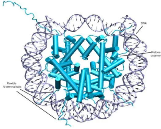
• In interphase (nondividing) cells, most of the chromatin (called euchromatin) is relatively decondensed and distributed. • During this period of the

Boltzmann disribution law states that the probability of finding the molecule in a particular energy state varies exponentially as the energy divided by k

Synthesis, Biological Evaluation, and Computer-Aided Drug Designing of New Derivatives of Hyperactive Suberoylanilide Hydroxamic Acid Histone Deacetylase Inhibitors. Chem Biol

When considering women empowerment, indicators in this thesis such as gender role attitude of women and controlling behavior of husbands, personal and relational

These compounds have been shown to possess promising histone deacetylase inhibition activities via in vitro fluorometric assay and molecular docking studies.. Key words:

In all 3 graphs (Figures 1a-1c), the bars on the negative side indicate that the compound either failed to dock to the enzyme or was bound very poorly. It is clear that the AutoDock