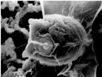
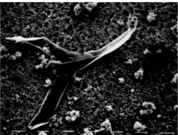
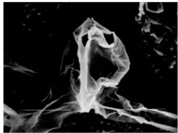
Scanning Electron Microscopic (SEM) observations on some members of actinosporean (Phylum Myxozoa) collective groups and possible usage of SEM in type description.
Tam metin
Şekil

![Fig. 4. Surface ornamentation on the spore body and three caudal processes of a aurantiactinomyxon type] spore](https://thumb-eu.123doks.com/thumbv2/9libnet/4027220.55948/3.892.442.787.103.365/fig-surface-ornamentation-spore-caudal-processes-aurantiactinomyxon-spore.webp)
Benzer Belgeler
Çalışmanın amacı, X, Y ve Z kuşaklarının kariyer algılarını, dört mevsim metaforu çerçevesinde ölçmeye çalışmak olduğundan, üç kuşakta bulunan
Bu araştırmada KUTEK'in geçerliğine ilişkin kanıtlar elde etmenin bir yolu olarak; bu envanterin uygulandığı öğrencilerin kendilerine ve onları tanıyan yakın
Türk mitolojisinde ve destanlarında cet/ata, koruyucu/hami, bilge ve yol gösterici gibi pek çok fonksiyonu olan at, Türk masallarında baĢlı baĢına bir masal tipi
Kilise ve devlet aynı kutsal otoritenin farklı yüzünü temsil etmektedir (s.. göre, çağdaş ulusal ve uluslararası siyasetin kaynağı ve arka planını oluşturduğunu
For reconstruction of orbital floor defects, an iliac bone graft was used in 14 patients, a conchal cartilage graft was used in 19 patients, an ultra thin porous polyethylene
Bu tarz süsleme elemanları Güney Batı Anadolu’da Sagalassos’da E1 Bazilikası, Antoninus Pius Tapınağı; Pergamon’da Traianeum, Dionysos Tapınağı; Ephesos’da Olympieion,
Open Access This article is distributed under the terms of the Creative Commons Attribution 4.0 International License ( http:// creativecommons.org/licenses/by/4.0/ ), which
Süreçte, öncelikle alt kriterlere göre oluşturulan karşılaştırma matrislerinin VZAHP ağırlıkları hesaplanmış ve Tablo 3’te maliyet ana kriterinin alt kriterlerine
