591 © The Author(s) 2019. Published by Oxford University Press on behalf of the European Orthodontic Society. All rights reserved.
For permissions, please email: [email protected]
Original article
Periodontal effects and survival rates of different
mandibular retainers: comparison of bonding
technique and wire thickness
Begüm Gökçe
1and Burçak Kaya
21Department of Orthodontics, Private Practice, Ankara, Turkey, 2Department of Orthodontics, Faculty of Dentistry, Başkent University, Ankara, Turkey
Correspondence to: Burcak Kaya, Baskent Universitesi, Dis Hekimligi Fakultesi, Ortodonti Anabilim Dali, 1. Cadde No: 107, 06490 Bahcelievler-Ankara, Turkey. E-mail: [email protected]
Summary
Objectives: This non-randomized prospective cohort study aimed to compare the periodontal effects and success rates of mandibular canine-to-canine fixed retainers having different bonding techniques and wire thicknesses.
Materials and methods: Hundred patients requiring retention after orthodontic treatment were assigned to five study groups (n = 20 in each group, 61 females/39 males, median age range 16.5–18.0 years). Retention was provided by 0.0215”/direct, 0.0215”/indirect, 0.0175”/direct, 0.0175”/indirect bonded multistranded wires and removable Essix appliances. The primary and secondary outcomes were periodontal effects and success rates. The patients were examined in 1 week, 1 month, 3 months and 6 months follow-up appointments. Plaque index, gingival index, probing depth, marginal recession, bleeding on probing, failure rate per retainer wire and survival of retainer wires were analysed by Kruskal–Wallis H, Mann–Whitney U and chi-square tests. Results: Significant differences were observed between the fixed retainer (FR) and Essix (E) groups in gingival index scores at 1 month [mean FR: 1.13 (95% confidence interval (CI): 0.81–1.44), mean E: 0.40 (95% CI: 0.14–0.69), mean difference: 0.73, P < 0.01], 3 months [mean FR: 0.97/1.01 (95% CI: 0.65–1.30/0.72–1.30), mean E: 0.52 (95% CI: 0.25–0.82), mean differences: 0.45/0.49, P < 0.05], 6 months [mean FR: 0.94 (95% CI: 0.62–1.27), mean E: 0.35 (95% CI: 0.15–0.58), mean difference: 0.59, P < 0.05] and in bleeding on probing scores at 1 month [mean FR: 3.05 (95% CI: 2.12–3.98), mean E: 1.15 (95% CI: 0.42–1.88), mean difference: 1.90, P < 0.01]. The survival rates of retainer wires were 85 per cent for the 0.0215” direct/indirect and 90 per cent for the 0.0175” direct/indirect groups for the 6 months follow-up.
Limitations: Six months follow-up period, which demonstrates only short-term outcomes.
Conclusions: The periodontal outcomes or survival rates of mandibular fixed retainers were not affected by bonding technique or wire thickness, whereas gingival health improved with Essix retainers but not with fixed retainers.
Introduction
Correction of crowded and rotated incisors requires long-term re-tention to prevent relapse (1). It is reported that 40–90 per cent of orthodontic patients display alignment problems of anterior teeth in
10 years post-retention period and only 10 per cent of orthodontic patients have acceptable mandibular alignment in 20 years post-retention period (2). Therefore, use of retention appliances for main-taining stability after orthodontic treatment is necessary particularly in mandibular anterior region (3). Fixed retention protocols have
European Journal of Orthodontics, 2019, 591–600
doi:10.1093/ejo/cjz060 Advance Access publication 1 August 2019
become increasingly popular since they were introduced in 1970s due to not requiring that much of patient compliance as removable reten-tion protocols do (4). Fixed retainers also have the advantage of being invisible from the front in addition to providing permanent retention more safely and predictably compared to removable plates (5).
Today, fixed retainers are routinely placed at the end of ortho-dontic treatment (6). However, some disadvantages are reported related with them such as their sensitive and time-consuming tech-nique of placement, potential for causing tooth movement due to dis-tortion and frequently observed bonding failures (7,8). Failure rates reported for mandibular fixed retainers show a wide range from 5.9 to 53 per cent in the literature (9–14). On the other hand, fixed re-tainers are shown to increase plaque and calculus accumulation.
A number of studies investigating survival rates or periodontal effects of fixed retainers are present in the current literature (1–20). Nevertheless, a comprehensive evaluation of fixed retainers based on the comparison of bonding technique and wire thickness is in-sufficient. Therefore, the primary aim of this prospective study was to compare the periodontal effects and success rates of mandibular lingual fixed retainers having different wire thicknesses, which are applied with indirect and direct bonding techniques. The H0 hy-pothesis was that differences in retainer wire thickness and bonding technique would not affect periodontal conditions or survival rates, whereas the H1 hypothesis was that changing the retainer wire thick-ness and bonding technique would affect periodontal conditions or survival rates. The secondary aim of this study was to compare the periodontal effects of fixed retainers with removable retainers.
Material and methods
This clinical research was organized as a non-randomized prospective cohort study with parallel group design and 1:1 allocation ratio. The sample size required for the study was calculated prior to the start of the study with power analysis by means of F-test by using data obtained from the literature (2,7). The primary and secondary out-comes of the study were designated as periodontal effects and success rates of fixed retainers, respectively. The primary outcome parameter determined for sample size calculation was gingival index which was measured in GI score. The anticipated mean difference between the groups for this parameter was 0.6 GI score, showing a standard de-viation of 0.6 GI score, having a baseline value of 0.8 GI score with large effect size estimation (f = 0.4). The power analysis revealed that a total sample size of 55 (n = 11 per group) was required to distinguish statistically significant differences between the five study groups with a fairly high power of 90 per cent at 0.05 significance level. Sample size estimation was performed by using NCSS and PASS 2000 soft-ware (NCSS LLC, Kaysville, Utah, USA). The study was approved by Başkent University Institutional Review Board and Ethics Committee with project number D-KA15/22 Assuming a potential dropout rate greater than 50 per cent during the follow-up period and for increas-ing the power of the study for other outcome parameters, the sample size was increased to 20 in each of the groups
Hundred consecutive patients who had completed orthodontic treatment between February 2016 and November 2016 in the post-graduate orthodontic clinic of Başkent University, Ankara, Turkey with fixed appliances and required retainers were included in the study. These patients were assigned to the five study groups consecu-tively in regular turn via alteration method by using a predetermined sequence of study groups to be assigned and through allocating only one patient into each study group at every turn, until 20 patients were assigned in each group. They may have presented with any
malocclusion prior to orthodontic treatment and their treatment may have involved extraction or expansion protocols. The inclusion criteria consisted of:
1. presence of six mandibular anterior teeth involving four incisors and two canines;
2. no caries, restorations, fractures and periodontal disease of man-dibular anterior teeth;
3. adequate oral hygiene and treatment compliance, determined through questioning and clinical examination.
Supragingival debridement was performed and dental impressions were taken following completion of orthodontic treatment in all groups. 0.0215” multistranded wire was applied with direct technique in the first group and with indirect technique in the second group. 0.0175” multistranded wire was applied with direct technique in the third group and with indirect technique in the fourth group. Retention was achieved with removable Essix appliances in the fifth group.
In the direct bonding groups, five-strand coaxial retainer wires (Orthoform II, 3M Unitek, Monrovia, California, USA) were bent and passively adapted to lingual surfaces of six mandibular an-terior teeth on plaster dental casts by the same laboratory technician (Supplementary Figure 1). In the clinic, lingual surfaces of these teeth were pumiced, rinsed with water and dried. Following positioning of the retainer wires, two pieces of ligature wire were attached, passing beneath the contact points of mandibular lateral and canine teeth through gingival embrasures and surrounding the retainer wires. The retainer wires were secured by twisting the ligature wires (Figure 1). Lingual surfaces of mandibular anterior teeth were etched with 37 per cent phosphoric acid for 30 seconds. The etched surfaces were rinsed thoroughly again, dried and bonding adhesive primer was ap-plied (Transbond XT primer, 3M Unitek). The retainer wires were bonded to lingual surfaces of six mandibular anterior teeth using light-cured adhesive resin (Transbond LR, 3M Unitek). Light curing was performed for 10 seconds per each tooth using LED device (Elipar S10, 3M ESPE, Monrovia, California, USA). The ligature wires securing the retainer wires were cut and removed after com-pletion of bonding.
In the indirect bonding groups, the same retainer wires were bent and passively adapted to lingual surfaces of six mandibular anterior teeth on plaster dental casts by the same laboratory tech-nician. Then, plaster dental casts were isolated with separation li-quid (Isolant, Dentsply, York, Pennsylvania, USA). The retainer wires were positioned and bonded to lingual surfaces of six mandibular anterior teeth on plaster dental casts with light-cured adhesive resin (Transbond LR, 3M Unitek). Light curing was performed for 10
Figure 1. Immobilization of the retainer wire by two pieces of ligature wires before direct bonding.
seconds per each tooth using the same LED device (Supplementary Figure 2). Transfer trays covering buccal, incisal and lingual surfaces of mandibular anterior teeth, including retainer wires and poly-merized adhesive pads, were constructed from transparent silicone (Memosil, Heraeus Kulzer, Wehrheim, Germany). The transfer trays were removed from plaster dental casts after being kept in warm water for 20 seconds (Figure 2). The uncovered surfaces of adhe-sive resin pads were sandblasted with 50 µ of aluminium oxide to remove the separation liquid and finally cleaned with acetone. In the clinic, lingual surfaces of mandibular anterior teeth were pumiced, rinsed with water, dried and then etched with 37 per cent phosphoric acid for 30 seconds. The etched surfaces were rinsed thoroughly again, dried and bonding adhesive primer was applied (Transbond XT primer, 3M Unitek). Low-viscosity light-cured indirect bonding adhesive (Transbond LV Supreme, 3M Unitek) was applied to the sandblasted surfaces of adhesive resin pads and the transfer trays were positioned on mandibular anterior teeth with a slight pressure (Supplementary Figure 3). Light curing was performed for 20 sec-onds per each tooth using the same LED device. The transfer trays were cut into two pieces of buccal and lingual using a scalpel and removed carefully after completion of bonding.
In the removable appliances group, Essix retainers of 1 mm thick-ness were fabricated on plaster dental casts with heat and vacuum applied by an Essix machine. Patients were instructed to use their Essix retainers all day long except while eating.
All fixed and removable retainers were applied; oral hygiene and retainer usage instructions to the patients were given and examin-ations were made by the same clinician (BG) to ensure the integ-rity and accuracy of the assessment procedures and to increase the quality of the study. Blinding was not possible due to the nature of this study. The following clinical variables were assessed at the
beginning of retention procedures and in 1 week, 1 month, 3 months and 6 months follow-up appointments:
1. Plaque index was assessed on the buccal and lingual surfaces for each tooth using a probe as described by Löe (7,21). The average for six mandibular anterior teeth was calculated and analysed. Plaque accumulation was categorized with the following scale: 0: no plaque formation;
1: plaque detectable with a probe along gingival margin; 2: visible plaque formation;
3: abundant amount of plaque.
2. Gingival index was assessed on the buccal, lingual, mesial and distal surfaces for each tooth as described by Löe (7,21). The average for six mandibular anterior teeth was calculated and ana-lysed according to the following scale:
0: no inflammation;
1: mild inflammation, slight discolouration, minor oedema and no bleeding on probing;
2: moderate inflammation, glazing, redness, oedema and bleeding on probing;
3: severe inflammation, marked redness, hypertrophy and spon-taneous bleeding
3. Probing depth was measured with a periodontal probe in milli-metres at three locations (mesiolingual, lingual and distolingual) for each tooth as the distance from gingival margin to the most apical part of sulcus. The average for six mandibular anterior teeth was calculated and analysed.
4. Marginal recession was measured with a periodontal probe in millimetres at three locations (mesiolingual, lingual and distolin-gual) for each tooth as the distance from cementoenamel junction to gingival margin. Scores were recorded when cementoenamel junction was visible due to recession. The average for six man-dibular anterior teeth was calculated and analysed.
5. Bleeding on probing was examined at six locations (mesiolin-gual, lin(mesiolin-gual, distolin(mesiolin-gual, mesiobuccal, lingual and distobuccal) for each tooth and recorded either positive (bleeding observed) or negative (bleeding not observed). Number of mandibular anterior teeth with positive recordings were summed and analysed. 6. Failure rate per retainer wire was assessed by examining
debond-ing of adhesive resin or breakage of retainer wires. Average number of detached teeth per retainer wire was calculated and analysed. 7. Distribution of failures per each tooth: debonding of adhesive resin
observed in each tooth was recorded and presented with numbers and percentages for making comparisons among different teeth. 8. Total success rate was assessed by examining survival or failure of
retainer wires. Absence of adhesive resin detachment or retainer wire breakage throughout 6 months follow-up period was defined as survival, whereas presence of any of these at any time was de-fined as failure. Adhesive remnants were removed and rebondings were undertaken at the chair side when detachments occurred.
Statistical analysis
Data analysis was performed using SPSS for Windows, version 20 (SPSS Inc., Chicago, Illinois, USA). Descriptive statistics were pre-sented with number and percentage for categorical variables such as gender and failure per tooth or retainer wire, whereas they were presented with mean and standard deviation for continuous vari-ables such as age, plaque index, gingival index, probing depth, mar-ginal recession, bleeding on probing and failure rate per retainer wire. P-value less than 0.05 was considered statistically significant. Figure 2. ( A) Preparation of the transparent silicone transfer tray on plaster
dental cast, which covers mandibular anterior teeth and contains the retainer wire with formerly polymerized adhesive pads arranged for indirect bonding technique. (B) Exposure of the uncovered surfaces of adhesive resin pads after removal of the transfer tray from plaster dental cast.
Shapiro–Wilk test was used to determine the normality of variable distribution. Skewness and kurtosis were also checked to understand the nature of the variables. The differences between the study groups were analysed by Kruskal–Wallis H-test for continuous variables not showing normal distribution and by chi-square test for categorical variables with frequencies. Mann–Whitney U-test with Bonferroni correction was used to distinguish the groups between which statis-tically significant difference was observed, if Kruskal–Wallis H-test revealed a significant difference. Monte Carlo simulation technique was used to analyse categorical variables when 50 per cent of the frequencies counted less than five.
Results
All of the 100 patients who were found to be eligible and included in the study completed the 6 months follow-up period without any dropouts. Comparisons of the groups according to gender and age are presented in Table 1. The differences between the groups were
not statistically significant for gender or age. None of the partici-pants had missing data for any of the variables.
Table 2 presents the comparison of plaque index scores between the groups. The mean plaque index scores ranged between 0.04 and 0.35 (95% CI ranged from −0.02–0.10 to 0.11–0.59) during the 6 months follow-up period and were smaller in the Essix retainer group starting from 1 month follow-up appointments. No significant difference was observed in plaque index scores between the groups in any evaluated time interval.
Table 3 reveals the comparison of gingival index scores between the groups. The mean gingival index scores ranged between 0.35 and 1.13 (95% CI ranged from 0.15–0.58 to 0.81–1.44) during the 6 months follow-up period and were smaller in the Essix retainer group starting from 1 month follow-up appointments. Significant differences were observed between the groups in gingival index scores at 1 month (P < 0.01), 3 months (P < 0.05) and 6 months (P < 0.05) follow-up appointments. These differences were detected between 0.0215” Indirect and Essix groups at 1 month, between 0.0215” Indirect and Essix and 0.0175” Indirect and Essix groups at 3 months and between 0.0215” Direct and Essix groups at 6 months.
Tables 4 and 5 show the comparisons of probing depth and mar-ginal recession scores between the groups. The mean probing depth scores ranged between 1.58 and 1.73 mm (95% CI ranged from 1.48–1.66 to 1.71–1.74) and the mean marginal recession scores ranged between 0.00 and 0.03 mm (95% CI ranged from 0.00–0.00 to −0.03–0.10) during the 6 months follow-up period. No significant differences were observed in probing depth and marginal recession scores between the groups in any evaluated time interval.
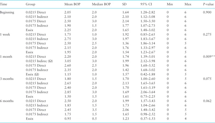
Table 6 reports the comparison of bleeding on probing scores between the groups. The mean bleeding on probing scores ranged between 0.95 and 3.05 (95% CI ranged from 0.37–1.53 to 2.12– 3.98) during the 6 months follow-up period and were smaller in the
Table 1. Comparison of gender and age between the groups by chi-square test and Kruskal–Wallis H-test, respectively.
n
Gender (n, %) Age (year)
Female/male P-value Median, min–max P-value
0.0215 Direct 20 12/8, 60/40% 17.5, 15.0–33.0 0.0215 Indirect 20 13/7, 65/35% 18.0, 15.0–24.0 0.0175 Direct 20 14/6, 70/30% 0.539 16.5, 14.0–40.0 0.187 0.0175 Indirect 20 9/11, 45/55% 17.5, 15.0–28.0 Essix 20 13/7, 65/35% 16.5, 13.0–24.0 P ≥ 0.05, non-significant.
Table 2. Comparison of plaque index (PI) between the groups by Kruskal–Wallis H-test. SD, standard deviation; CI, confidence interval.
Time Group Mean PI Median PI SD 95% CI Min Max P-value
Beginning 0.0215 Direct 0.04 0 0.13 –0.02–0.10 0 0.5 0.661 0.0215 Indirect 0.11 0 0.25 0.00–0.23 0 1.0 0.0175 Direct 0.05 0 0.17 –0.02–0.12 0 0.7 0.0175 Indirect 0.08 0 0.23 –0.03–0.18 0 1.0 Essix 0.09 0 0.22 0.00–0.19 0 0.7 1 week 0.0215 Direct 0.06 0 0.23 –0.04–0.17 0 1.0 0.524 0.0215 Indirect 0.17 0 0.27 0.04–0.28 0 0.7 0.0175 Direct 0.11 0 0.24 0.00–0.21 0 0.7 0.0175 Indirect 0.14 0 0.32 0.00–0.29 0 1.0 Essix 0.13 0 0.26 0.00–0.24 0 0.8 1 month 0.0215 Direct 0.15 0 0.29 0.01–0.29 0 1.0 0.534 0.0215 Indirect 0.30 0 0.49 0.07–0.53 0 1.5 0.0175 Direct 0.35 0 0.52 0.11–0.59 0 2.0 0.0175 Indirect 0.22 0 0.36 0.05–0.39 0 1.0 Essix 0.11 0 0.24 0.00–0.22 0 1.0 3 months 0.0215 Direct 0.18 0 0.33 0.03–0.34 0 1.0 0.418 0.0215 Indirect 0.25 0 0.38 0.07–0.42 0 1.0 0.0175 Direct 0.28 0 0.44 0.07–0.48 0 1.3 0.0175 Indirect 0.14 0 0.28 0.01–0.27 0 1.0 Essix 0.12 0 0.31 –0.02–0.26 0 1.0 6 months 0.0215 Direct 0.26 0 0.38 0.09–0.45 0 1.0 0.121 0.0215 Indirect 0.16 0 0.33 0.01–0.32 0 1.0 0.0175 Direct 0.25 0 0.40 0.06–0.44 0 1.0 0.0175 Indirect 0.12 0 0.27 0.00–0.24 0 1.0 Essix 0.05 0 0.22 –0.05–0.16 0 1.0 P ≥ 0.05, non-significant.
Essix retainer group starting from 1 month follow-up appointments. Significant difference (P < 0.01) was observed between the groups in bleeding on probing scores at 1 month follow-up appointments. This difference was detected between 0.0215” Indirect and Essix groups.
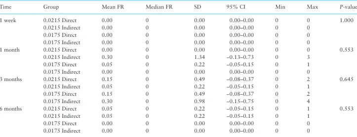
Table 7 displays the comparison of failure rate (number of de-tached teeth) per retainer wire between the groups. The mean failure rate per retainer wire scores ranged between 0.00 and 0.30 (95% CI ranged from 0.00–0.00 to −0.15–0.75) during the 6 months
Table 3. Comparison of gingival index (GI) between the groups by Kruskal–Wallis H-test. SD, standard deviation; CI, confidence interval.
Time Group Mean GI Median GI SD 95% CI Min Max P-value
Beginning 0.0215 Direct 0.79 0.7 0.58 0.51–1.05 0 2.0 0.959 0.0215 Indirect 0.78 0.7 0.75 0.44–1.13 0 2.0 0.0175 Direct 0.94 1.0 0.75 0.59–1.29 0 2.0 0.0175 Indirect 0.82 1.0 0.60 0.56–1.14 0 2.0 Essix 0.83 0.7 0.62 0.50–1.07 0 2.0 1 week 0.0215 Direct 0.54 0.3 0.64 0.25–0.85 0 2.0 0.139 0.0215 Indirect 1.02 1.1 0.67 0.71–1.34 0 2.0 0.0175 Direct 0.81 0.7 0.53 0.56–1.06 0 1.8 0.0175 Indirect 0.83 0.7 0.55 0.55–1.08 0 2.0 Essix 0.71 0.7 0.56 0.43–0.99 0 1.7 1 month 0.0215 Direct 0.75 0.7 0.61 0.48–1.04 0 2.0 0.005** 0.0215 Indirec (α) 1.13 1.2 0.67 0.81–1.44 0 2.0 0.0175 Direct 0.93 1.0 0.65 0.62–1.23 0 2.0 0.0175 Indirect 0.86 0.9 0.51 0.63–1.10 0 2.0 Essix (α) 0.40 0.3 0.56 0.14–0.69 0 1.8 3 months 0.0215 Direct 0.63 0.5 0.6 0.36–0.91 0 1.7 0.047* 0.0215 Indirect (β) 0.97 0.7 0.70 0.65–1.30 0 2.0 0.0175 Direct 0.87 0.7 0.61 0.57–1.14 0 2.0 0.0175 Indirect (θ) 1.01 1.0 0.62 0.72–1.30 0 2.0 Essix (β)(θ) 0.52 0.5 0.58 0.25–0.82 0 2.0 6 months 0.0215 Direct (ϒ) 0.94 0.9 0.70 0.62–1.27 0 2.0 0.049* 0.0215 Indirect 0.64 0.5 0.65 0.34–0.95 0 2.0 0.0175 Direct 0.86 1.2 0.73 0.52–1.20 0 1.7 0.0175 Indirect 0.60 0.5 0.57 0.33–0.87 0 1.7 Essix (ϒ) 0.35 0.2 0.44 0.15–0.58 0 1.3
P ≥ 0.05, non-significant. *P < 0.05; **P < 0.01. Values with the same symbols (α, β, θ and ϒ) show statistically significant difference (P < 0.05).
Table 4. Comparison of probing depth (PD) between the groups by Kruskal–Wallis H-test. SD, standard deviation; CI, confidence interval.
Time Group Mean PD Median PD SD 95% CI Min Max P-value
Beginning 0.0215 Direct 1.58 1.7 0.20 1.48–1.66 1.0 1.7 0.111 0.0215 Indirect 1.61 1.7 0.20 1.50–1.68 1.1 1.8 0.0175 Direct 1.66 1.7 0.15 1.58–1.71 1.2 1.9 0.0175 Indirect 1.64 1.7 0.24 1.52–1.74 1.1 2.0 Essix 1.72 1.7 0.07 1.66–1.73 1.6 1.9 1 week 0.0215 Direct 1.60 1.7 0.18 1.51–1.66 1.2 1.8 0.340 0.0215 Indirect 1.65 1.7 0.15 1.56–1.70 1.2 1.9 0.0175 Direct 1.66 1.7 0.13 1.59–1.70 1.3 1.8 0.0175 Indirect 1.64 1.7 0.18 1.55–1.72 1.1 1.9 Essix 1.72 1.7 0.08 1.67–1.76 1.6 2.0 1 month 0.0215 Direct 1.69 1.7 0.05 1.64–1.71 1.6 1.8 0.690 0.0215 Indirect 1.67 1.7 0.21 1.56–1.74 1.0 2.1 0.0175 Direct 1.69 1.7 0.08 1.63–1.71 1.4 1.8 0.0175 Indirect 1.71 1.7 0.13 1.63–1.75 1.3 1.9 Essix 1.69 1.7 0.02 1.68–1.70 1.6 1.7 3 months 0.0215 Direct 1.71 1.7 0.02 1.69–1.72 1.6 1.8 0.224 0.0215 Indirect 1.71 1.7 0.09 1.64–1.73 1.5 2.0 0.0175 Direct 1.69 1.7 0.11 1.63–1.73 1.3 1.9 0.0175 Indirect 1.73 1.7 0.06 1.71–1.74 1.7 1.9 Essix 1.70 1.7 0.03 1.66–1.72 1.6 1.8 6 months 0.0215 Direct 1.72 1.7 0.06 1.66–1.74 1.6 1.9 0.392 0.0215 Indirect 1.67 1.7 0.19 1.58–1.75 1.1 2.0 0.0175 Direct 1.71 1.7 0.04 1.66–1.72 1.6 1.8 0.0175 Indirect 1.72 1.7 0.06 1.68–1.74 1.6 1.9 Essix 1.66 1.7 0.13 1.59–1.69 1.3 1.8 P ≥ 0.05, non-significant.
Table 5. Comparison of marginal recession (MR) between the groups by Kruskal–Wallis H-test. SD, standard deviation; CI, confidence
interval.
Time Group Mean MR Median MR SD 95% CI Min Max P-value
Beginning 0.0215 Direct 0.00 0 0.00 0.00–0.00 0 0.0 0.905 0.0215 Indirect 0.02 0 0.09 −0.02–0.06 0 0.4 0.0175 Direct 0.01 0 0.04 −0.01–0.02 0 0.2 0.0175 Indirect 0.03 0 0.13 −0.03–0.08 0 0.6 Essix 0.03 0 0.11 −0.02–0.07 0 0.5 1 week 0.0215 Direct 0.00 0 0.00 0.00–0.00 0 0.0 0.905 0.0215 Indirect 0.02 0 0.09 −0.02–0.06 0 0.4 0.0175 Direct 0.01 0 0.04 −0.01–0.02 0 0.2 0.0175 Indirect 0.03 0 0.13 −0.03–0.08 0 0.6 Essix 0.03 0 0.11 −0.02–0.07 0 0.5 1 month 0.0215 Direct 0.00 0 0.00 0.00–0.00 0 0.0 0.905 0.0215 Indirect 0.02 0 0.09 −0.02–0.06 0 0.4 0.0175 Direct 0.01 0 0.04 −0.01–0.02 0 0.2 0.0175 Indirect 0.03 0 0.13 −0.03–0.08 0 0.6 Essix 0.03 0 0.11 −0.02–0.07 0 0.5 3 months 0.0215 Direct 0.00 0 0.00 0.00–0.00 0 0.0 0.905 0.0215 Indirect 0.02 0 0.09 −0.02–0.06 0 0.4 0.0175 Direct 0.01 0 0.04 −0.01–0.02 0 0.2 0.0175 Indirect 0.03 0 0.15 −0.03–0.10 0 0.7 Essix 0.03 0 0.13 −0.03–0.09 0 0.6 6 months 0.0215 Direct 0.00 0 0.00 0.00–0.00 0 0.0 0.905 0.0215 Indirect 0.03 0 0.11 −0.02–0.07 0 0.5 0.0175 Direct 0.01 0 0.04 −0.01–0.02 0 0.2 0.0175 Indirect 0.03 0 0.15 −0.03–0.10 0 0.7 Essix 0.03 0 0.15 −0.03–0.10 0 0.7 P ≥ 0.05, non-significant.
Table 6: Comparison of bleeding on probing (BOP) between the groups by Kruskal–Wallis H-test. SD, standard deviation; CI, confidence
interval.
Time Group Mean BOP Median BOP SD 95% CI Min Max P-value
Beginning 0.0215 Direct 2.05 2.0 1.64 1.28–2.82 0 6 0.900 0.0215 Indirect 2.10 2.0 2.10 1.12–3.08 0 6 0.0175 Direct 2.50 3.0 2.14 1.50–3.50 0 6 0.0175 Indirect 1.90 1.5 1.77 1.07–2.73 0 6 Essix 2.25 2.0 1.65 1.48–3.02 0 6 1 week 0.0215 Direct 1.75 1.0 1.92 0.85–2.65 0 6 0.275 0.0215 Indirect 2.75 3.0 1.97 1.83–3.67 0 6 0.0175 Direct 2.50 2.5 1.36 1.86–3.14 0 5 0.0175 Indirect 2.15 2.0 1.76 1.33–2.97 0 6 Essix 1.95 2.0 1.54 1.23–2.67 0 5 1 month 0.0215 Direct 2.20 2.0 1.74 1.39–3.01 0 6 0.009** 0.0215 Indirec (Ω) 3.05 3.0 1.99 2.12–3.98 0 6 0.0175 Direct 2.60 2.5 1.96 1.68–3.52 0 6 0.0175 Indirect 2.35 2.0 1.42 1.68–3.02 0 6 Essix (Ω) 1.15 1.0 1.57 0.42–1.88 0 5 3 months 0.0215 Direct 1.80 1.5 1.70 1.00–2.60 0 5 0.075 0.0215 Indirect 2.65 2.0 2.13 1.65–3.65 0 6 0.0175 Direct 2.40 2.0 1.70 1.61–3.19 0 6 0.0175 Indirect 2.85 3.0 1.69 2.06–3.64 0 6 Essix 1.50 1.5 1.61 0.75–2.25 0 6 6 months 0.0215 Direct 2.50 2.0 1.99 1.57–3.43 0 6 0.062 0.0215 Indirect 1.85 1.5 1.73 1.04–2.66 0 6 0.0175 Direct 2.45 3.5 2.06 1.48–3.42 0 5 0.0175 Indirect 1.75 1.5 1.65 0.98–2.52 0 5 Essix 0.95 0.5 1.23 0.37–1.53 0 4
P ≥ 0.05, non-significant. **P < 0.01. Values with the same symbols (Ω) show statistically significant difference (P < 0.05).
follow-up period. All failures occurred due to debonding at adhe-sive–enamel interface and none of the retainer wires were completely detached, deformed or broken. No significant difference was ob-served in failure rate scores between the four fixed retainer groups in any evaluated time interval.
Distribution of failures per each tooth is presented in Supplementary Table 1 and total success rates of retainer wires for 6 months follow-up period is presented in Table 8. The survival rates of retainer wires were 85 per cent for the 0.0215” direct and indirect groups, whereas they were 90 per cent for the 0.0175” direct and indirect groups. The difference between the groups in total success rates was not statistically significant.
Discussion
Fixed lingual retainers are frequently used to prevent relapse after orthodontic treatment. Therefore, their influence on periodontal health, ability to maintain post-treatment stability and factors af-fecting their survival rate has been investigated by a great number of studies (1-4,7-20,22–24). Nonetheless, a study evaluating the influence of both wire thicknesses and bonding technique of fixed retainers in comparison with a removable retainer does not exist in the literature. For this reason, this prospective study intended to compare the periodontal outcomes and failure rates of mandibular lingual fixed retainers having different wire thicknesses and applied with different bonding techniques in conjunction with removable Essix retainers.
The five-strand coaxial retainer wire design and adhesive primer/ resin system used in this study were the same in both thick/thin wire and direct/indirect bonded fixed retainer groups to eliminate the influence of wire design and adhesive material differences on the parameters evaluated. Additionally, all retainer wires were bent by the same laboratory technician and all supragingival debridements were performed, retainers were bonded, instructions to the patients were given and examinations were made by the same clinician to eliminate the influence of technician- and clinician-related factors.
The variances observed between the groups for gender and age were both statistically insignificant and clinically unimportant, with a maximum difference of 25 per cent for gender and 1.5 years for
age. Hence, the groups were comparable according to demographic variables.
The plaque index assessment indicated that only minimal de-posits of plaque were observed and oral hygiene was good in man-dibular anterior region in all study groups. Very slight increases were observed in the plaque index scores starting at 1 week and continuing until 3 months follow-up appointments in the fixed retainer groups, which were clinically unimportant. This small increase in deposits of plaque, which shows an increasing trend only in the first 3 months, can be explained with the delayed adaptation period of the patients that probably ended as a result of the ongoing instructions given to them in the follow-up appointments. No significant difference was observed in plaque index scores between the five groups in any evaluated time interval. This result is comparable with the literature, but the plaque index scores observed in this study were smaller and no other study compared the outcomes of different bonding tech-niques or retainer wire thicknesses (1,2,7,23). Therefore, this result is unique for this study. The smaller plaque index scores observed in this study may be attributed to the standardized detailed instructions given to the patients at the beginning of the retention phases by the same clinician.
The gingival index assessment indicated that gingival health con-ditions in mandibular anterior region did not change during the re-tention phases in the fixed retainer groups but improved in the Essix retainer group. The gingival index scores remained approximately the same in all fixed retainer groups during the 6 months follow-up
Table 7. Comparison of failure rate per retainer wire (FR) between the groups by Kruskal–Wallis H-test. SD, standard deviation; CI, confidence
interval.
Time Group Mean FR Median FR SD 95% CI Min Max P-value
1 week 0.0215 Direct 0.00 0 0.00 0.00–0.00 0 0 1.000 0.0215 Indirect 0.00 0 0.00 0.00–0.00 0 0 0.0175 Direct 0.00 0 0.00 0.00–0.00 0 0 0.0175 Indirect 0.00 0 0.00 0.00–0.00 0 0 1 month 0.0215 Direct 0.00 0 0.00 0.00–0.00 0 0 0.553 0.0215 Indirect 0.30 0 1.34 −0.13–0.73 0 3 0.0175 Direct 0.05 0 0.22 −0.05–0.15 0 1 0.0175 Indirect 0.00 0 0.00 0.00–0.00 0 0 3 months 0.0215 Direct 0.15 0 0.49 −0.08–0.37 0 2 0.645 0.0215 Indirect 0.05 0 0.22 −0.05–0.15 0 1 0.0175 Direct 0.15 0 0.49 −0.08–0.37 0 2 0.0175 Indirect 0.30 0 0.98 −0.15–0.75 0 4 6 months 0.0215 Direct 0.05 0 0.22 −0.05–0.15 0 1 0.553 0.0215 Indirect 0.05 0 0.22 −0.05–0.15 0 1 0.0175 Direct 0.00 0 0.00 0.00–0.00 0 0 0.0175 Indirect 0.00 0 0.00 0.00–0.00 0 0 P ≥ 0.05, non-significant.
Table 8. Total success rates of retainer wires for the 6 months follow-up period.
Group
Total Survival Failure
P-value n n % n % 0.0215 Direct 20 17 85 3 15 0.928 0.0215 Indirect 20 17 85 3 15 0.0175 Direct 20 18 90 2 10 0.0175 Indirect 20 18 90 2 10
P ≥ 0.05, non-significant. Chi-square test and Monte Carlo simulation tech-nique is used for analysis.
period, whereas a clinically detectable decrease was observed in the Essix retainer group starting at 1 month follow-up appointments. Hence, significant differences were observed between the groups in gingival index scores at the 1, 3 and 6 months follow-up appoint-ments. This result suggests that fixed retention appliances prevent improvement of gingival health conditions during retention phase, which is independent from wire thickness or bonding technique. This finding is similar to the literature, but the gingival index scores observed in this study were slightly smaller and the outcomes of different bonding techniques or retainer wire thicknesses were not evaluated in other studies (1,2,7,23). Thus, this finding is also unique for this study. The smaller gingival index scores observed in this study may also be attributed to the standardized instructions given to the patients at the beginning of the retention phases.
Assessment of probing depth and marginal recession indicated that probing depth remained within normal limits and no reces-sion occurred in gingival margin during the retention phases. The probing depth and marginal recession scores did not show any change in fixed retainer or Essix retainer groups during the 6 months follow-up period. Therefore, no significant differences were observed in probing depth and marginal recession scores between the five groups in any evaluated time interval. These results reveal that nei-ther fixed lingual nor removable Essix retainers cause detrimental effects in periodontal tissues that lead to increase in probing depth or marginal recession. This study examined the changes that oc-curred in periodontal conditions due to usage of fixed or removable retainers only for short term, which was limited with 6 months. The probing depth and marginal recession results obtained in this study were compatible with the short-term results but incompatible with the long-term results of other studies, as they observed increase in probing depth and marginal recession within 5 to 11 years retention phases (2,7). Besides different bonding techniques or retainer wire thicknesses were not of concern in those studies, which makes these results also unique for this study.
Bleeding on probing showed a gradual decrease in the Essix re-tainer group, whereas irregular changes were observed in the fixed retainer groups during the retention phases. Thus, significant dif-ference was observed between the groups in bleeding on probing scores at the 1 month follow-up appointments. This finding suggests that removable Essix retainers cause decline in gingival bleeding and improvement of gingival health conditions during retention phase, whereas fixed retention appliances do not cause a change, inde-pendent from wire thickness or bonding technique. This finding is comparable with the finding of Dietrich et al. (2); however, their study evaluated the periodontal outcomes of 0.016 × 0.016” maxil-lary retainer wires within a 7 years retention phase retrospectively. Hence, no similar study design evaluating bleeding on probing is present in the literature.
No detachments were observed in the 1 week follow-up ap-pointments. On the other hand, seven detachments were observed in the 1 month follow-up appointments and 13 detachments were observed in the 3 months follow-up appointments. Number of de-tachments was decreased to only two in the 6 months follow-up appointments. The decrease observed in the number of detachments after the first 3 months may be explained with the adaptation period of the patients. These findings are consistent with the findings of other studies having different methods, which stated that failure rate was inversely associated with survival time and the highest num-bers of failures were observed soon after bonding of fixed retainers (8-10,12-14,16,22,24,25). The greatest number of detachments were observed in the right central (n = 6), left central (n = 5) and
left lateral (n = 6) teeth, whereas the sum of the detachments ob-served in the right and left canines was only four. This result is also compatible with other studies, although the retainer wire designs, bonding techniques and observation periods in those studies are dif-ferent (3,8,16).
Numbers of detached teeth per retainer wire were similar in all fixed retainer groups during the retention phases. Therefore, no sig-nificant difference was observed in failure rate per retainer wire be-tween the four fixed retainer groups in any evaluated time interval. On the other hand, the survival rates of retainer wires were 85 per cent for the 0.0215” direct and indirect bonding groups, whereas 90 per cent for the 0.0175” direct and indirect bonding groups for the 6 months follow-up period. The difference between the groups was not statistically significant for the survival rates of retainer wires. These findings are similar with the findings of other studies, which compared the survival and failure rates of direct/indirect bonded (3,8,16) or thick/thin wired (10,13) fixed retainers, separately. Nevertheless, a study comparing the failure rate of direct and indirect bonding together with thick and thin fixed retainers having the same wire design is not present in the literature and this is another unique finding of this study. The survival rates of retainer wires were higher and the failure rates were lower in this study compared to the other studies; however, the wire designs, bonding techniques, observation periods and study methods were not comparable (1,3,4,8-10, 12-14,16,22,26). The higher survival and lower failure rates obtained in this study may again be attributed to the very detailed instructions given to the patients at the beginning of the retention phases.
The prospective design of this study allowed for more compre-hensive examination of the time-related changes in periodontal con-ditions in addition to failure patterns and frequencies compared to retrospective studies in which data are generally collected from pa-tient files and are less detailed. On the other hand, dropouts are one of the biggest problems of prospective retention studies, because pa-tients are often unwilling to attend follow-up appointments as they consider active treatment is completed. Nonetheless, no dropouts were observed during the follow-up periods in any of the group of this study, as the follow-up appointments were reminded to the pa-tients in advance and either confirmed or rescheduled.
The 6 months follow-up period is one of the limitations of this study, since it can demonstrate only the short-term periodontal out-comes and failure rates of mandibular fixed retainers along with removable Essix retainers. Lack of blinding is the other limitation of this study, since it can cause the evaluator to make measure-ments in favour of one of the methods assessed. Another limitation of the study could be anticipating a standard deviation of 0.6 GI score for the primary outcome parameter during sample size calcu-lation, which is equal to the anticipated mean difference between the groups for this parameter. This limitation was tried to be eliminated by increasing the sample size to n = 20 per group, despite the power analysis revealing that n = 11 was required. Being unable to know the real usage time for removable retainers may also be a limitation of the study, which makes the results obtained in the Essix retainer group unclear whether they are due to the retainer itself or due to lack of patient compliance with the advised retainer usage protocol. This situation was tried to be minimized by encouraging the pa-tients to write down the duration of Essix retainer wear each day and by motivating them when lack of regular usage was observed. A possible confounding factor could be unreported supragingival de-bridements performed by other dentists throughout the 6 months follow-up period, which could damage the study outcomes. This situation was avoided by explaining the detrimental effects of this
procedure during the study period and asking the patients to post-pone it until the end of the study period. On the other hand, the generalizability of the findings of this study is limited to the operator experience, retainer designs, materials and placement techniques. Both the periodontal outcomes and the survival rates could be dif-ferent if the same procedures were applied by an experienced oper-ator in private practice rather than a postgraduate student working in a university clinic.
Conclusions
1. Retainer wire thickness did not make a difference in the peri-odontal outcomes or survival rates of mandibular fixed retainers. 2. Direct or indirect bonding technique did not make a difference in
the periodontal outcomes or survival rates of mandibular fixed re-tainers.
3. The H0 hypothesis suggesting that differences in retainer wire thickness and bonding technique does not affect periodontal con-ditions or survival rates was accepted, whereas the H1 hypothesis suggesting that changing the retainer wire thickness and bonding technique affects periodontal conditions or survival rates was re-jected.
4. Gingival health improved and bleeding on probing decreased with mandibular Essix retainers but not with fixed retainers.
5. Lower incisors were more susceptible to detachments compared to canines.
6. The survival rates were 85 per cent for 0.0215” and 90 per cent for 0.0175” retainer wires for 6 months follow-up period.
Supplementary material
Supplementary Table 1: Distribution of failures per each tooth. Supplementary Figure 1: The retainer wire passively adapted on plaster dental cast, which is prepared for direct bonding technique.
Supplementary Figure 2: The retainer wire passively adapted and bonded to mandibular anterior teeth on plaster dental cast, which is prepared for indirect bonding technique.
Supplementary Figure 3: The transfer tray positioned on man-dibular anterior teeth after applying low viscosity indirect bonding adhesive to the uncovered surfaces of adhesive resin pads for indirect bonding technique.
Supplementary Data: The complete data set of the study. Supplementary data are available at the European Journal of
Orthodontics online.
Acknowledgements
We would like to thank our laboratory technician Ebru Güven and our biostatistician Dr Ahmet Gül for their contributions in this work.
Funding
This work was supported by Başkent University Research Fund (Project No: D-KA15/22).
Conflict of interest
None to declare.References
1. Al-Nimri, K., Al Habashneh, R. and Obeidat, M. (2009) Gingival health and relapse tendency: a prospective study of two types of lower fixed re-tainers. Australian Orthodontic Journal, 25, 142–146.
2. Dietrich, P., Patcas, R., Pandis, N. and Eliades, T. (2015) Long-term follow-up of maxillary fixed retention: survival rate and periodontal health. European Journal of Orthodontics, 37, 37–42.
3. Egli, F., Bovali, E., Kiliaridis, S. and Cornelis, M.A. (2017) Indirect vs direct bonding of mandibular fixed retainers in orthodontic patients: Comparison of retainer failures and posttreatment stability. A 2-year follow-up of a single-center randomized controlled trial. American Journal of Orthodontics and Dentofacial Orthopedics, 151, 15–27.
4. Pandis, N., Fleming, P.S., Kloukos, D., Polychronopoulou, A., Katsaros, C. and Eliades, T. (2013) Survival of bonded lingual retainers with chemical or photo polymerization over a 2-year period: a single-center, randomized controlled clinical trial. American Journal of Orthodontics and Dentofa-cial Orthopedics, 144, 169–175.
5. Zachrisson, B.U. (2007) Long-term experience with direct-bonded re-tainers: update and clinical advice. Journal of Clinical Orthodontics, 41, 728–737; quiz 749.
6. Zachrisson, B.U. (2015) Multistranded wire bonded retainers: from start to success. American Journal of Orthodontics and Dentofacial Ortho-pedics, 148, 724–727.
7. Pandis, N., Vlahopoulos, K., Madianos, P. and Eliades, T. (2007) Long-term periodontal status of patients with mandibular lingual fixed retention. European Journal of Orthodontics, 29, 471–476.
8. Taner, T. and Aksu, M. (2012) A prospective clinical evaluation of man-dibular lingual retainer survival. European Journal of Orthodontics, 34, 470–474.
9. Dahl, E.H. and Zachrisson, B.U. (1991) Long-term experience with direct-bonded lingual retainers. Journal of Clinical Orthodontics, 25, 619–630. 10. Artun, J., Spadafora, A.T. and Shapiro, P.A. (1997) A 3-year follow-up
study of various types of orthodontic canine-to-canine retainers. European Journal of Orthodontics, 19, 501–509.
11. Andrén, A., Asplund, J., Azarmidohkt, E., Svensson, R., Varde, P. and Mohlin, B. (1998) A clinical evaluation of long term retention with bonded retainers made from multi-strand wires. Swedish Dental Journal, 22, 123–131.
12. Lumsden, K.W., Saidler, G. and McColl, J.H. (1999) Breakage incidence with direct-bonded lingual retainers. British Journal of Orthodontics, 26, 191–194.
13. Störmann, I. and Ehmer, U. (2002) A prospective randomized study of different retainer types. Journal of Orofacial Orthopedics, 63, 42–50.
14. Lie Sam Foek, D.J., Ozcan, M., Verkerke, G.J., Sandham, A. and Dijk-stra, P.U. (2008) Survival of flexible, braided, bonded stainless steel lingual retainers: a historic cohort study. European Journal of Orthodontics, 30, 199–204.
15. Demling, A., Demling, C., Schwestka-Polly, R., Stiesch, M. and Heuer, W. (2009) Influence of lingual orthodontic therapy on microbial parameters and periodontal status in adults. European Journal of Orthodontics, 31, 638–642.
16. Bovali, E., Kiliaridis, S. and Cornelis, M.A. (2014) Indirect vs direct bonding of mandibular fixed retainers in orthodontic patients: a single-center randomized controlled trial comparing placement time and failure over a 6-month period. American Journal of Orthodontics and Dentofa-cial Orthopedics, 146, 701–708.
17. Littlewood, S.J., Millett, D.T., Doubleday, B., Bearn, D.R. and Worthing-ton, H.V. (2006) Orthodontic retention: a systematic review. Journal of Orthodontics, 33, 205–212.
18. Westerlund, A., Daxberg, E.L., Liljegren, A., Oikonomou, C., Ransjö, M., Samuelsson, O. and Sjögren, P. (2014) Stability and side effects of ortho-dontic retainers-a systematic review. Dentistry, 4, 258.
19. Iliadi, A., Kloukos, D., Gkantidis, N., Katsaros, C. and Pandis, N. (2015) Failure of fixed orthodontic retainers: a systematic review. Journal of Den-tistry, 43, 876–896.
20. Al-Moghrabi, D., Pandis, N. and Fleming, P.S. (2016) The effects of fixed and removable orthodontic retainers: a systematic review. Progress in Orthodontics, 17, 24.
21. Löe, H. (1967) The gingival index, the plaque index and the retention index systems. Journal of Periodontology, 38, 610–616.
22. Forde, K., Storey, M., Littlewood, S.J., Scott, P., Luther, F. and Kang, J. (2018) Bonded versus vacuum-formed retainers: a randomized controlled trial. Part 1: stability, retainer survival, and patient satisfaction outcomes after 12 months. European Journal of Orthodontics, 40, 387–398. 23. Storey, M., Forde, K., Littlewood, S.J., Scott, P., Luther, F. and Kang, J.
(2018) Bonded versus vacuum-formed retainers: a randomized controlled
trial. Part 2: periodontal health outcomes after 12 months. European Journal of Orthodontics, 40, 399–408.
24. Jin, C., Bennani, F., Gray, A., Farella, M. and Mei, L. (2018) Survival analysis of orthodontic retainers. European Journal of Orthodontics, 40, 531–536.
25. Segner, D. and Heinrici, B. (2000) Bonded retainers–clinical reliability. Journal of Orofacial Orthopedics, 61, 352–358.
26. Tang, A.T., Forsberg, C.M., Andlin-Sobocki, A., Ekstrand, J. and Hägg, U. (2013) Lingual retainers bonded without liquid resin: a 5-year follow-up study. American Journal of Orthodontics and Dentofacial Orthopedics, 143, 101–104.