Introduction
Idiopathic granulomatous mastitis (IGM) is a rare benign inflammatory breast entity characterized by lobulocentric granulomas (1). IGM has a persistent or recurrent disease course and affects premenopausal women with a history of lactation. The clinical and radiologic features of IGM are very similar to those of breast carcinoma. The most common clinical manifestation is a unilateral, tender, painful, extra-areolar breast lump (2, 3). Although ethnic predisposition has not been proven precisely, the high prevalence of IGM has been observed in certain racial populations (4, 5). It may be confused with other breast lesions that have radiologically or histologically similar features to IGM. Lesions of similar characteristics include breast cancer (BC), infective mastitis, foreign body injection granulomas, mam-mary duct ectasia, diabetic fibrous mastopathy, and systemic granulomatous processes (6).
Mainly ultrasonography (US) and mammography (MG), and to a lesser extent, magnetic resonance imaging (MRI), are used for the diagnosis of IGM (7, 8). Imaging findings of this condition have a wide spectrum between benign and malignant features (8, 9). A core-needle biopsy is necessary to differentiate IGM from BC and other benign inflammatory breast lesions. Patients with IGM have excellent prognosis when they are appropriately treated with oral steroids or second-line immunosuppressive and prolactin-lowering medications. Surgical treatment may be an option for patients who fail drug therapy (6).
The etiology and pathogenesis of IGM remain unclear. An association with pregnancy, lactation, local autoimmune processes, infection, hyperprolactinemia, and chemical reaction induced by oral contraceptive pills has been reported in the literature (10, 11). To our knowledge, there are no studies about the seasonal relationship with IGM in the literature. We have observed that these patients were successive at certain times and that we encountered the diagnosis of IGM more frequently at certain times of the year. Accordingly, the current study aimed to investigate if there was a seasonal frequency in this condition. In addition, etiologic factors and radiologic findings were also reviewed.
Is There any Relationship Between Granulomatous
Mastitis and Seasons? An Analysis of Seasonal
Frequency, Clinical, and Radiologic Findings
Leyla Tekin
1, Funda Dinç Elibol
21Department of Pathology, Muğla Sıtkı Koçman University Faculty of Medicine, Muğla, Turkey 2Department of Radiology, Muğla Sıtkı Koçman University Faculty of Medicine, Muğla, Turkey
Corresponding Author :
Funda Dinç Elibol; [email protected] Accepted: 18.08.2020Received: 24.06.2020
DOI: 10.5152/ejbh.2020.5897
235
ABSTRACT
Objective: Idiopathic granulomatous mastitis (IGM) is a rare, resistant, and recurrent benign disease of the breast. IGM can be clinically and
radiologically confused with breast carcinoma, and core needle biopsy is needed to diagnose. The etiology and pathogenesis of IGM have not been fully explained. This premenopausal disease may be associated with pregnancy, breastfeeding, autoimmune processes, inflammation, and oral contra-ceptives. However, there is no study on whether there is a seasonal relationship.
Materials and Methods: From January 2015 to January 2020, the seasonal relationship of IGM was evaluated in 37 females aged between 25-49. Results: Although all cases were distributed between September and May, there was no statistically significant result in the relationship with the
season. US is the main modality in the diagnosis of this condition which only provides an accurate pre-diagnosis approach with the typical USG appearance features. Some MRI features may help us to distinguish IGM from breast malignities.
Conclusion: IGM is a rare chronic non-specific inflammatory lesion of the breast, which can be confused with benign and malignant breast diseases
in both clinical and radiologic aspects. To understand the etiology of this condition better, the seasonal connection needs to be evaluated in larger patient groups.
Keywords: Granulomatous, mastitis, radiological findings
Cite this article as: Tekin L, Dinç Elibol F. Is There any Relationship Between Granulomatous Mastitis and Seasons? An Analysis of Seasonal Frequency, Clinical, and Radiologic Findings. Eur J Breast Health 2020; 16(4): 235-243.
of diagnosis of the disease because information about how long the symptoms have been present in patients is often absent in our records and the retrospective history evaluations for patients are inconsistent. Therefore, in the evaluation of monthly and seasonal frequencies, the date of the biopsy was used.
Histopathologic evaluation
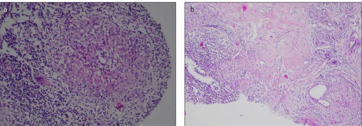
Hematoxylin and eosin-stained paraffin histologic sections were evalu-ated in detail. IGM was defined as ‘perilobular granulomatous inflam-mation, accompanied by infiltration centered on lobules with lympho-cytes, plasma cells, epithelioid histiolympho-cytes, multinucleated giant cells, and neutrophils with or without intralobular micro abscess formation (Figure 1). Tuberculosis mastitis was excluded using polymerase chain reaction or Ziehl-Neelsen staining for all cases.
Radiologic evaluation
The imaging modalities used in diagnosis, and the imaging features of lesions in each modality were noted. Also, if preliminary diagnosis or suspicion of IGM was reported in radiology reports, it was recorded. Lesions were classified in accordance with the American College of Radiology Breast Imaging Reporting and Data System (BI-RADS) Atlas 5th edition (12). The frequencies of quadrant and retroareolar involvement was noted. Lymph node status was also assessed. USG examinations of bilateral breast and axilla were performed using a 7-12–MHz probe (Toshiba Aplio 500, Toshiba Medical System Cor-poration, Tokyo, Japan). Mammography examination was performed in standard craniocaudal and mediolateral oblique positions (Giotto Tomo, IMS Bologna, Italy), and MRI was performed using a 3T MR (Siemens Magnetom Skyra, Erlangen, Germany). USG reports and images, mammography images, and MRI were evaluated again. Ki-netic curve measurement on dynamic contrast-enhanced series was performed. Also, diffusion coefficient measurements were made twice by a radiologist for each patient on the ADC maps (b values = 50, 400, 800 s/mm2), and means of the measurements were used.
Date:02/06/2020). Informed consent was taken from all patients be-fore the biopsy.
Results
Histopathologically, 37 patients with IGM with perilobular non-ca-seous granulomatous inflammation along with infiltration of neutro-phils were evaluated. The average age of the 37 patients with IGM was 37.56±7.41 (range, 24-59) years. A total of 29 (78.4%) patients were aged ≤40 years, and 35 (94.6%) had a history of pregnancy. The date of the last delivery for eight patients was within the last 5 years. One patient was pregnant (43-year-old) and one patient was lactat-ing (37-year-old) durlactat-ing the histopathologic diagnosis. A breast lump with pain was observed in all patients. Nipple retraction was observed in six patients (Figure 2). Fistula tract was observed in seven (18.9%) patients during diagnosis. Two patients had galactorrhea. Radiologi-cally, ipsilateral axillary lymph node enlargement was observed in 14 (37.8%) patients (Table 1, 2).
Monthly frequencies were recorded as shown in Table 3. Although most cases were diagnosed in May and November, no significant dif-ference was observed as compared with the other months. Seasonal categories were created with the months that belonged to the specific season (e.g. season 1 represents the months of December, January, and February), but no seasonal differences were observed p=0.392. Age was categorized within two groups based on the mean, which was recorded as 37.88±7.19 years. The seasonal differences were compared with age categories, but no statistically significant relationship was observed p=0.427 (Table 4).
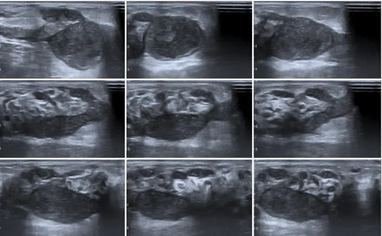
All patients underwent a USG evaluation in our center before the biopsy procedure. The sonographic results of the patients accord-ing to the BI-RADS lexicon were predominantly BI-RADS 3 and BI-RADS 4a (Table 5). In 21 (56.8%) patients, the initial diagnosis of GM was noted in USG reports. Only one patient was reported as suspicious for inflammatory breast cancer and categorized as BI-RADS 5 lesion. The distribution of lesions by quadrants is shown in Table 6. In our study group, only one quadrant involvement (48.6%) was the most frequent involvement, followed by retroareolar space involvement (43.2%). The most common USG features were hy-poechoic mass or masses with tubular extensions (total 54%) (Table 7, Figure 3).
Thirteen of the 37 patients had mammography. Mammography was performed in seven of the 25 patients aged under 40 (between 30-38, mean age 31.4) years, and six of 12 patients aged 40 years and over (age 44-59, mean age 49.3). The most common mammographic
Key Points
• Although the most frequent diagnosis of IGM is in May and Octo-ber in our patient population, there was no statistically significant difference.
• In more than half of IGM cases, US could provide an accurate pre-diagnosis approach of IGM.
• Besides MRI has a very limited role in discriminating malignancies from IGM, MRI enhancement kinetics may help in distinguishing this condition from malignancies.
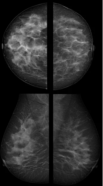
finding of IGM was asymmetrically increased density in our study population (Figure 4). Mammographic features were normal in one
(7.7%) patient, focal asymmetrically increased density in six (46.2%), diffuse asymmetrically increased density in four (30.7%), both diffuse asymmetrically increased density and parenchymal distortion in one (7.7%), and mass in one (7.7%) patient.
Figure 1. a, b. (a) x200 (b) x100 magnification hematoxylin and eosin stain (H&E) demonstrating perilobular inflammation and granuloma
formation in the background of diffuse lympho-histiocytic infiltration with giant cells
a
b
Figure 2. Unilateral breast erythema with retraction of the nipple
and also draining sinus tract in the left breast of a 38-year-old- patient
Table 1. Clinical characteristics of patients
Characteristics (n=37)
Age, mean (range), years 37.56±7.41 (24-59) ≤40 years, n (%) 29 (78.4) >40 years, n (%) 8 (21.6) Pregnancy history, n (%) 35 (94.5) Delivery, n (%) 34 (91.8) Number of births 1.48 (1-3) Years postpartum ≤5, n (%) 8 (22.9) >5, n (%) 27 (77.1) Lactation, n (%) 1 (2.7) Abortion history, n (%) 9 (24.3)
Table 2. Local manifestation of IGM
Characteristics IGM (n=37) Side, n (%) Right 12 (32.4) Left 25 (67.5) Bilateral 0 Nipple Retraction, n (%) 6 (16.2) Galactorrhea, n (%) 2 (5.4)
The diameter of mass, (cm)
Mean 5.8
Range 3.0-10.0 Pain, n (%) 37 (100) Lymph node enlargement, n (%) 14 (37.8)
IGM: idiopathic granulomatous mastitis
Table 3. Number of cases over the months
Month Number of cases % in overall2 5 13.5 3 5 13.5 5 7 18.9 6 1 2.7 9 4 10.8 10 3 8.1 11 7 18.9 12 5 13.5
237
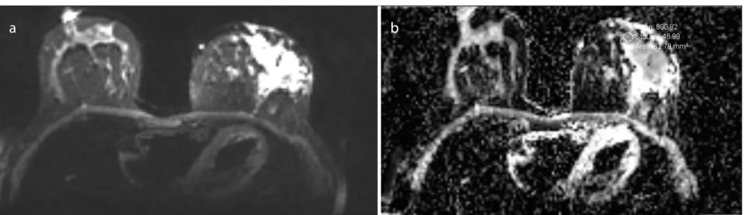
Breast MRI was performed in 10 patients; non-mass enhancement (NME) was observed in five of these patients, mass was found in three, and both NME and mass were detected in two patients (Table 8). In kinetic mea-surements, one lesion showed a type 2 curve, and nine lesions had a type 1 curve (Figure 5). Diffusion-weighted imaging was performed in six pa-tients and all lesions showed diffusion restriction (Figure 6). The mean ADC values were 0.78 mm2/s.10-3 and ranged from 0.6-0.92 mm2/s.10-3.
There was no follow-up information of 22 (59.4%) patients in our center. The follow-up time of 15 patients with follow-up data in our center ranged between one and 26 months and the average follow-up period was 11.8 months. In the radiologic follow-up, complete recov-ery was observed in six (40%) patients, regression in four (26.6%), progression in three (20%), recurrence after recovery in one (6.7%), and one (6.7%) patient’s USG findings were stable. Surgical excision was performed in two patients because of an insufficient response to medical treatment.
Discussion and Conclusion
IGM is considered as a rare chronic non-specific inflammatory lesion of the breast (1). Histopathologically, it is characterized by the presence
Table 7. Sonographic features of IGM
Sonographic features n %
Multiple irregular hypoechoic 11 29.7 masses and collections with tubular
connection with internal echoes
A large irregular hypoechoic 9 24.3 parallel mass with tubular extensions
Focal hypoechoic heterogeneity 6 16.2 with indistinct border
An irregular hypoechoic mass with 3(+2) 13.5 internal echoes (+signs of inflammation Total 5 around the mass)
Collection areas with low-level internal 4 10.8 echoes consistent with abscesses
The hypoechoic heterogeneous masses 2 5.4 within ducts and inflammation signs around
the ducts
IGM: idiopathic granulomatous mastitis
% within month group 100.00% 0.00% % within age group 5.26% 0.00%
Season 4 (9,10,11) Cases 8 6
% within month group 57.14% 42.86% % within age group 42.10% 33.33%
Table 5. BI-RADS categorization of the lesions
BI-RADS category n % 3 14 38.7 4A 15 40.5 4B 4 10.8 4C 3 8.1 5 1 2.7
BI-RADS: Breast Imaging-Reporting and Data System
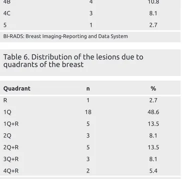
Table 6. Distribution of the lesions due to
quadrants of the breast
Quadrant n % R 1 2.7 1Q 18 48.6 1Q+R 5 13.5 2Q 3 8.1 2Q+R 5 13.5 3Q+R 3 8.1 4Q+R 2 5.4
Q: quadrant; R: retroareolar region
of epithelioid and multinucleated giant cell non-caseating granulomas accompanied by neutrophils around the lobules (13). Although it has
been reported that the age of patients with IGM may range from 11 to 80 years (14), the high-risk group is women, aged between 30 and 40 years (15). In our study population, eight patients were aged over 40 years and five patients were aged under 30 years, the mean age of the study population was 37.56±7.41 years, correlating with the literature. The etiology and pathogenesis of IGM remain unclear. An association with pregnancy, lactation, a locally autoimmune process, infection, hyperprolactinemia, and chemical reaction induced by oral contracep-tive pills have been reported in previously published articles (10, 11). After reviewing the literature, it is revealed that the majority of patients are of Mediterranean (Turkey and Jordan) and Asian (Arabia, China, and Malaysia) origin (4). Although no obvious ethnic predisposition has been previously reported, the prevalence of IGM in specific ethnic populations has been mentioned in several reports (5, 16). Previous studies supported the conclusion that patients with IGM were usu-ally parous women with a recent history of pregnancy and delivery (9, 14). In our study group, 94.6% of patients had a history of pregnancy and one was pregnant at the time of diagnosis. It has been published that extravasation lactational secretions may spontaneously produce a granulomatous inflammatory response (5, 17). Also, that high serum prolactin levels and subsequent overexcitation and lactation change can potentially cause IGM (18). In our study population, two patients have galactorrhea and one patient was lactating at the time of diag-nosis.
The most common clinical symptoms of IGM include erythema, edema, variable sized-sensitive-palpable unilateral breast mass, nipple retraction, ulceration, discharge, and axillary lymphadenopathy (19). The presence of a fistula tract in patients is an important clinical clue for the referral diagnosis of IGM (20, 21). In our study, fistula was present in approximately 20% of patients. In some studies, the fistula
Figure 3. Ultrasound images of a patient with hypoechoic masses containing internal echoes with tubular extensions
Table 8. MRI features of IGM (5 cases shows
non-mass enhancement (NME), 3 cases non-mass and, 2
cases both NME and mass)
MRI lesion type and imaging feature n
Non-mass enhancement 7 Patterns: Clustered ring 3 Heterogeneous 3 Clumped 1 Distribution: Regional 6 Diffuse 1 Mass 5 Shape: Round 3 Irregular 2 Margin: Circumscribed 1 Irregular 4
IGM: idiopathic granulomatous mastitis; MRI: magnetic resonance imaging
presentation was found as 30-50% (8, 9, 22). In our study group, axillary lymph node enlargement was observed in 37.8% of patients, and studies have shown unilateral lymph node involvement as between 20-60% in IGM (9, 23).
To our knowledge, there is no article discussing seasonal frequency in IGM. Most of our patients presented to the hospital and biopsied in May and November (n=14, 37.8%), but there was no significant differ-ence when compared with the other months. Of these two months when the most frequent cases were seen, May corresponds to the end of spring, and November to the end of autumn. Most of the patients presented to hospital in autumn and spring. Only one patient’s diagnosis was in sum-mer. Although there was no statistically significant difference between the months, the distribution of months may show us seasonal proximity in this condition and this feature may help to understand the etiopathogen-esis of IGM. Our population was very limited, so to investigate the sea-sonal relationship in this condition there is a need for studies with much larger numbers of patients. We think that the evaluation of the onset of symptoms rather than the biopsy date will lead to more accurate results.
extensions (54%), which allows us to consider the preliminary diagno-sis of granulomatous mastitis (9, 24, 25).
In our cases, single quadrant involvement (48.6%) was the most frequent involvement, followed by retroareoler space involvement (43.2%). In a study that evaluated 30 patients, lesion extension to the retroareoler space was found in 50% of patients (9). In another study with 37 patients, retroareoler involvement was found in 66.7% of pa-tients, and all-quadrant involvement (38.1%) was the most frequent (22). In this study, the reason that the retroareoler area involvement and 3 and more quadrant involvement was more frequent than our study may be due to the population of the other study comprising patients who had MRI for further investigations. It may also be due to the better determination of the extent of lesions with MRI. Retroar-eoler involvement may be related to the process and progression of lesions. In a study, it was shown that patients with retroareoler space involvement had poorer treatment success (26). Therefore, it may be clinically important to document whether retroareoler site involve-ment is present.
Although mammographic sensitivity is low due to the young age group having a dense breast pattern, the most common mammographic find-ing in our study was asymmetrically increased density (10/13). In pre-vious studies, almost half of the mammograms were negative and the most common finding was an asymmetric density, which was a non-specific finding (9, 21, 25, 27).
There are a few studies about the MRI features of IGM, and this mo-dality has a very limited role in discriminating malignancies from IGM in the initial diagnosis of this condition. MRI findings have a wide spectrum (25, 28, 29). Although NME was seen in all MRIs in one study, in our study NME was observed in 70% of MRIs (28). Aslan et al. (29) showed NME in 92.3% of patients, and Yilmaz et al. (22) reported NME in 55% of patients.
One of the most striking findings of this condition in MRI was that the kinetic curve was seen as a type 1 curve in most cases. In contrast to our findings, Chu et al. (28) found wash-out in all of lesions. Yilmaz et al. (22) reported that 64% of patients had type 1 enhancement, and 36% of patients had type 2 enhancement. In MRI, enhancement kinetics may help in distinguishing this condition from malignancies. Although IGM is a benign condition, in our study, all lesions showed diffusion restriction, similar to a study by Aslan et al. (29) However, in our study, the mean ADC values were found lower than in that study. In a study in terms of ADC values, there was no difference in IGM
Figure 4. In the mammography of a patient asymmetrically increased
density in upper quadrant central portion of the right breast
and malignancies (25). In contrast to that study, a recent study dem-onstrated that in non-mass enhancement without rim-enhancement, using texture analysis in diffusion images was useful in the differentia-tion of IGM from malignancies (30). In a study evaluating mastitis, it was shown that ADC values could be used to classify mastitis subtypes (31). There are conflicting MRI findings in different studies and in our study, MRI findings were non-specific, similar to the literature. Besides the clinical features, radiologic findings of IGM may also be confused with malignant pathologies of the breast. The disease must
be diagnosed through a pathologic evaluation. Fine needle aspiration is not satisfactory in distinguishing malignant and other benign inflam-matory disorders. A core biopsy should be preferred for this purpose (5, 7, 23). In our patient group, all patients were diagnosed after hav-ing a core needle biopsy. A diagnostic excisional biopsy is not preferred due to substantial scratching, loss of breast symmetry, breast deformity, and the possibility of unhealed ulcers or sinus tract formation (7, 32). Treatment of IGM should be initiated after the exclusion of infective causes. It involves non-surgical management including surveillance,
Figure 5. a-d. Non-mass enhancement on the right breast with type 1 kinetic curve
a
c
b
d
Figure 6. a, b. Diffusion restriction (ADC value: 0.90 mm2/s.10-3) of granulomatous mastitis on diffusion-weighted imaging
a
b
in patients who had follow-up information was 26.6%.
The main limitation of the study is the low number of patients. An-other important limitation of the current study is that there are no data on how long after symptoms developed in patients the biopsy was performed. The date of the biopsy was accepted as the date of illness. Patients may have presented at different periods after symptoms devel-oped, and so, the time between symptoms and biopsy may vary from case to case. For this reason, considering the date of biopsy as the date of disease in our study may have caused some errors and bias. Studies with a larger series focusing on the onset of symptoms may shed light on the IGM season relationship.
In conclusion, IGM is a rare chronic non-specific inflammatory le-sion of the breast, which can be confused with benign and malignant breast diseases in both clinical and radiologic aspects. To understand the etiology of this condition better, the seasonal connection needs to be evaluated in larger patient groups.
Ethics Committee Approval: Ethics committee approval was received for this study from the ethics committee of Muğla Sıtkı Koçman University (Number: 74, Date:02/06/2020).
Informed Consent: Written informed consent was obtained from patients be-fore the biopsy.
Peer-review: Externally peer-reviewed.
Author Contributions: Concept – L.T., F.D.E.; Design – F.D.E., L.T.; Su-pervision – F.D.E.; Resources – L.T., F.D.E.; Materials – L.T., F.D.E.; Data Collection and/or Processing – L.T., F.D.E.; Analysis and/or Interpretation – F.D.E., L.T.; Literature Search – F.D.E., L.T.; Writing Manuscript – F.D.E., L.T.; Critical Review – F.D.E., L.T.
Conflict of Interest: The authors have no conflicts of interest to declare.
Financial Disclosure: The authors declared that this study has received no fi-nancial support.
References
1. Sheybani F, Sarvghad M, Naderi HR, Gharib M. Treatment for and clini-cal characteristics of granulomatous mastitis. Obstet Gynecol 2015; 125: 801-807. (PMID: 25751209) [Crossref]
2. Gurleyik G, Aktekin A, Aker F, Karagulle H, Saglamc A. Medical and surgical treatment of idiopathic granulomatous lobular mastitis: A benign inflammatory disease mimicking invasive carcinoma. J Breast Cancer 2012;15: 119-123. (PMID: 22493638) [Crossref]
(PMID: 29528819) [Crossref]
8. Dursun M, Yilmaz S, Yahyayev A, Salmaslioglu A, Yavuz E, Igci A, Acu-nas G, Tunacı M. (2012) Multimodality imaging features of idiopathic granulomatous mastitis: outcome of 12 years of experience. Radiol Med 2012; 117: 529-538. (PMID: 22020426) [Crossref]
9. Yildiz S, Aralasmak A, Kadioglu H, Toprak H, Yetis H, Gucin Z, Kocakoc E. Radiologic findings of idiopathic granulomatous mastitis. Med Ultra-son 2015; 17: 39-44. (PMID: 25745656) [Crossref]
10. Altintoprak F, Kivilcim T, Ozkan OV. Aetiology of idiopathic granuloma-tous mastitis World J Clin Cases 2014; 2: 852-858. (PMID: 25516860)
[Crossref]
11. Uysal E, Soran A, Sezgin E. Factors related to recurrence of idiopathic granulomatous mastitis: what do we learn from a multicentre study? ANZ J Surg 2018; 88: 635-639. (PMID: 28749045) [Crossref]
12. American College of Radiology. Breast Imaging Reporting & Data Sys-tem, BI-RADS: Mammography. 5th ed. Reston, VA: 2013.
13. Tuli R, O'Hara BJ, Hines J, Rosenberg AL. Idiopathic granulo-matous mastitis masquerading as carcinoma of the breast: A case report and review of the literature. Int Semin Surg Oncol 2007; 4: 21. (PMID: 17662130)
[Crossref]
14. Bani-Hani KE, Yaghan RJ, Matalka II, Shatnawi NJ. Idiopathic granulo-matous mastitis: Time to avoid unnecessary mastectomies. Breast J 2004; 10: 318-322. (PMID: 15239790) [Crossref]
15. Pereira FA, Mudgil AV, Macias ES, Karsif K. Idiopathic granulomatous lobular mastitis. Int J Dermatol 2012; 51: 142-151. (PMID: 22250621)
[Crossref]
16. Centers for Disease Control and Prevention (CDC). Idiopathic granu-lomatous mastitis in Hispanic women -Indiana, 2006-2008. MMWR Morb Mortal Wkly Rep 2009; 58: 1317-1321. (PMID: 19959984) 17. Marriott DA, Russell J, Grebosky J, Wallace AM, Joste N, Royce ME.
Idiopathic granulomatous lobular mastitis masquerading as a breast ab-scess and breast carcinoma. Am J Clin Oncol 2007; 30: 564-565. (PMID: 17921721) [Crossref]
18. Cserni G, Szajki K. Granulomatous lobular mastitis following drug-in-duced galactorrhea and blunt trauma. Breast J 1999; 5: 398-403. (PMID: 11348321) [Crossref]
19. Ocal K, Dag A, Turkmenoglu O, Kara T, Seyit H, Konca K. Granuloma-tous mastitis: clinical, pathological features, and management. Breast J 2010; 16: 176-182. (PMID: 20030652) [Crossref]
20. Almasad, Jamal K. Mammary duct fistulae: classification and manage-ment. ANZ J Surg 2006, 76: 149-152. (PMID: 16626355) [Crossref]
21. Ozel L, Unal A, Unal E, Kara M, Erdoğdu E, Krand O, Güneş P, Karagül H, Demiral S, Titiz MI. Granulomatous mastitis: is it an autoimmune disease? Diagnostic and therapeutic dilemmas. Surg Today 2012; 42: 729-733. (PMID: 22068681) [Crossref]
22. Yilmaz R, Demir AA, Kaplan A, Sahin D, Ozkurt E, Dursun M, Acu-nas G. Magnetic resonance imaging features of idiopathic granuloma-tous mastitis: is there any contribution of diffusion-weighted imaging in the differential diagnosis?. Radiol Med 2016; 121: 857-866. (PMID: 27406630) [Crossref]
23. Sripathi S, Ayachit A, Bala A, Kadavigere R, Kumar S. Idiopathic granulo-matous mastitis: a diagnostic dilemma for the breast radiologist. Insights Imaging 2016; 7: 523-529. (PMID: 27164916) [Crossref]
24. Engin G, Acunaş G, Acunaş B. Granulomatous mastitis: gray-scale and color Doppler sonographic findings. J Clin Ultrasound 1999; 27: 101-106. (PMID: 10064406) [Crossref]
25. Yilmaz E, Lebe B, Usal C, Balci P. Mammographic and sonographic find-ings in the diagnosis of idiopathic granulomatous mastitis. Eur Radiol 2001; 11: 2236-2240. (PMID: 11702165) [Crossref]
26. Altunkeser A, Arslan FZ, Eryılmaz MA. Magnetic resonance imaging findings of idiopathic granulomatous mastitis: can it be an indirect sign of treatment success or fail?. BMC Med Imaging 2019; 19: 94. (PMID: 31842782) [Crossref]
27. Memis A, Bilgen I, Ustun EE, Ozdemir N, Erhan Y, Kapkac M. Granulo-matous mastitis: imaging findings with histopathologic correlation. Clin Radiol 2002; 57: 1001-1006. (PMID: 12409111) [Crossref]
28. Chu AN, Seiler SJ, Hayes JC, Wooldridge R, Porembka JH. Magnetic resonance imaging characteristics of granulomatous mastitis. Clin Imag-ing 2017; 43: 199-201. (PMID: 28364724) [Crossref]
29. Aslan H, Pourbagher A, Colakoglu T. Idiopathic granulomatous mastitis: magnetic resonance imaging findings with diffusion MRI. Acta Radiol 2016; 57: 796-801. (PMID: 2650879) [Crossref]
30. Zhao Q, Xie T, Fu C, Chen L, Bai Q, Grimm R, Peng W, Wang S. Differ-entiation between idiopathic granulomatous mastitis and invasive breast carcinoma, both presenting with non-mass enhancement without rim-enhanced masses: The value of whole-lesion histogram and texture analy-sis using apparent diffusion coefficient. Eur J Radiol 2020; 123: 108782. (PMID: 31864142) [Crossref]
31. Zhang L, Hu J, Guys N, Meng J, Chu J, Zhang W, Liu A, Wang S, Song Q. Diffusion-weighted imaging in relation to morphology on dynamic contrast enhancement MRI: the diagnostic value of characterizing non-puerperal mastitis. Eur Radiol 2018; 28: 992-999. (PMID: 28956122)
[Crossref]
32. Kok KY, Telisinghe PU. Granulomatous mastitis: presentation, treat-ment and outcome in 43 patients. Surgeon 2010; 8: 197-201. (PMID: 20569938) [Crossref]
33. Freeman CM, Xia BT, Wilson GC, Lewis JD, Khan S, Lee SJ, Lower EE, Edwards MJ, Shaughnessy EA. Idiopathic granulomatous mastitis: A diagnostic and therapeutic challenge. Am J Surg 2017; 214: 701-706. (PMID: 28739122) [Crossref]
34. Atak T, Sagiroglu J, Eren T, Alimoglu O. Strategies to treat idiopathic granulomatous mastitis: retrospective analysis of 40 patients. Breast Dis 2015; 35: 19-24. (PMID: 24989362) [Crossref]