CASE REPORT
119
1Department of Otolaryngology, Medipol University, İstanbul, Turkey
2Department of Otolaryngology, Erciyes University School of Medicine, Kayseri, Turkey 3Department of Radiology, Erciyes University School of Medicine, Kayseri, Turkey
4Department of Otolaryngology, Kayseri Training and Research Hospital, Kayseri, Turkey
Received 07.09.2015 Accepted 25.01.2016 Correspondence Dr. Alperen Vural, Erciyes Üniversitesi Tıp Fakültesi, Kulak Burun Boğaz Anabilim Dalı, Kayseri, Turkey Phone: +90 533 236 86 16 e.mail: alperenvural@yahoo.com
©Copyright 2016 by Erciyes University School of Medicine - Available online at www.erciyesmedj.com
Zygomatic Abscess with Temporomandibular
Joint Effusion Complicating Acute Otitis Media
Hanifi Kaya
1, Alperen Vural
2, Mehmet Akif Somdaş
2, Mustafa Öztürk
3, Murat Doğan
4ABSTRACT The incidences of extracranial and intracranial complications of acute otitis media (AOM) in children have markedly de-creased in the postantibiotic era. Zygomatic abscesses are the rarest type of abscesses originating from mastoiditis. This paper presents a case with a zygomatic abscess as a complication of acute coalescent mastoiditis in a 7-year-old girl who underwent cortical mastoidectomy and myringotomy–ventilation tube insertion.
Keywords: Acute otitis media, complications, subperiosteal abscess, zygomatic abscess Erciyes Med J 2016; 38(3): 119-21 • DOI: 10.5152/emj.2016.150046
INTRODUCTION
Acute coalescent mastoiditis (ACM) and a mastoid subperiosteal abscess are extracranial complications of acute otitis media (AOM). Subperiosteal abscesses are the most frequent complications of ACM (1-6). Zygomatic ab-scesses are a form of mastoid subperiosteal abab-scesses. There are three types of mastoid abab-scesses: i) posterior sub-periosteal (postauricular), ii) inferior subsub-periosteal [a-sternocleidomastoid (Bezold’s abscess) and b-digastric (Citelli’s abscess)], and iii) anterior subperiosteal (zygomatic) (7). Zygomatic abscesses are the rarest of these. There are few case reports in the literature in which the abscess causes some degree of myositis, but temporomandibular joint arthritis is poorly known to further associate with complication (7-11).
Despite modern antibiotic therapy, ACM can still be a potentially dangerous situation due to possible extracranial and intracranial spread (1). In the past, in 25–50% of patients, AOM resulted in ACM; by 1950 the reported inci-dence of surgical mastoidectomy because of AOM was below 10% (12, 13). Antibiotics reduced the complication rate to 0.02–0.15%, yet the mortality rate is still high (approximately 20%), particularly in populations with lower socioeconomic conditions (1, 12-14). However, complications of AOM might be expected to increase with the escalation of antibiotic resistance (15). ACM is the most common complication of AOM in children, but recently, it has been reduced to an almost nonexistent event with an incidence between 0.2 and 2% (2, 5). ACM may occur in the absence of an obvious middle ear infection, when an aditus obstruction segregates the middle ear and mastoid cells. This dangerous condition is known as silent otits media, where the tympanic membrane is almost normal in 10-20% of ACM cases (6, 16).
The aim of this report is to present the clinical findings and progress of a patient with a zygomatic abscess, which is a rare complication of AOM.
CASE REPORT
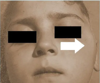
A 7-year-old girl was admitted to the Department of Otolaryngology with ear pain, hearing loss, otorrhea, and swelling of the zygomatic region on the left side. She had a history of otitis media with left ear pain, hearing loss, fever, and otorrhea and had been intramuscularly administered 1 g of ceftriaxone for the previous 5 days. She then developed a swelling on the left side of her face, which drove the family to our hospital. On her physical examination, a purulent material was aspirated from the external auditory channel. The tympanic membrane was irregular and macerated. A painful, diffuse, nonerythematous swelling on the zygomatic region was noted (Figure 1). Facial nerve functions were normal, and trismus and nystagmus were not observed. She was hospitalized, and intravenous antibiotics (ceftriaxone 1x1 g and clindamycin 3x200 mg) were started, but no improvement was observed in the following 48 h. The leukocyte count was 7500/mm³. The patient’s aerobic culture was negative. A conductive hearing loss of 40 dB on the left side was found on audiometry, and a type B trace was observed on tympanometry. The examination of the right ear was normal. A computed tomography (CT) scan of the temporal bones showed an inflammatory infiltration of the left zygomatic region and opacification of pneumatic cells of the
left mastoid. Cranial magnetic resonance imaging (MRI) showed an abscess formation, 3x1 cm in size, in the left zygomatic region. There was an exantion to the temporomandibular joint and an incrase in the intra-articular fluid amount. The inflammation also extended to the muscles (myositis) (Figure 2).
The patient underwent surgical treatment. Cortical mastoidectomy through the postauricular approach and myringotomy–ventila-tion tube insermyringotomy–ventila-tion was performed. The zygomatic region was not opened but was squeezed instead, and the purulent material that drained from the zygomatic region to the mastoid cells was aspirat-ed. The granulation tissue blocking the aditus and all other patho-logic tissue were removed. A transmastoid drain was placed in the mastoid cavity. The mastoid cavity was aspirated daily for the fol-lowing 7 days. The patient’s postoperative recovery was quick;
fe-ver, swelling of the zygomatic region, pain, and purulent discharge from left ear disappeared. On the 7th day after the surgery, no
secretion was present, and the drain from the mastoid cavity was removed. Her histopathological examination result was reported as inflammation with granulation and fibrin tissue. The patient was discharged from the hospital 9 days after the surgery with no ear complaints and no swelling of the left zygomatic region. Her pure tone audiometry test was totally normal on the first control which was performed 1 week after discharge. The tube was removed from the ear channel 6 months later. Informed consent was taken from the patient for this case report.
DISCUSSION
Acute coalescent mastoiditis is the most common complication of otitis media in all pediatric age groups (15, 17). During the last 60 years, antibiotics and public health care systems have significantly decreased the incidence and modified the treatment for these com-plications. The incidence of ACM is less than 0.1% (18), and sub-periosteal abscesses are very rare. Zygomatic abscesses, which are the rarest type of the mastoid subperiosteal abscesses, can lead to periosteal elevation under the temporalis muscle with pain, tender-ness, and swelling in the region of the zygomatic process. Middle ear infection spreading to the mastoid can extend through the tym-panomastoid suture or along vascular channels in the cribriform area. Direct erosion of the mastoid cortex by inflammation is also possible (19). In our case the progression pathway of the mastoid abscess to the subperiosteal area was unclear.
The most common symptoms of a subperiosteal abscess are fever (100%), tenderness (85%), erythema (79%), swelling and protrusion of the auricle (73%), spontaneous tympanic membrane perforation (24%), and facial palsy (9%) (6); in our case, all, except facial palsy, were present in our case. Tympanic membrane perforation on its own was too small to be detected; however, obviously, there was a perforation where the drainage came through. An elevated C-reactive protein (CRP) level (>100) and leukocyte count (>15,000/ mm3) are expected in case of a zygomatic abscess (6); these were
normal in our case. Previously used antibiotics might be the reason for this situation.
A diagnosis of complicated AOM is based on clinical and radio-logical findings and laboratory assessment. Coalescent mastoiditis is difficult to diagnose due to both the infrequency of presenta-tion and inconsistency of signs or symptoms. A laboratory evalua-tion is usually not helpful; most patients are afebrile, and they lack systemic complaints because of previously used antibiotics. Thus, there must be a high index of suspicion during the management of these patients, and early CT scans of the temporal bone are es-sential. If a complication is found, MRI should be the next imaging technique to help surgical planning (20). In our case, an abscess was not detected through a CT scan but was detected through MRI. Bacteria cultured from complicated middle ear and mastoid ef-fusions are more resistant to regularly applied antibiotics than bacteria found in uncomplicated AOM (21). Streptococcus
pneu-moniae is the most frequently cultured bacteria in cases of both
AOM and its complications (21). In addition, Pseudomonas
ae-ruginosa, Streptococcus pyogenes, and Staphylococcus aureus
are frequently found in cultures of mastoid effusion in case of acute Figure 2. Picture showing a left-sided zygomatic abscess in
the coronal section of MRI
Figure 1. Picture showing the patient with swelling in the zygomatic region (arrow)
mastoiditis (3, 15). Anaerobic otitis media or mastoiditis tends to follow a subacute and subclinical course in comparison with the course of aerobic illness (22). This is shown by an afebrile course, a lack of periauricular pain or tenderness, a noninflamed tympanic membrane, and a near-normal leukocyte count (23). In our case, the clinical course was rather serious, but anaerobic microorgan-ism might be the reason for the negative culture. Previously used antibiotics might have had an influence as well.
Coalescent mastoiditis without further complications usually re-sponds to more conservative treatment (2, 17). However, myrin-gotomy or tympanostomy tube insertion is recommended to en-sure adequate drainage of the middle ear. Treatment is supposed to open the blocked aditus; otherwise, cortical mastoidectomy should be performed. Cholesteatoma and purulent otorrhea and/ or granulation tissue resistant to topical and systemic antibiotics for more than 2 weeks are indications for mastoidectomy (24, 25). Additionally, in case of coalescent mastoiditis in children where signs and symptoms do not subside within 48 h, mastoidectomy should be performed (24, 25). All other complications also require mastoidectomy.
CONCLUSION
Zygomatic abscesses are the rarest type of the mastoid subperi-osteal abscesses. The diagnosis of subperisubperi-osteal abscesses is diffi-cult due to both the infrequency of presentation and inconsistency of signs or symptoms. Early diagnosis, adequate medication, and mastoidectomy are necessary to treat subperiosteal abscesses.
Informed Consent: Written informed consent was obtained from the
pa-tient.
Peer-review: Externally peer-reviewed.
Authors’ Contributions: Conceived and designed the experiments or
case: MAS, HK, AV. Performed the experiments or case: MAS, HK, AV. Analyzed the data: HK, AV, MÖ, MD. Wrote the paper: HK, AV. All au-thors have read and approved the final manuscript.
Conflict of Interest: No conflict of interest was declared by the authors.
Financial Disclosure: The authors declared that this study has received no
financial support.
REFERENCES
1. Kangsanarak J, Fooanant S, Ruckphaopunt K, Ruckphaopunt K. Extracranial and intracranial complications of suppurative otitis me-dia, report of 102 cases. J Laryngol Otol 1993; 107(11): 999-1004.
[CrossRef]
2. Palva T, Virtanen H, Makinen J. Acute and latent mastoiditis in chil-dren. J Laryngol Otol 1985; 99(2): 127-36. [CrossRef]
3. Leskinen K, Jero J. Complications of acute otitis media in children in southern Finland. Int J Pediatr Otorhinolaryngol 2004; 68(3): 317-24
[CrossRef]
4. Spratley J, Silveira H, Alvarez I, Pais-Clemente M. Acute mastoiditis in children: review of the current status. Int J Pediatr Otorhinolaryngol 2000; 56(1): 33-40. [CrossRef]
5. Kuczkowski J, Mikaszewski B. Intracranial complications of acute and chronic mastoiditis: report of two cases in children. Int J Pediatr Oto-rhinolaryngol 2001; 60(3): 227-37. [CrossRef]
6. Gliklich RE, Eavey RD, Iannuzzi RA, Camacho AE., A contemporary analysis of acute mastoiditis. Arch Otolaryngol Head Neck Surg 1996; 122: 135-9. [CrossRef]
7. Spiegel JH, Lustig LR, Lee KC, Murr AH, Schindler RA. Contem-porary presentation and management of a spectrum of mastoid ab-scesses. Laryngoscope 1998: 822-8. [CrossRef]
8. Kuczkowski J, Narozny W, Stankiewicz C, Mikaszewski B, Izycka-Swieszewska E. Zygomatic abscess with temporal myositis - a rare extracranial complication of acute otitis media. Int J Pediatr Otorhino-laryngol 2005; 69(4): 555-9. [CrossRef]
9. Tsai CJ, Guo YC, Tsai TL, Shiao AS. Zygomatic abscess complicating a huge mastoid cholesteatoma with intact eardrum. Otolaryngol Head Neck Surg 2003; 128(3): 436-8. [CrossRef]
10. Warnaar A, Snoep G, Stals FS. A swollen cheek: an unusual course of acute mastoiditis. Int J Pediatr Otorhinolaryngol 1989; 17(2): 179-83. [CrossRef]
11. Gurgel RK, Woodson EA, Lenkowski PW, Gubbels SP, Hansen MR. Zygomatic Root Abscess: A Rare Complication of Otitis Media. Otol Neurotol 2010; 31(5): 856-7. [CrossRef]
12. Matin MA, Khan AH, Khan FA, Haroon AA. A profile of 100 compli-cated cases of chronic suppurative otitis media. J R Soc Health 1997; 117(3): 157-9. [CrossRef]
13. Osma U, Cureoglu S, Hosoglu S. The complications of chronic otitis me-dia: report of 93 cases. J Laryngol Otol 2000; 114: 97-100. [CrossRef]
14. Shamboul KM. An unusual prevalence of complications of chronic suppurative otitis media in young adults. J Laryngol Otol 1992; 106: 874-7. [CrossRef]
15. Butbul-Aviel Y, Miron D, Halevy R, Koren A, Sakran W. Acute mas-toiditis in children: Pseudomonas aeruginosa as a leading pathogen. Int J Pediatr Otorhinolaryngol 2003; 67(3): 277-81. [CrossRef]
16. Vera-Cruz P, Farinha RR and Calado V. Acute mastoiditis in children—our experience. Int J Pediatr Otorhinolaryngol 1999; 50(2): 113-7. [CrossRef]
17. Go C, Bernstein JM, de Jong AL, Sulek M, Friedman EM. , Intracra-nial complications of acute mastoiditis. Int J Pediatr Otorhinolaryngol 2000; 52(2): 143-48. [CrossRef]
18. Casselbrant ML, and Mandel EM. Epidemiology. In: Rosenfeld MR and Bluestone CD (Editors). Evidence-Based Otitis Media. Ontario BC Decker, Hamilton 1999; pp.117-36.
19. Hawkins DB and Dru D. Mastoid subperiosteal abscess. Arch Otolar-yngol Head Neck Surg 1989; 109: 369-71. [CrossRef]
20. Vazquez E, Castellote A, Piqueras J, Mauleon S, Creixell S, Pumarola F, et al. Imaging of complications of acute mastoiditis in children. Ra-diographics 2003; 23(2): 359-72. [CrossRef]
21. Zapalac JS, Billings KR, Schwade ND, Roland PS. Suppurative com-plications of acute otitis media in the era of antibiotic resistance. Arch Otolaryngol Head Neck Surg 2002; 128(6): 660-3. [CrossRef]
22. Moloy PJ. Anaerobic mastoiditis: a report of two cases with complica-tions. Laryngoscope 1982; 92(11): 1311-5. [CrossRef]
23. Babin E, Lemarchand V, Moreau S, Goullet de Rugy M, Valdazo A, Bequignon A. Failure of antibiotic therapy in acute otitis media. J Laryngol Otol 2003; 117: 173-6. [CrossRef]
24. Michalski G, Hocke T, Hoffmann K, Goullet de Rugy M, Valdazo A, Bequignon A., Therapy of acute mastoiditis. Laryngorhinootologie 2002; 81(12): 857-60. [CrossRef]
25. Taylor MF and Berkowitz RG. Indications for mastoidectomy in acute mastoiditis in children. Ann Otol Rhinol Laryngol 2004; 113(1): 69-72. [CrossRef]
