ABSTRACT
Objective: Evaluation of the effectiveness of ultrasound (US)-guided erector spinae plane block (ESPB) and thoracic paravertebral block (TPVB) compared to no intervention control group for postoperative pain management in video assisted thoracic surgery (VATS) patients.
Method: Three groups - Group ESPB, Group TPVB and the control group (n=30 per group) were included in this prospective, randomized, controlled study. The US-guided blocks were performed preoperatively in the ESPB and TPVB groups. Intravenous patient-controlled postoperative anal-gesia via fentanyl was administered in all of the patients. The patients were evaluated using visual analogue scale (VAS) scores, opioid consumption, and adverse events.
Results: At all time intervals fentanyl consumption and VAS scores were significantly lower both in ESPB and TPVB groups compared to the control group (p<0.001). Block procedure time was significantly lower and success of one time puncture was higher in Group ESPB as compared with that in Group TPVB (p<0.001).
Conclusion: ESPB and TPVB provide more effective analgesia compared to control group in patients who underwent video-assisted thoracic surgery. ESPB had a shorter procedural time and higher success of single-shot technique compared to TPVB.
Keywords: Erector spinae plane block, thoracic paravertebral block, postoperative analgesia, video-assisted thoracic surgery
ÖZ
Amaç: Video yardımcılı torakal cerrahi yapılan hastalarda postoperatif analjezi yönetimi için ultrasonografi (US) eşliğinde yapılan erektor spina plan bloğu (ESPB) ve torakal paravertebral bloğun (TPVB) kontrol grubuna göre etkinliğinin değerlendirilmesi amaçlanmıştır.
Yöntem: Bu çalışmaya her grup için 30 hasta olmak üzere toplam 90 hasta dahil edilmiştir. Çalışma 3 gruptan oluşmaktadır; Grup ESPB, Grup TPVB ve Kontrol Grubu. ESPB ve TPVB grupla-rındaki hastalara preoperatif olarak US eşliğinde blok yapıldı. Tüm gruplardaki hastalara fentanil içeren hasta kontrollü analjezi (HKA) uygulandı. Hastalar vizuel analog skala (VAS), opioid tüketi-mi ve yan etkiler kaydedilerek değerlendirildi.
Bulgular: Tüm zaman aralıklarında fentanil tüketimi ve VAS Grup ESPB ve Grup TPVB de kontrol grubuna göre anlamlı olarak daha düşüktü (p<0.001). Blok işlem süresi ESPB grubunda anlamlı olarak daha kısaydı ve iğne ile tek giriş başarısı ESPB grubunda TPVB grubuna göre anlamlı olarak daha yüksekti (p<0.001).
Sonuç: ESPB ve TPVB, video yardımcılı torakal cerrahi yapılan hastalarda kontrol grubuna göre etkili analjezi oluşturmaktadır. ESPB, TPVB’ye göre daha kısa işlem süresi ve tek iğne girişi ile daha yüksek başarı oranına sahiptir.
Anahtar kelimeler: Erektor spina plan bloğu, torakal paravertebral blok, postoperatif analjezi, video yardımcılı torakal cerrahi
ID
Ultrasound-Guided Erector Spinae Plane Block
and Thoracic Paravertebral Block for
Postoperative Analgesia Management
Following Video-Assisted Thoracic Surgery:
A Prospective, Randomized, Controlled Study
Video Yardımcılı Torakal Cerrahi Sonrası
Postoperatif Analjezi Yönetimi için Ultrasonografi
Rehberliğinde Yapılan Erektor Spina Plan Bloğu
ve Torakal Paravertebral Blok Etkinliği:
Prospektif, Randomize, Kontrollü Çalışma
M. Ekinci 0000-0002-5580-5960 İ.C. Tukac 0000-0001-5578-6889 Y.O. Atalay 0000-0001-9928-9956
İstanbul Medipol Üniversitesi Tıp Fakültesi, Anesteziyoloji ve Reanimasyon Ana Bilim Dalı, İstanbul, Türkiye
E.C. Çelik 0000-0002-7773-9562
Erzurum Atatürk Üniversitesi Araştırma Hastanesi, Anesteziyoloji ve Reanimasyon Ana Bilim Dalı, Erzurum, Türkiye
B.E. Gölboyu 0000-0002-2011-2574
Manisa Şehir Hastanesi, Anesteziyoloji ve Reanimasyon Bilim Dalı, Manisa, Türkiye
M.Z. Günlüoğlu 0000-0002-6952-3470
İstanbul Medipol Üniversitesi Tıp Fakültesi, Göğüs Cerrahisi Ana Bilim Dalı, İstanbul, Türkiye
Bahadır Çiftçi Mürsel Ekinci Erkan Cem Çelik İsmail Cem Tukac Birzat Emre Gölboyu Mehmet Zeki Günlüoğlu Yunus Oktay Atalay
Bahadır Çiftçi Medipol Mega Üniversite Hastanesi Anesteziyoloji ve Reanimasyon Kliniği, İstanbul - Türkiye
✉
[email protected] ORCID: 0000-0002-3245-6614© Telif hakkı Anestezi ve Reanimasyon Uzmanları Derneği. Logos Tıp Yayıncılık tarafından yayınlanmaktadır. Bu dergide yayınlanan bütün makaleler Creative Commons Atıf-GayriTicari 4.0 Uluslararası Lisansı ile lisanslanmıştır. © Copyright Anesthesiology and Reanimation Specialists’ Society. This journal published by Logos Medical Publishing. Licenced by Creative Commons Attribution-NonCommercial 4.0 International (CC BY-NC 4.0)
Cite as: Çiftçi B, Ekinci M, Çelik EC, et al.
Ultrasound-guided erector spinae plane block and thoracic pa-ravertebral block for postoperative analgesia mana-gement following video-assisted thoracic surgery: A prospective, randomized, controlled study. JARSS 2020;28(3):170-8.
Received: 03 December 2019 Accepted: 04 May 2020 Publication date: 30 July 2020
ID ID ID ID ID ID
INTRODUCTION
Video-assisted thoracic surgery (VATS) has become the standard procedure for minor and major lung surgeries. Compared with open thoracotomy, VATS has the advantages of lower postoperative pulmo-nary complications, shorter length of hospital stay, better short-term outcome, and milder postoperati-ve pain (1,2). However, patients may complain of
moderate or severe postoperative and chronic pain
(3). For the management of postoperative pain,
opioid-sparing multimodal analgesic approaches such as thoracic epidural analgesia (TEA), intercostal nerve block, thoracic paravertebral block (TPVB),and, more recently, erector spinae plane block (ESPB) have been preferred (3,4). Among these, TEA has been
a gold standard pain management approach. Since several studies have demonstrated comparable pos-toperative analgesia and fewer side effects (e.g., hypotension, urinary retention, and nausea and vomiting) with TPVB (5,6), ultrasound-guided TPVB for
postoperative pain management after VATS has been more frequently performed (7,8). However, TPVB
has also some potential risks, such as hematoma, pneumothorax, neuraxial injury, and intercostal artery puncture (9,10). Another promising analgesic
approach after VATS is ESPB. In the literature, there are increasing number of case reports and a pros-pective randomized study demonstrating the analge-sic efficacy of ESPB for pain management following thoracoscopy (4,11,12). Therefore, this present study
aimed to evaluate the analgesic effectiveness of TPVB and US-guided ESPB compared to no- interven-tion control group following VATS. The primary out-come was to compare perioperative and postopera-tive (48 h) opioid consumption. The secondary out-comes were to evaluate the patients’ postoperative pain scores using Visual Analogue Scale (VAS), block performance time, success of one-time puncture, rescue analgesic usage, and adverse events related with opioid consumption (itching, nausea, and vomi-ting etc).
MATERIAL and METHOD
After approval of the local ethics committee was obtained (Istanbul Medipol University, ethics decisi-on number: 66291034-604.01.01-E.4419), the Consolidated Standards of Reporting Trials
(CONSORT) flow diagram was used for recording and distribution of patients (Figure 1). Written informed consent was obtained from all patients for the study. Ninety adult patients with American Society of Anesthesiologists (ASA) classification of I-II and aged between 18 and 65 years, and scheduled for VATS lobectomies/wedge resections were included in the study. The exclusion criteria for this study were blee-ding diathesis, pregnancy or breastfeeblee-ding, receiving anticoagulant treatment, a known history of allergy to the study drugs (local anesthetic or opioids), infections at the region of the blocks, and refusal to take part in the procedure. The study included three groups of 30 patients each: an ESPB group, TPVB group and a control group. The patients were divi-ded into these groups using a randomizing computer program. A pain nurse anesthetist, who was blinded to the study, evaluated and recorded the postopera-tive pain and opioid consumption.
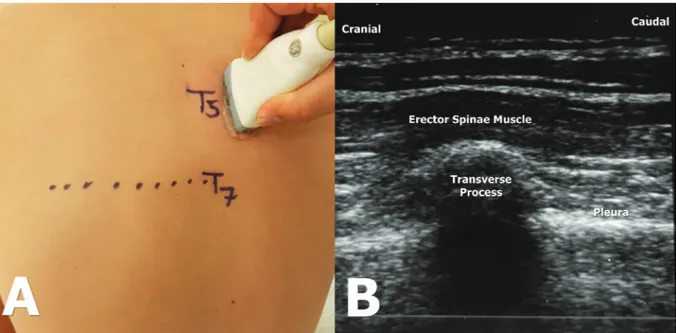
All the patients were sedated with 2 mg of midazo-lam intravenously (IV) in the preoperative room. After a standardized ASA monitorization and perfor-ming aseptic conditions, the Vivid q US device (GE Healthcare, Wauwatosa, WI, USA), a high frequency 12 MHz linear probe, and a 22G, 50 mm block need-le (Stimupneed-lex Ultra 360; B. Braun, Melsungen, Germany) were used for the blocks. All the blocks were performed unilaterally, with the patients in a sitting position, at the level of the T5 vertebra. We did not perform any intervention in the control group.
ESPB technique
In the ESPB group, the probe was placed longitudi-nally at the level of the T5 transverse process, 2-3 cm lateral from the midline. The muscles were visualized superior to the transverse process (Figure 2); then, the needle was inserted in the craniocaudal directi-on using the in-plane technique. A dose of 2 mL normal saline were injected into the two layers of the interfacial area under the erector spinae muscle, and the proper injection site was confirmed. After visualizing the linear spread of saline in the fascial plane, 20 mL of 0.25% bupivacaine was injected there for the block.
TPVB technique
laterally and vertically of the T5 spinous process. Once the transverse process, corresponding paraver-tebral space, internal intercostal membrane, and pleura was identified (Figure 3), the needle was inserted in the lateromedial direction using the in-plane technique. After confirmation of pleural displacement with 2 mL saline, 20 mL of 0.25% bupi-vacaine was administered for the block.
General Anesthesia
In the operating room, all of the patients were moni-tored via ECG, measurements of noninvasive blood pressure, and SpO2. General anesthesia and muscle relaxation were performed using IV propofol (2-2.5 mg kg-1), fentanyl (1-1.5 µg kg-1), and rocuronium
bromide (0.6 mg kg-1). A left-sided double-lumen
tube was usually used for orotracheal intubation. The position of the double lumen tube was confir-med via a fiberoptic bronchoscopy. The patients were placed in a lateral decubitus position for the surgery. The mechanical ventilation was adjusted using a one-lung mechanical ventilation model. The maintenance of anesthesia was performed via gas inhalation (a mixture of sevoflurane, oxygen and fresh air) and a remifentanil infusion (0.01-0.1 µg kg-1
min). Additional analgesia during surgery was provi-ded with 1 mcg kg-1 fentanyl if heart rate and mean
arterial pressure of the patient could be raised 20% above the baseline. Invasive artery monitoring was performed through the radial artery with a 20 gauge cannula. A dose of 4 mg IV ondansetron was injected at the end of the surgery for preventing postoperati-ve nausea and vomiting. VATS lobectomy/wedge resection was performed by the same surgical team using the same technique. A standard three-port VATS approach (at 5th, 8th, and 9th intercostal space)
was used and none of the incisions were required to be extended. A 24 F chest tube was placed at 8th
intercostal space in midaxillary line. A dose of 400 mg IV ibuprofen and 100 mg IV tramadol was admi-nistered at the end of the surgery. The patients were extubated after exhibiting sufficient spontaneous respiration and transferred to the intensive care unit (ICU) for further monitoring.
Standard postoperative analgesia protocol and measurements of pain
Management of postoperative analgesia was perfor-med using classical IV PCA protocol of our
depart-ment (2 mL-10 µg mL-1-bolus dose of fentanyl, no
infusion, 20-min lock out time, and 4-hour limit of 200 µg). At the PACU, a fentanyl PCA device was attached to the patients, and in the presence of pain (VAS score ≥ 4), the patient was asked to push the PCA button. As a part of multimodal analgesia, all patients received NSAID (400 mg ibuprofen in every 8 hours).
A nurse anesthetist blinded to the study evaluated and recorded the opioid consumption and the pati-ents’ pain scores using the visual analogue scale (VAS: 0 = no pain, 10 = the most severe pain). Pain at rest and during coughing (active and passive VAS scores) was assessed at postoperative 0, 2, 4, 8, 16, 24 and 48 hours. If the VAS was ≥ 4 despite the admi-nistraion of ibuprofen and fentanyl PCA bolus, IV meperidine (0.5 mg kg-1) was administered as a
res-cue analgesic drug. Sedation levels were evaluated using the Michigan Sedation Scale (0 = awake and eyes open, 1 = sleepy but responds to verbal stimu-lus, 2 = hard to wake up, and 3 = sleepy and not aroused by shaking). The adverse effects, block pro-cedure time, and success of single- shot technique were recorded. Our aim of comparing single-shot technique with the other method was to compare the easy applicability of both techniques. In this puncture technique block is performed through only a single entrance into the tissues to be blocked and the needle directly goes into the target.
Sample size and statistical analyses
In the power analysis performed with the total opio-id consumption variable that was the primary outco-me of the study, it was determined that the effect size was 1.97 in the 95% confidence interval and the power was 0.99 in the significance level. This result shows that the study sample is sufficient. Descriptive statistics was expressed as mean±SD and frequency. The Kolmogorov-Smirnov test was used to evaluate the distribution of data. Categorical data was compa-red between groups using the Pearson Chi-square test. We used the One-way ANOVA followed by Tukey’s tests to check differences among groups, at a significance level of 5% for normally distributed con-tinuous variables. Independent samples t-test was used to compare two groups. The statistical data was analyzed by the IBM SPSS 20.0 software.
RESULTS
The CONSORT flow diagram shows the enrollment of the patients for the study (Figure 1). This study comprised 90 patients, with 30 in each group. There were no statistical differences between groups in
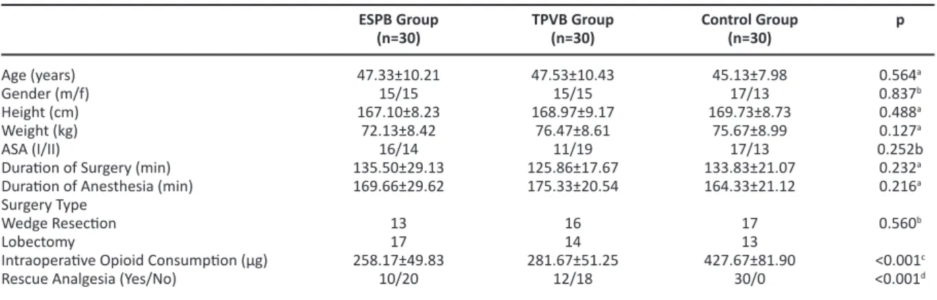
terms of age, weight, height, ASA status, duration of anesthesia, length of surgery, and the type of surgi-cal procedure (wedge vs lobectomy) (p>0.05). The intraoperative opioid consumption was 258.17±49.83 µg in the ESPB group, 281.67±51.25 µg in the TPVB group, and 427.67±81.90 µg in the Control group.
Figure 1. CONSORT flow diagram of the study
Assessed for eligibility (n=90)
Randomized (n=90)
Excluded (n=0)
• Not meeting inclusion criteria (n=0) • Declined to participate (n=0) • Other reasons (n=0)
Allocated to intervention Control Group (n=30) • Received allocated intervention (n=30) • Did not receive allocated intervention (give reasons) (n=0)
Lost to follow-up (give reasons) (n=0) Discontinued intervention (give reasons) (n=0)
Analysed (n=30)
• Excluded from analysis (give reasons) (n=0)
Allocated to intervention ESPB group (n=30) • Received allocated intervention (n=30) • Did not receive allocated intervention (give reasons) (n=0)
Lost to follow-up (give reasons) (n=0) Discontinued intervention (give reasons) (n=0)
Analysed (n=30)
• Excluded from analysis (give reasons) (n=0)
Allocated to intervention TPVB group (n=30) • Received allocated intervention (n=30) • Did not receive allocated intervention (give reasons) (n=0)
Lost to follow-up (give reasons) (n=0) Discontinued intervention (give reasons) (n=0)
Analysed (n=30)
• Excluded from analysis (give reasons) (n=0)
Table I. Demographic data, comparison of operative procedures, intraoperative opioid consumption and rescue analgesia between groups
Age (years) Gender (m/f) Height (cm) Weight (kg) ASA (I/II)
Duration of Surgery (min) Duration of Anesthesia (min) Surgery Type
Wedge Resection Lobectomy
Intraoperative Opioid Consumption (µg) Rescue Analgesia (Yes/No)
ESPB Group (n=30) 47.33±10.21 15/15 167.10±8.23 72.13±8.42 16/14 135.50±29.13 169.66±29.62 13 17 258.17±49.83 10/20 TPVB Group (n=30) 47.53±10.43 15/15 168.97±9.17 76.47±8.61 11/19 125.86±17.67 175.33±20.54 16 14 281.67±51.25 12/18 Control Group (n=30) 45.13±7.98 17/13 169.73±8.73 75.67±8.99 17/13 133.83±21.07 164.33±21.12 17 13 427.67±81.90 30/0
Values are expressed mean ± standard deviation or frequency, ASA; American Society of Anesthesiologist, m; male, f; female, cm; centimeter, kg; kilogram, min; minutes,
aOne Way ANOVA between groups bChi-square test between groups.
cOne Way ANOVA, between ESPB & TPVB and Control group dChi-square test was used, between ESPB & TPVB and Control group
p 0.564a 0.837b 0.488a 0.127a 0.252b 0.232a 0.216a 0.560b <0.001c <0.001d
There was a significantly difference between ESPB, TPVB and Control groups (p<0.001), however there was no difference between the ESPB and TPVB gro-ups (p>0.05). Rescue analgesia (meperidine) was used in 10 patients in the ESPB group, 12 patients in the TPVB group, and 30 patients in the Control group. The use of rescue analgesia was significantly lower in ESPB and TPVB groups compared to the control group (p<0.001) (Table I).
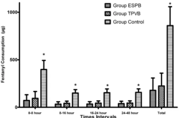
Postoperative mean total fentanyl consumption was 178.66±129.39 µg in the ESPB group, 224.66±134.59 µg in the TPVB group, and 859.33±198.99 µg in the Control group. There was a significantly difference between ESPB/TPVB and Control groups (p<0.001), however there was no difference between the ESPB and TPVB groups (p>0.05) in terms of postoperative total fentanyl consumption. The fentanyl consumpti-on at all time intervals was significantly lower both in ESPB and TPVB groups compared to the control
Figure 2A. Probe localization in a longitudinal direction during ESPB performing at 2-3 cm lateral to the T5 transverse process. The patient is in sitting position during the procedure.The bold black lines indicate the vertebral column. 2B. Corresponding sonographic anatomy of the block. The erector spinae muscle, transverse process and pleura are seen
Figure 3A. Patient position and probe localization during TPVB performing at T5 vertebrae level. 3B. Corresponding sonographic anatomy of the block. The paravertebral space, transverse process and pleura are seen
group. There were no statistical difference between the ESPB and TPVB groups for fentanyl consumption at any time interval (Figure 4).
There was no statistical difference between the ESPB and TPVB groups in terms of the active and passive VAS scores in any time interval (p>0.05 for each
interval). The active and passive VAS was signifi-cantly higher in Control group compared to the ESPB and TPVB groups at all time intervals (p<0.001) (Figure 5 and 6).
Five patients in the ESPB, six patients in the TPVB and 18 patients in the Control group had postopera-tive nausea (p<0.001). Six patients in the ESPB group, seven patients in the TPVB group and 17 patients in the Control group had postoperative itching (p=0.002). There was no significant difference bet-ween groups in terms of the other adverse effects (Table II). No block related complications such as pneumothorax, vascular or neuraxial injury were seen in both of the groups.
Block procedure time was significantly lower in the ESPB group (7.13±1.59 min) compared to the TPVB group (13±2.49 min) (p<0.001) (Table III). Success of single- shot technique was significantly higher in the ESPB group than in the TPVB group (in 25 vs 10 pati-ents (p<0.001) (Table III).
DISCUSSION
This study was designed to evaluate the analgesic
Figure 4. The Comparison of opioid consumption in time inter-vals between groups
Figure 5. Comparisons of passive VAS assessment between groups
Figure 6. Comparisons of active VAS assessment between groups
Table II. Comparison of the adverse effect incidences between groups Respiratory depression Sedation/Confusion Nausea Vomiting Itching Constipation Dyspepsia ESPB Group (n=30) 0 0 5 4 6 0 0 TPVB Group (n=30) 0 0 6 5 7 0 0 Control Group (n=30) 0 0 18 7 17 0 0
Values are expressed as frequency. Chi-square test was used
aControl group was significant than other groups
p -<0.001a 0.587 0.004a
-Table III. Comparison of the block procedure time and success of one time puncture between group ESPB and TPVB
Block procedure time (min) Success of one time puncture (yes) Group ESPB (n=30) 7.13± 1.59 25 Group TPVB (n=30) 13 ±2.49 10 p <0.001a <0.001b
aIndependent samples t test was used. bChi-square testwas used.
efficacy of US-guided TPVB with US-guided ESPB compared to control group following thoracoscopic surgery. The results showed that both single-shot ESPB and TPVB provided similar effective analgesia compared to the control group during the first 48 hours. ESPB and TPVB significantly reduced the intra-operative and postintra-operative opioid consumption with lower VAS scores than the control group. However, ESPB had a shorter performance time and higher success of single-shot technique compared to TPVB.
The US-guided ESPB was described by Forero et al.
(13) in 2016 for the treatment of thoracic neuropathic
pain. With increasing number of case reports and randomized controlled trials, the application area of the ESPB has been widened from cervicothoracic region to lumbar spine (14-18). Based on its injection
site (over the vertebral transverse process) and the spread of injectate, ESPB is a type of paraspinal block
(14), and it is described as “paravertebral block by
proxy” (19,20). In this respect, the ESPB is similar to
TPVB, but it offers the benefit of lesser technical difficulty with comparable efficacy (18). Local
anesthe-tic spread to the paravertebral space in ESPB has been also shown in some cadaveric studies (19,21,23),
and the analgesic effectiveness of ESPB for thoracic surgery as an alternative to TPVB and TEA has been shown in numerous case reports (12,24-29). The first
randomized, controlled study comparing the efficacy of ESPB and TPVB for postoperative analgesia was performed in patients undergoing breast surgery. In this study, the authors demonstrated that both TPVB and US-guided ESPB reduced 24-hour postoperative morphine consumption without any statistically sig-nificant differences between the two groups (30). In
another randomized clinical trial, Chen et al. (31)
investigated US-guided intercostal nerve block, single-shot ESPB, and multiple-shot TPVB after tho-racoscopic surgery. While they reported superior analgesia with multiple-injection TPVB over intercos-tal nerve block and single-shot ESPB, intercosintercos-tal nerve block and single injection ESPB were equally effective in reducing pain after thoracoscopic sur-gery. However they compared single-shot ESPB with multiple-shot TPVB which may not indicated that TPVB has a superiority over ESPB in terms of analge-sia after VATS. To reach this conclusion it would be better to compare multiple- shot ESPB with multiple-
shot TPVB. Single level ESPB provided good analgesia in our current study. In a study comparing US-guided, preoperative, single-dose ESPB with TPVB after tho-racotomy, Fang et al. (32) found that ESPB provided
similar, satisfying postoperative pain control in the first postoperative 48 hours. It also offered advanta-ges as technical simplicity, a shorter puncture time, a higher success of one-time puncture, and higher patient satisfaction, which resemble the results of the present study. Fang et al. (32) also demonstrated
that, in the TPVB group, the incidence of adverse events, such as bradycardia and hypotension, were higher than in the ESPB group. In the present study, there were no differences between both groups in terms of adverse events. Although the volumes used for TPVB were the same (20 mL), this may be due to the difference in degree of sympathetic blockage for TPVB in both studies. Of course, ESPB is not without complications. In the literature, local anesthetic spread to the epidural space and the pneumothorax have been reported (33). ESPB has a technical
simpli-city and a better risk profile compared to TPVB. However, ESPB may also be risky if the needle tip is not seen during procedure. In addition, it may be risky if the procedure is being done by less skilled hands (21).
As VATS is a widespread surgical technique, and since fast-track surgery is also an important issue for thoracoscopy, multimodal, opioid-sparing analgesic techniques should become part of this fast-track program. Therefore, peripheral nerve blocks may be preferred for this procedure (3). The analgesic
effecti-veness of TPVB and its role in fast-track surgery has already been proven (7). With increasing number of
case reports and studies about ESPB, this block may be a good option for fast-track thoracoscopic sur-gery. ESPB is performed on the same side of surgery, which involves a chest tube placement. This makes ESPB quite safe. In the literature, randomized studies about ESPB are limited. Therefore, meta-analyses and further studies with larger sample sizes are necessary.
This study did have some limitations. First, dermato-mal sensory testing could be done to explore the dermatomal distribution of these two blocks. Second, the researchers utilized 20 mL of 0.25% bupivacaine, as in other studies. Therefore, it is still unknown
whether 20 mL of local anesthetic is the minimum effective dose. In our previous study we evaluated the efficacy of single-shot ESPB for VATS and found single shot ESPB with 20 mL volume as an effective analgesic technique (4). In this study we wanted to
compare this effectiveness with TPVB. May higher volumes or different concentrations have longer analgesic effects? Lastly, a block catheter may be used, but we didn’t use it to see the effectiveness of the single-shot blocks.
CONCLUSION
To summarize, the results of the trial showed that US-guided, single-shot ESPB and TPVB provided more effective analgesia than IV fentanyl PCA after VATS. In addition, single-shot ESPB offers the advan-tages of technical simplicity that ESPB had a shorter performance time and higher success of single-shot technique compared to TPVB.
Ethics Committee Approval: Ethics decisi on number: 66291034-604.01.01-E.4419
Conflict of Interest: The authors report no conflicts of interest.
Funding: There is no funding for the research. Informed Consent: Written informed consent was obtained from all patients for the study
REFERENCES
1. Agostini P, Lugg ST, Adams K et al. Postoperative pul-monary complications and rehabilitation requirements following lobectomy: a propensity score matched study of patients undergoing video-assisted thoracos-copic surgery versus thoracotomy. Interactive Cardiovascular and Thoracic Surgery. 2017;24:931-7. https://doi.org/10.1093/icvts/ivx002
2. Desai H, Natt B, Kim S, Bime C. Decreased in-hospital mortality after lobectomy using video-assisted thora-coscopic surgery compared with open thoracotomy. Annals of the American Thoracic Society. 2017;14:262-6.
https://doi.org/10.1513/AnnalsATS.201606-429OC 3. Umari M, Carpanese V, Moro V et al. Postoperative
analgesia after pulmonary resection with a focus on video-assisted thoracoscopic surgery. European Journal of Cardio-Thoracic Surgery. 2017;53:932-8.
https://doi.org/10.1093/ejcts/ezx413
4. Ciftci B, Ekinci M, Celik EC et al. Efficacy of an Ultrasound-Guided Erector Spinae Plane Block for Postoperative Analgesia Management After Video-Assisted Thoracic Surgery: A Prospective Randomized Study. Journal of Cardiothoracic and Vascular Anesthesia. 2019;00:1-6.
https://doi.org/10.1053/j.jvca.2019.04.026
5. Davies RG, Myles PS, Graham JM. A comparison of the analgesic efficacy and side-effects of paravertebral vs epidural blockade for thoracotomy-a systematic revi-ew and meta-analysis of randomized trials. BJA: British Journal of Anaesthesia. 2006;96:418-26.
https://doi.org/10.1093/bja/ael020
6. Conlon NP, Shaw AD, Grichnik KP. Postthoracotomy paravertebral analgesia: will it replace epidural analge-sia?. Anesthesiology Clinics. 2008;26:369-80. https://doi.org/10.1016/j.anclin.2008.01.003
7. Komatsu T, Kino A, Inoue M et al. Paravertebral block for video-assisted thoracoscopic surgery: analgesic effectiveness and role in fast-track surgery. International Journal of Surgery. 2014;12:936-9.
https://doi.org/10.1016/j.ijsu.2014.07.272
8. Okajima H, Tanaka O, Ushio M et al. Ultrasound-guided continuous thoracic paravertebral block provides com-parable analgesia and fewer episodes of hypotension than continuous epidural block after lung surgery. Journal of Anesthesia. 2015;29:373-8.
https://doi.org/10.1007/s00540-014-1947-y
9. Naja Z, Lönnqvist PA. Somatic paravertebral nerve blockade incidence of failed block and complications. Anaesthesia. 2001;56:1181-1201.
https://doi.org/10.1111/j.1365-2044.2001.2084-2.x 10. Song L, Zhou Y, Huang D. Inadvertent posterior
inter-costal artery puncture and haemorrhage after ultrasound-guided thoracic paravertebral block: a case report. BMC Anesthesiology. 2018;18:196.
https://doi.org/10.1186/s12871-018-0667-5
11. Wilson JM, Lohser J, Klaibert B. Erector spinae plane block for postoperative rescue analgesia in thoracos-copic surgery. Journal of Cardiothoracic and Vascular Anesthesia. 2018;32:e5-e7.
https://doi.org/10.1053/j.jvca.2018.06.026
12. Luis-Navarro JC, Seda-Guzmán M, Luis-Moreno C, López-Romero JL. The erector spinae plane block in 4 cases of video-assisted thoracic surgery. Revista Española de Anestesiología y Reanimación (English Edition). 2018;65:204-8.
https://doi.org/10.1016/j.redare.2018.02.002 13. Forero M, Adhikary SD, Lopez H, Tsui C, Chin KJ. The
erector spinaeplane block: a novelan algesic technique in thoracic neuropathic pain. Reg Anesth Pain Med. 2016;41:621-7.
https://doi.org/10.1097/AAP.0000000000000451 14. Chin KJ, Adhikary SD, Forero M. Erector Spinae Plane
(ESP) Block: a New Paradigm in Regional Anesthesia and Analgesia. Current Anesthesiology Reports. 2019;1-10.
https://doi.org/10.1007/s40140-019-00333-0
15. Yayik AM, Cesur S, Ozturk F, et al. Postoperative Analgesic Efficacy of the Ultrasound-Guided Erector Spinae Plane Block in PatientsUndergoing Lumbar Spinal Decompression Surgery: A Randomized Controlled Study. World Neurosurgery. 2019;126:779-85.
https://doi.org/10.1016/j.wneu.2019.02.149
16. Ahiskalioglu A, Tulgar S, Celik M, Ozer Z, Alici HA, Aydin ME. Lumbar Erector Spinae Plane Block as a Main Anesthetic Method for Hip Surgery in High Risk Elderly Patients: Initial Experience with a Magnetic Resonance Imaging. Eurasian J Med. 2020;52:16-20.
https://doi.org/10.5152/eurasianjmed.2020.19224 17. Tulgar S, Ahiskalioglu A, De Cassai A, Gurkan Y. Efficacy
of bilateral erector spinae plane block in the manage-ment of pain: current insights. J Pain Res. 2019;12:2597-2613. Published 2019 Aug 27.
https://doi.org/10.2147/JPR.S182128
18. Altiparmak B, Toker MK, Uysal Aİ, Kuşçu Y, Demirbilek SG. Eficácia do bloqueio do plano do músculo eretor da espinha guiado por ultrassom para analgesia após colecistectomia laparoscópica: um estudo controlado randômico [Efficacy of ultrasound-guided erector spi-nae plane block for analgesia after laparoscopic cho-lecystectomy: a randomized controlled trial]. Rev Bras Anestesiol. 2019;69:561-8.
https://doi.org/10.1016/j.bjan.2019.09.001
19. Yang HM, Choi YJ, Kwon HJ et al. Comparison of injecta-te spread and nerve involvement between retrolaminar and erector spinae plane blocks in the thoracic region: a cadaveric study. Anaesthesia. 2018;73:1244-50. https://doi.org/10.1111/anae.14408
20. Costache I, Pawa A, Abdallah FW. Paravertebral by proxy-time to redefine the paravertebral block. Anaesthesia. 2018;73:1185-8.
https://doi.org/10.1111/anae.14348
21. Greenhalgh K, Womack J, Marcangelo S. Injectate spre-ad in erector spinae plane block. Anaesthesia. 2019;74:126-7.
https://doi.org/10.1111/anae.14523
22. Adhikary SD, Bernard S, Lopez H, Chin KJ. Erector spi-nae plane block versus retrolaminar block: a magnetic resonance imaging and anatomical study. Reg Anesth Pain Med. 2018;43:756-62.
https://doi.org/10.1097/AAP.0000000000000798 23. Ivanusic J, Konishi Y, Barrington MJ. A cadaveric study
investigating the mechanism of action of erector spi-nae blockade. Reg Anesth Pain Med. 2018; 43:567-71. https://doi.org/10.1097/AAP.0000000000000789 24. Muñoz F, Cubillos J, Bonilla AJ, Chin KJ. Erector spinae
plane block for postoperative analgesia in pediatric oncological thoracic surgery. Canadian Journal of Anesthesia Journal canadiend’anesthésie. 2017;64:880-2.
https://doi.org/10.1007/s12630-017-0894-0
25. Forero M, Rajarathinam M, Adhikary S, Chin KJ. Continuous erector spinae plane block for rescue anal-gesia in thoracotomy after epidural failure: a case report. A&A Practice. 2017;8:254-6.
https://doi.org/10.1213/XAA.0000000000000478 26. Raft J, Chin KJ, Belanger ME et al. Continuous Erector
Spinae Plane Block for thoracotomy analgesia after epidural failure. Journal of Clinical Anesthesia. 2018;54:132.
https://doi.org/10.1016/j.jclinane.2018.10.024 27. Ciftci B, Ekinci M, Demiraran Y. Ultrasound-guided
single-shot preemptive erector spinae plane block for postoperative pain management. Journal of Cardiothoracic and Vascular Anesthesia. 2019;33:1175-6.
https://doi.org/10.1053/j.jvca.2018.10.040
28. Hernandez MA, Palazzi L, Lapalma J et al. Erector spi-nae plane block for surgery of the posterior thoracic wall in a pediatric patient. Regional Anesthesia & Pain Medicine. 2018;43:217-9.
https://doi.org/10.1097/AAP.0000000000000716 29. Adhikary SD, Pruett A, Forero M, Thiruvenkatarajan V.
Erector spinae plane block as an alternative to epidural analgesia for post-operative analgesia following video-assisted thoracoscopic surgery: a case study and a literature review on the spread of local anaesthetic in the erector spinae plane. Indian Journal of Anaesthesia. 2018;62:75.
https://doi.org/10.4103/ija.IJA_693_17
30. Gürkan Y, Aksu C, Kuş A et al. Ultrasound guided erec-tor spinae plane block reduces postoperative opioid consumption following breast surgery: a randomized controlled study. Journal of Clinical Anesthesia. 2018;50:65-8.
https://doi.org/10.1016/j.jclinane.2018.06.033 31. Chen N, Qiao Q, Chen R et al. The effect of
ultrasound-guided intercostal nerve block, single-injection erector spinae plane block and multiple-injection paraverteb-ral block on postoperative analgesia in thoracoscopic surgery: A randomized, double-blinded, clinical trial. Journal of clinical anesthesia. Ahead of print.
https://doi.org/10.1016/j.jclinane.2019.07.002 32. Fang B, Wang Z, Huang X. Ultrasound-guided
preope-rative single-dose erector spinae plane block provides comparable analgesia to thoracic paravertebral block following thoracotomy: a single center randomized controlled double-blind study. Annals of Translational Medicine. 2019;7:174.
https://doi.org/10.21037/atm.2019.03.53
33. Schwartzmann A, Peng P, Maciel MA, Forero M. Mechanism of the erector spinae plane block: insights from a magnetic resonance imaging study. Canadian Journal of Anesthesia/Journal Canadiend’ Anesthésie. 2018;65:1165-6.