638
http://journals.tubitak.gov.tr/medical/ © TÜBİTAK
doi:10.3906/sag-2005-287
COVID-19: pathogenesis, genetic polymorphism, clinical features and laboratory findings
Recep ÖZTÜRK1,*, Yeşim TAŞOVA2, Akif AYAZ3
1Department of Infectious Diseases and Clinical Microbiology, Medical School, İstanbul Medipol University, İstanbul, Turkey 2Department of Infectious Diseases and Clinical Microbiology, Faculty of Medicine, Çukurova University, Adana, Turkey
3Department of Medical Genetics, Medical School, Istanbul Medipol University, İstanbul, Turkey
* Correspondence: [email protected]
1. Introduction
The COVID-19 pandemic caused by SARS-CoV-2 virus continues to be effective around the world. Although it is a novel agent and disease with a background of nearly 5 months, the research studies conducted on this subject continue intensively, and the information produced is rapidly shared via open access publications.
This paper will discuss the pathogenesis, genetic polymorphism, clinical and laboratory findings related to COVID-19.
2. Pathogenesis
The pathogenesis of COVID-19 caused by SARS-CoV-2, which is a novel virus, is being updated and evolving as part of various discussions.
SARS-CoV-2 is mainly transmitted through respiratory droplets and contact and potentially by fecal-oral routes. In addition, the aerosols generated during some procedures, particularly in hospitals, are another mode of transmission [1].
SARS-COV-2 spike proteins bind to angiotensin-converting enzyme 2 (ACE2) receptors on the surface of a target cell. The type II transmembrane serine protease (TMPRSS2) binds to the ACE2 receptor and makes it cleaved. In this proteolytic process, the spike protein is activated and viral entry into the cell takes place [1,2].
It is reported that CD147 (a transmembrane glycoprotein that belongs to the immunoglobulin superfamily) is the other receptor bound by the virus; CD147 exists in epithelial cells and is expressed at very high levels in inflamed tissues, pathogen-infected cells and tumor tissues [3].
For the pathogenesis of COVID-19, this is the very first step of the complex pathogenesis process [1].
It is assumed that primary viral replication develops in the mucosal epithelium of the upper respiratory tract (nasal cavity and pharynx), with further multiplication in lower respiratory tract and gastrointestinal mucosa, which results in a mild viremia [1,4]. At this point, a small number of infections are checked and remain asymptomatic [1]. A Abstract: COVID-19 caused by a novel agent SARS-CoV-2 progressed to a pandemic condition and resulted in a major public health concern worldwide, leading to social and economic issues at the same time. The pathogenesis of COVID-19 starts with the bonding of the virus to ACE2 receptors expressed in many tissues, and the triggered excessive immune response plays a critical role in the course of the disease. The cytokine storm that occurs upon excessive production of pro-inflammatory cytokines is considered responsible for the severe progression of the disease and the organ damage. However, the accurate pathophysiological mechanism of the disease, which progresses with various clinical presentations, is still substantially unknown. While various studies have been conducted on the effect of genetic polymorphism on the course and severity of the disease, the presence of a significant effect has not been proven yet. The clinical course of the disease is variable, with clinical representation ranging from 81% mild course to 14% severe course along with 5% critical course in patients. Asymptomatic course is considered to be higher than expected, although its frequency is not known exactly. Older adults and those with comorbidities are exposed to a more severe disease course. The disease progress with various symptoms, such as fever, cough, dyspnea, malaise, myalgia, taste and smell dysfunctions, diarrhea, and headache. A range of complications (acute respiratory distress syndrome, thromboembolic conditions, arrhythmia and cardiac events, secondary infections) could be seen during the course of the disease. Varied laboratory tests are vital to determine these verity and prognosis of the disease, along with the condition and exposure of the affected systems during thecourse of COVID-19.
Key words: SARS-CoV-2, COVID-19, pathogenesis, genetic polymorphism, clinical features, laboratory findings Received: 22.05.2020 Accepted/Published Online: 06.06.2020 Final Version: 21.04.2020
number of recent studies indicate that the asymptomatic course may be at a higher rate [1,5].
The pathogenesis is basically described as binding of the SARS-CoV2 to the receptors (ACE2) in the type II pneumocytes in the lungs and its triggering of a cascade of inflammation in the lower respiratory tract [1].
Some patients also exhibit nonrespiratory symptoms, such as acute liver and heart injury, kidney failure and diarrhea, which are indicative of multiple organ involvement [6,7,8].
ACE2 is broadly expressed in nasal mucosa, bronchus, lung, heart, esophagus, kidney, stomach, bladder, and ileum, and all these human organs are vulnerable to SARS-CoV-2 [1,2,9]. Recently, the potential pathogenicity of SARS-CoV-2 to testicular tissues was also proposed, which has raised concerns over fertility issues in young patients [10]. Biophysical and structural evidence suggest that SARS-CoV-2 S protein binds to human ACE2 with 10- to 20-fold higher affinity than SARS-CoV S protein [1,11].
The critical steps of pathogenesis of the SARS-CoV-2 infection based on the data compiled to date are shown in Figure.
It was suggested that 1 of the factors that determine the clinical course of COVID-19 is associated with the viral load transmitted to the person. In general, the disease is more severe in people, who are exposed to higher risk of viral load transmission, such as healthcare professionals. It was reported that the COVID-19 patients having more severe diseases, who should be admitted to intensive care unit, had high a viral RNA load at 10 days and beyond, after symptom onset. A mild clinical or asymptomatic course is usually observed in patients infected with a lower viral load; there are further contradictory reports on this issue [1, 12,13].
The course of the disease is linked to the body’s immune response to the virus and the patient’s comorbidities, apart from the viral load. If the immune system is not activated, the virus reproduces faster. If the immune system is clearly activated, it causes a cytokine storm and induces a multisystemic effect [1,14].
The development mechanisms of severe cases, other than asymptomatic or mild clinical cases, during the pathogenesis process will be summarized below.
Fast viral replication and cellular damage, virus-induced ACE2 downregulation and shedding and antibody dependent enhancement (ADE) are responsible for the aggressive inflammation caused by SARS-CoV-2 [1,15] (see Figure).
2.1. Immune response
The active host immune reaction, including innate and adaptive immune to SARS-CoV-2, is essential for controlling and resolving viral infection. Both innate
and adaptive immune cells are synergistically involved in the antiviral response during infection. A considerable increase in the number of leukocytes and neutrophils and the neutrophil to lymphocyte ratio (NLR) was found in severe cases than in mild cases [16].
Viral entry and cellular infection trigger the immune response of the host cell, and the inflammatory cascade is initiated by antigen-presenting cells (APC). The process starts with ACP performing 2 functions: (1) presentation of the foreign antigen to CD4 + -T-helper (Th1) cells and (2) release of interleukin-12 to further stimulate Th1 cell. Th1 cells stimulate CD8 + -T-killer (Tk) cells that will target the cells containing foreign antigen. In addition, activated Th1 cells stimulate B cells to produce antigen-specific antibodies[1].
The inflammatory process can accelerate the apoptosis of lymphocytes while stimulating the synthesis of neutrophils. This irregular response given by the immune system and immunological abnormality may result in conditions leading to death [1].
The studies carried out demonstrated that neutrophil to lymphocyte ratio in patients with COVID-19 may be a significant marker in explaining disease severity and mortality [1].
It is not clear yet whether an immune response effective against SARS-CoV-2 has developed. Several studies showed the existence of a significant relationship between the disease severity and the levels of proinflammatory cytokines and subsets of immune cells. Immune dysregulation and high levels of proinflammatory cytokines were potentially suggested to be the root cause of tissue damage during response to SARS-CoV-2 [1,16].
For COVID-19, the production of proinflammatory cytokines and the jointly functioning innate and adaptive immune response that makes CD4+/CD8+ T cells activated are vital to control viral replication, restrain the spread of virus and clear the infected cells [1,16].
It was suggested that survival from COVID-19 could rely on the ability to replenish lymphocytes killed by the virus. Therefore, the count of lymphocytes, especially of CD4 cells, could serve as a clinical predictor of disease severity and prognosis [17].
2.2. Antibody response and ADE
As with SARS, a small proportion of patients, especially those who produce neutralizing antibodies early, experience persistent inflammation, ARDS and sudden death, while most patients survive the inflammatory responses and clear the virus. For the SARS-CoV-2 infection, antibody-dependent enhancement (ADE) is considered to be 1 of the potential underlying mechanisms as in some cases of other viral infections [1]. Interaction of FcR with the virus-anti-S protein neutralizing antibodies (anti-S-IgG) complex may catalyze both inflammatory
responses and persistent viral replication in the lungs [15].
2.3. Immune dysfunction
Peripheral CD4 and CD8 T cells were reduced and hyperactivated in severe patients. In addition, high concentrations of proinflammatory CD4 T cells and cytotoxic granules CD8 T cells were identified, which suggests antiviral immune responses and hyperactivation of T cells. Furthermore, many studies indicate that lymphopenia, a common feature of COVID-19 is a critical factor regarding disease severity and mortality [1, 18, 19].
2.4. Cytokine storm
Cytokine storm is an extreme and severe reaction (hyperreaction) given by the immune system, where large amounts of cytokines are rapidly released into the systemic circulation [1,15].
High levels of cytokines in critical COVID-19 patients are considered to be important in explaining the pathogenesis of the disease due to the effect of the inflammatory storm [16].
Exuberant inflammatory responses occur during the clinical course of the SARS-CoV-2 infection,
which subsequently results in uncontrolled pulmonary inflammation. This is probably the leading cause of case fatality [15].
The initial onset of rapid viral replication causes mass epithelial and endothelial cell death and vascular permeability, potentially triggering the production of exuberant proinflammatory cytokines and chemokines [20].
COVID-19 mainly targets the respiratory system; the main pathogenesis is the resulting severe pneumonia, RNAaemia and acute cardiac injury [6].
SARS-CoV-2 binds to the receptors in alveolar and gastrointestinal epithelial cells and activates the innate and adaptive immune system, leading to the secretion of a vast number of cytokines, including IL-6 [21].
In some severe cases referred to intensive care units, high levels of cytokines and chemokines were noted in COVID-19 patients, such as IL1-β, IL1RA, IL7, IL8, IL9, IL10, basic FGF2, GCSF, GMCSF, IFNγ, IP10, MCP1, MIP1α, MIP1β, PDGFB, TNFα, and VEGFA [18]. The markers related to hyperinflammation
(hs-Mild viremia
Figure. The critical steps of pathogenesis of the SARS-CoV-2 infection [1]. ADE: Antibody-dependent enhancement; ACE2: Angiotensin-convertingenzyme 2; RAS: Renin-angiotensin system; ARDS: Acute respiratory distress syndrome.
CRP, procalcitonin, IL-2R, IL-6, IL-8, IL-10, TNF-alpha, inducible protein (IP10), monocyte chemoattractant protein-1 (MCP1), macrophage inflammatory protein-1α (MIP1A)) were noted significantly higher in patients who died than survivors [22]. These cytokines and chemokines are positively linked to the SARS-CoV-2 viral load [16]. The high level of IL-6 may be considered a marker of severe disease or progressive disease [23].
IL-6 levels in admission are a predictor of the need for mechanical ventilation [24]. In addition, those with high IL-6 levels experience RNAaemia, with the longer virus RNA spread [23-26]. RNAaemia is associated with more severe clinical courses [25,26]. IL-6 induces intracellular signaling, antibody secretion, acute phase protein secretion, platelet formation with effect on megakaryocytes, angiogenesis with increment in vascular endothelial growth factor (VEGFR), mesenchymal cell proliferation, and cardiomyopathy. Additionally, IL-6 plays a role in activating the complement and coagulation system in DIC pathophysiology. For these properties, IL-6 is the key molecule for cytokine storm [21]. The increased plasma level of IL-6 that is regarded a significant cytokine contributing to MAS is noted in mild and severe patient groups of COVID-19: severe patients have a significantly higher level of IL-6 than mild patients or patients without fever [16].
Excessive increase in inflammatory cytokines such as IL-6, which may result in “cytokine storm”, is not only likely to be a driver behind acute lung injury and ARDS, but also may cause other tissue damage that progresses to multiple organ failure (MOF). In addition, high levels of interleukin 10 (IL-10) were noted in severe patients. It was noted that this could be associated with the compensatory antiinflammatory response syndrome (CARS) that may be responsible for a greater number of secondary infections (50%) and sepsis (100%) reported in survivors [27].
Tissue damage caused by the virus may induce excessive production of proinflammatory cytokines, and recruitment of proinflammatory macrophages and granulocytes. This gives rise to the cytokine storm (CS) referred to as macrophage activation syndrome (MAS) or secondary hemophagocytic lymphohistiocytosis (sHLH), thus causing further damage to tissues [1, 16].
Several mechanisms may contribute to hyperactivation of monocyte-induced macrophages observed in patients with COVID-19 [28]. The inflammatory response resulting from excessive cytokine secretion observed with the activation of T cells and monocytes/macrophages increases vascular permeability, causing exudative effusion in the alveoli and resulting in respiratory failure. Cytokines play a key role in pathophysiology and clinical findings. INF is secreted from activated T cells, which then activates macrophages that produce proinflammatory and
antiinflammatory cytokines, such as IL-6, TNFα, IL-18, IL-2, IL-1α, IL-1β and IL-10 [1,28 ].
The delayed production of type I interferon, which results in enhanced cytopathic effects and increased sensing of microbial threats, boosts the enhanced release of monocyte chemoattractants by alveolar epithelial cells (and probably further by macrophages and stromal cells), giving rise to sustained recruitment of blood monocytes into the lungs [1,28].
Monocytes distinctly change into proinflammatory macrophages mediated by the activation of Janus kinase (JAK), such as signal transducer and activator of transcription (STAT) pathways. The activated natural killer (NK) cells and T cells further stimulate the recruitment and activation of monocyte-induced macrophages with the help of the production of granulocyte-macrophage colony-stimulating factor (GM-CSF), tumor necrosis factor (TNF) and interferon-γ (IFNγ). Accumulation of oxidized phospholipids (OxPLs) occurs in infected lungs, which activates monocyte-induced macrophages through the Toll-like receptor 4 (TLR4)–TRAF6–NF-κB pathway. Virus sensing has the potential to trigger TLR7 activation through the recognition of viral single-stranded RNA. The expression of severe acute respiratory syndrome coronavirus 2 (SARS-CoV-2) entry receptors, which is induced by type I interferons, causes the virus to reach the cytoplasm of macrophages and to activate the NLRP3 inflammasome, giving rise to the secretion of mature IL-1β and/or IL-18. IL-1β may enhance the activation of monocyte-derived macrophages in an autocrine or paracrine manner, but also may lead to reduction in type I interferon production in infected lungs [28 ].
The activation of NLRP3 inflammasome in macrophages, epithelial cells and even endothelial cells results in the release of proinflammatory cytokines that contribute to pathogenic inflammation responsible for the severity of COVID-19 symptoms [28].
The interaction of Fcg receptors with antispike protein IgG immune complexes may contribute to the amplified inflammatory activation of monocyte-induced macrophages. The activated monocyte-monocyte-induced macrophages release large amounts of proinflammatory cytokines and contribute to the COVID-19 cytokine storm [28].
These conclusions show that SARS-CoV-2 is responsible for immune irregularity resulting from the change in the entire lymphocyte subset level, which may lead to cytokine storm and further tissue damage, by inducing abnormal cytokine and chemokine response[1,28].The excessive inflammatory response caused by cytokine storm results in severe disease course and aggravates the prognosis in COVID-19[1,28].
2.5. Acute respiratory distress syndrome (ARDS)
Earlier studies indicate that genetic vulnerability and inflammatory cytokines are closely related to the occurrence of ARDS. ACE2 is considered to be linked to the development or outcome of over 40 candidate genes, including interleukin 10 (IL-10), tumor necrosis factor (TNF) and vascular-endothelial growth factor (VEGF) [1,29].
The data from patients infected with SARS-CoV-2 indicated that severe cases characterized by a cytokine storm inevitably progress to acute respiratory distress syndrome (ARDS) [16].
In addition, the increased plasma level of IL-6 and IL-8 was shown to be associated with negative outcomes of ARDS [1]. Considering pulmonary infiltration in patients with ARDS, the large area of lung injury (≥50%) is closely related to the increased level of IL-6 and the subset of lymphocytes in the peripheral blood [1,16].
Pulmonary damage such as ARDS is noted in 50% of the patients developing MAS. During the course of COVID-19, a cytokine profile resembling MAS is also associated with enhanced disease severity. These patients were noted for the increased level of serum CRP, lactate dehydrogenase (LDH) ferritin, serum creatinine, creatine kinase (CK) and IL-6 and D-dimer [1,16].
2.6. ACE2 downregulation and shedding
It was suggested that the loss of pulmonary ACE2 function is related to acute lung injury due to the potential of ACE2 downregulation and shedding to cause dysfunction of the renin-angiotensin system (RAS) and to further enhance inflammation and cause vascular permeability [1,15].
Fast viral replication and cellular damage, virus-induced ACE2 downregulation and shedding and antibody dependent enhancement (ADE) are responsible for the aggressive inflammation caused by SARS-CoV-2 [1,15].
Initially, rapid viral replication causes mass epithelial and endothelial cell death and vascular permeability, potentially triggering the production of exuberant proinflammatory cytokines and chemokines [1,15].
2.7. Other effects in pathogenesis: Effects on the coagulation system:
Activation and interactions of macrophages, monocytes, endothelial cells, platelets and lymphocytes play a critical role in the procoagulant effect of viral infections [1,30].
Increased venous and arterial thrombosis (including small vessels) resulting from the overactivation of the coagulation system are shown to affect the clinical course adversely [30,31].
Irregularity of the coagulation cascade, followed by the formation of intraalveolar or systemic fibrin clots, is 1 of the distinct symptoms of severe respiratory disease caused by COVID-19 [30,31].
Both thrombocytopenia and high D-dimer level can be explained by overactivation of the coagulation cascade and platelets. Viral infections cause the systemic inflammatory response and result in an imbalance between procoagulant and anticoagulant homeostatic mechanisms. There are many pathogenetic mechanisms, including endothelial dysfunction, von Willebrand factor elevation, Toll-like receptor activation and tissue-factor pathway activation. Dysfunction of endothelial cells induced by infection results in excessive thrombin production and fibrinolysis inhibition, which indicates hypercoagulability in a COVID-19 infected patient [1,30,31].
COVID-19 may make patients prone to both venous and arterial thromboembolism owing to excessive inflammation, hypoxia, immobilization and DIC. Acute pulmonary embolism (PE), deep-vein thrombosis (DVT), ischemic stroke, myocardial infarction or systemic arterial embolism are frequently noted in these patients. Actual studies have shown that patients with severe COVID-19 are usually complicated by coagulopathy, with DIC accompanying most deaths. Patients severely infected with COVID-19 may develop DIC through the activation of fulminant coagulation, leading to depletion of common microvascular thrombosis and coagulation factors. As a consequence, the presentation turns out to be thrombocytopenia, prolonged PT/INR, PTT, D-dimer elevation and reduced fibrinogen level. Cystocytes can be observed in the peripheral smear of patients [30,31].
The prolonged immobility of patients or the long inpatient treatment duration increases the risk of venous thromboembolism in severe COVID-19 disease. The RAAS pathway is activated upon binding of the virus to ACE2 and leading to reduced enzyme expression. RAAS activation bears the risk of platelet adhesion and aggregation and, theoretically, of pulmonary embolism, pulmonary hypertension and fibrosis. The increased level of lactate dehydrogenase, ferritin, C-reactive protein, D-dimer and interleukin seen in COVID-19 indicates that the disease predisposes to a proinflammatory and hypercoagulable condition [31,32].
The recently emerging various data, some part of which are described above, support the argument that patients infected with the SARS-CoV-2 virus are at risk of developing disseminated intravascular coagulation (DIC) [33,34].
Cardiac effects
Cardiac physiopathology: 1) failure of the limited cardiac reserve to meet the increased metabolic needs due to infection, 2) capacity of viral infections to trigger acute coronary syndromes, arrhythmias and heart failure (this presentation is associated with the localized vascular inflammation at arterial plaque level as well as the severity of needs imbalance, and systemic inflammatory response)
and 3) rapid and severe downregulation developing in myocardial and pulmonary ACE2 pathways enhance the risk of myocardial injury. In addition, cytokine storm, severe immunopathological cases mediated by interferon, and hypoxemia associated with respiratory dysfunction act on myocardial injury [22, 35].
In the systematic review and meta-analysis, there is an increased risk of acute cardiac/myocardial injury associated with a more severe COVID-19 infection (exaggerated inflammatory response), and this acute cardiac injury is associated with death [36].
Effects on the gastrointestinal system
Patient reports and biopsies showed that the infection may also affect the gastrointestinal system that is rich in ACE2 receptors. Diarrhea is noted in approximately 20% of the patients. The mechanism is not fully known. Viral infection alters intestinal permeability, leading to enterocyte malabsorption [37].
Additionally, the proposition was that intestinal ACE2 is involved in the uptake of dietary amino acids, regulates the expression of antimicrobial peptides and stimulates the homeostasis of gut microbiome [37,38]. Mouse models demonstrated that the presence of ACE2 alterations is linked to colitis, which indicates that virus activity is likely to result in enzyme modifications and to enhance the vulnerability to intestinal inflammation and diarrhea [38].
Liver damage
The cause of liver damage induced by COVID-19 is not fully understood, and the elevation of liver enzymes may result from cytokine storm or drug-induced liver damage. The SARS-CoV-2 infection is considered to be capable of infecting endothelial cells in the bile duct of the liver and causing inflammatory damage to the liver; the cellular damage in the liver may be the direct consequence of viral infection [39].
Kidney damage
Kidney damage is common in very severe cases, and contributes to mortality. The virus may attack the kidneys directly or kidney damage may be a part of systemic conditions such as low blood pressure [40].
Neurological involvement
The conditions of stroke, epilepsy and encephalitis noted in some patients with COVID-19 E indicate that the virus also affects nerve cells. The olfactory nerve endings of some patients are affected, and they completely lose their sense of smell. Coronaviruses can cause damage to the nervous system through direct infection pathways (bloodstream and neuronal pathways), hypoxia (enhanced anaerobic metabolism and acid metabolites), immune injury, ACE2 and other mechanisms. Additionally, coronaviruses cause hypoxia as they attack the lung tissue [1,41].
Coronaviruses can penetrate the nervous system directly through the olfactory nerve and also cause
neurological disorders (infectious toxic encephalopathy, viral encephalitis, acute cerebrovascular disease) by penetrating through neuronal pathways [42].
2.8. Conclusion
The pathogenesis is summarized based on the available information given above. There are still several unknown and controversial issues regarding the pathogenesis of COVID-19, although a considerable amount of information has been obtained in a short time in this respect.
Both the viral particle and the virus-induced exaggerated immune response play an important role in the pathogenesis of COVID-19. The cytokine storm that occurs upon excessive production of proinflammatory cytokines is considered responsible for the severe progression of the disease and the organ damage. However, the accurate pathophysiological mechanism of the disease, which progresses with various clinical presentations, is still substantially unknown. Having an understanding of the pathogenesis will bring benefits for identifying prognostic biomarkers and developing therapies for patients at high risk of developing ARDS or multiple organ failure.
3. Polymorphisms and infectious diseases
With the completion of the human genome project and the discovery of new technologies, the variants, such as short tandem repeat (STR), variable number tandem repeat (VNTR) and single nucleotide polymorphism (SNP), which are more common than mutations in the genome, began to be identified more rapidly. Today, many genetic polymorphisms are known to be effective in pathways that play an important role in the attachment of the microbiological agent to the host cell, the host’s resistance to disease, susceptibility to disease and severity of diseases. This is supported by community-based genetic epidemiological analyses. For a long time, the best known example is the protection provided by HbS against the malaria caused by falciparum [43]. A variant in the Duffy antigen gene promoter region inhibits the expression of the protein bound by the Plasmodium vivax Duffy binding protein on the erythrocyte surface [44]. Amongst others, the best documented genetic polymorphism regulating disease susceptibility is the CCR5-Δ32 HIV resistance allele [45]. To enter the cell, HIV-1 uses several chemokine co-receptors such as CCR5. Depending on whether this polymorphism is homozygous or heterozygous, the disease does not occur or progression of the disease is delayed.
Humanity has been exposed to significant epidemics of coronavirus types over the past 20 years. The first and third of these are caused by the severe acute respiratory syndrome (SARS), coronavirus (CoV) and SARS-CoV-2 (COVID-19). Angiotensin-converting enzyme 2 (ACE2) is the cellular receptor for both coronavirus types.
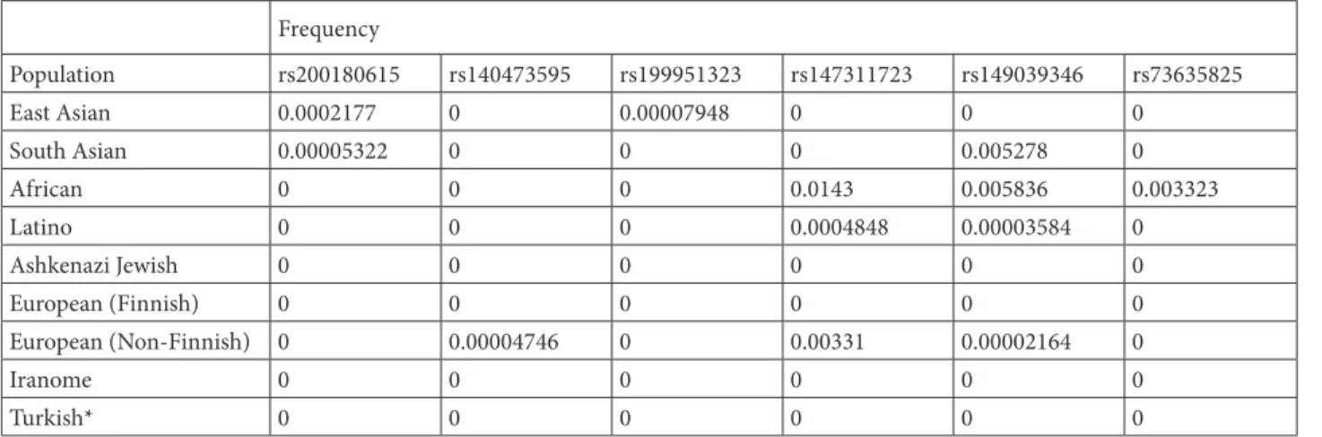
There are some theories of ACE2 gene polymorphisms and low expression levels to protect the host against and to fight these viruses [46]. According to the publication by Stawiski et al., although human ACE2 variants S19P, I21V, E23K, K26R, T27A, N64K, T92I, Q102P and H378R are predicted to increase susceptibility, especially T92I, part of a consensus NxS/T N-glycosylation motif and other ACE2 variants K31R, N33I, H34R, E35K, E37K, D38V, Y50F, N51S, M62V, K68E, F72V, Y83H, G326E, G352V,D355N, Q388L and D509Y are putative protective variants predicted to show decreased binding to SARS-CoV-2 S-protein [47]. In Italy, 1 of the countries having highest mortality rate from SARS-CoV-2 infections, no significant correlation was found between the disease severity and gender susceptibility of ACE2 [48]. However, in the same study, TMPRSS2 levels and genetic variants proved to be possible candidate disease modulators, contributing to the observed epidemiological data among Italian patients. It has been reported that not only the ACE2 gene, but also the ACE1 gene may be important for SARS-CoV-2 infections in terms of affecting the clinical course [49]. However, according to the gnomAD database, there is no significant difference in frequencies of the 6 ACE2 variants (rs200180615, rs140473595, rs199951323, rs147311723, rs149039346, rs73635825) mentioned in the literature [50] among South Asian, East Asian, African, Latin, Ashkenazi Jews, European (Finnish and non-Finnish) races. At the same time, based on the data from İstanbul Medipol University Genetic Diagnosis Center, there is no significant difference in frequency of these variants when comparing between Turkish society and Iranome with gnomAD databases (see Table 1). ICAM-3, OAS1 and MxA gene polymorphisms are also among other candidate factors associated with the protection against
SARS-CoV infections [51,52]. The second epidemic is Middle East respiratory syndrome coronavirus (MERS-CoV), which emerged in Saudi Arabia in 2012 and spread to 27 countries. The MERS-CoV spike (S) glycoprotein mediates viral entry into target cells [53]. Some genetic polymorphisms that adversely affect the entry of the MERS-CoV virus into the cell have been reported to be present in the DPP4.
Genetic polymorphisms are used in medicine for many purposes today. As the mechanisms caused by genetic polymorphisms are better understood, this is expected to allow for new treatments and discover of preventive drugs.
4. Clinical features
The novel coronavirus (COVID-19) infection is a respiratory system infection caused by SARS-CoV-2. This novel infectious agent, which was striking for the successive pneumonia cases occurring in Wuhan province of China in December 2019, is a betacoronavirus that is closely correlated to the SARS virus in genetic terms [54].
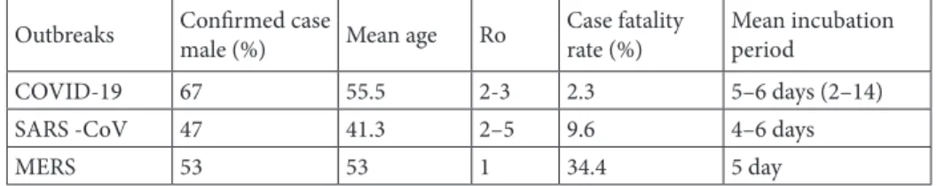
COVID-19 is a highly contagious infection. Various studies indicated the basic reproduction number (R0) as 2.6 to 4.7. When all 3 coronavirus infections leading to epidemic are compared, the mean age and sex distribution is similar. Table 2 shows the comparison of COVID-19 with SARS and MERS. Disease burden doubles every 7.7 days and 1 person infects an average of 2 to 3 people [55– 58].
The incubation duration of COVID-19 infection ranges from 2 to 14 days, and various studies demonstrate that symptoms are seen within an average of 4 to 7 days [56]. The frequency of asymptomatic infection is uncertain. The initial data from China indicate this frequency as 1%–2%, while a modeling study suggests that this ratio Table 1. Frequency of ACE2 variants in different population.
Frequency Population rs200180615 rs140473595 rs199951323 rs147311723 rs149039346 rs73635825 East Asian 0.0002177 0 0.00007948 0 0 0 South Asian 0.00005322 0 0 0 0.005278 0 African 0 0 0 0.0143 0.005836 0.003323 Latino 0 0 0 0.0004848 0.00003584 0 Ashkenazi Jewish 0 0 0 0 0 0 European (Finnish) 0 0 0 0 0 0 European (Non-Finnish) 0 0.00004746 0 0.00331 0.00002164 0 Iranome 0 0 0 0 0 0 Turkish* 0 0 0 0 0 0
is minimum 18% [5,55]. Additionally, abnormal lung tomography findings were found in 67% of asymptomatic cases identified by contact tracing [59].
The recovery takes about 2 weeks in mild cases and 3 to 6 weeks in severe cases. The duration from the onset of symptoms to death ranges from 2 to 8 weeks. Virus transmission peaks at the early stage of the disease. Transmission starts 24 to 48 h before symptoms and generally takes 7 to 12 days in mild/moderate cases and > 2 weeks in severe cases. PCR might be positive even after the patient is relieved of symptoms [60,61].
The COVID-19 clinical presentation is mild, moderate, severe and critical (Table 3) [62]. 81% of the confirmed cases in China were mild, while 14% (accompanied by hypoxemia, dyspnea and tachypnea) involved severe disease that required hospitalization and oxygen therapy. However, 5% were critical (with respiratory failure, septic shock and/or multiple organ dysfunction) and required admission to intensive care units [54]. No pneumonia was found in approximately 40% of the cases. In mild cases, the clinical presentation is dominated by the symptoms of viral upper respiratory tract infection, with likely symptoms of mild pneumonia in some cases. There are some further symptoms, such as low-grade fever, nasal
congestion, sore throat, headache, dry cough, fatigue and muscle pain. However, mild cases may still rapidly become critical. The conditions in moderate cases may worsen more frequently, therefore this group of patients should be monitored by exercising greater care [60–62]. The rapidly developing ARDS, septic shock, metabolic acidosis, coagulation dysfunction and multiple organ failure (acute kidney damage, cardiac damage, etc.) are important in critical cases [5,57].
Renal damage may occur depending on the virus or antivirals. Additionally, the virus may result in liver damage and testicular damage at varying levels [62].
Acute respiratory distress syndrome (ARDS) is diagnosed on a mild, moderate or critical basis in the intensive care setting considering PaO2/FiO2 parameters used to evaluate hypoxia as well as given the manifestation of the new-onset or worsening respiratory failure in the patient. In this case, it is better to perform diagnosis using chest X-ray, lung CT scan and ultrasound [60,61].
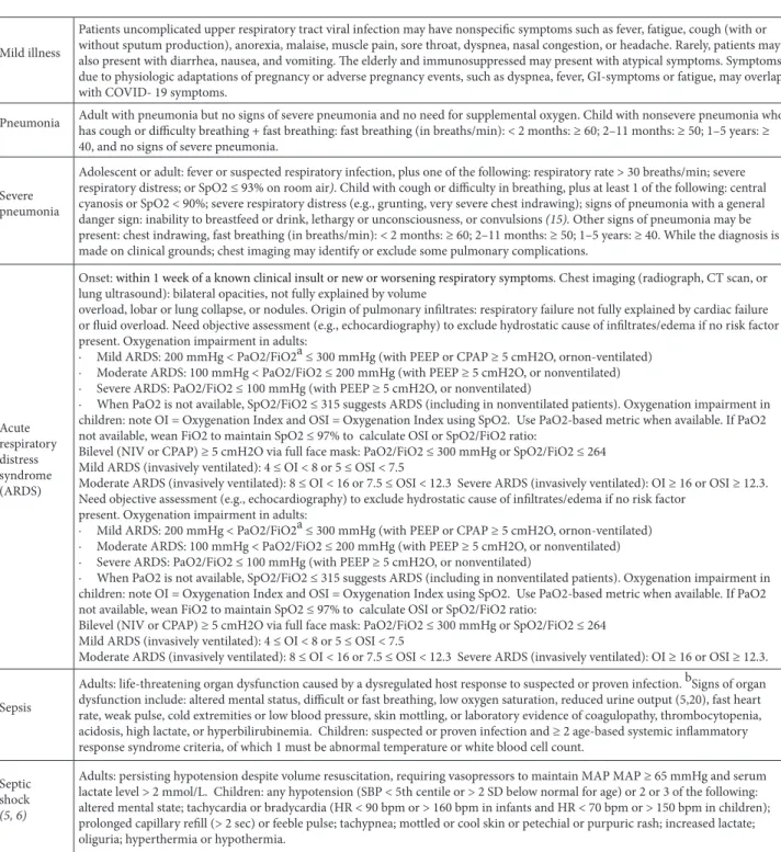
The clinical syndromes related to COVID-19, which are published and updated, as required, by the World Health Organization to help handling and monitoring of critical cases as clinical information about cases piles up, are exactly shown in Table 4 [1].
Table 2. Comparison of some characteristics between SARS-CoV, SARS- CoV-2, and MERS-CoV. (Adapted from resources 5, 57 and 58)
Outbreaks Confirmed case male (%) Mean age Ro Case fatalityrate (%) Mean incubation period
COVID-19 67 55.5 2-3 2.3 5–6 days (2–14)
SARS -CoV 47 41.3 2–5 9.6 4–6 days
MERS 53 53 1 34.4 5 day
*SARS-CoV-2: Severe acute respiratory syndrome coronavirus 2; SARS-CoV: Severe acute respiratory syndrome coronavirus; MERS- CoV: Middle East respiratory syndrome coronavirus; R0: Reproduction number.
Table 3. Clinical types of COVID-19 [62].
Mild The clinical symptoms were mild, and there was no or mild sign of pneumonia on imaging Moderate Showing fever and respiratory symptoms with radiological findings of pneumonia. Severe
In accordance with any of the following: a. Shortness of breath (RR ≧ 30 breaths/min); b. In resting state, oxygen saturation ≤ 93%;
c. Arterial partial pressure of oxygen (PaO2)/fraction of inspired oxygen (FiO2) ≦ 300 mmHg (l mmHg = 0.133kPa). d. Cases with chest imaging showed obvious lesion progression more than 50% within 24–48 h.
Critical
One of the following:
a. Respiratory failure, requiring mechanical ventilation; b. Septic shock;
Table 4. Clinical syndromes associated with COVID-19.
Mild illness
Patients uncomplicated upper respiratory tract viral infection may have nonspecific symptoms such as fever, fatigue, cough (with or without sputum production), anorexia, malaise, muscle pain, sore throat, dyspnea, nasal congestion, or headache. Rarely, patients may also present with diarrhea, nausea, and vomiting. The elderly and immunosuppressed may present with atypical symptoms. Symptoms due to physiologic adaptations of pregnancy or adverse pregnancy events, such as dyspnea, fever, GI-symptoms or fatigue, may overlap with COVID- 19 symptoms.
Pneumonia Adult with pneumonia but no signs of severe pneumonia and no need for supplemental oxygen. Child with nonsevere pneumonia who has cough or difficulty breathing + fast breathing: fast breathing (in breaths/min): < 2 months: ≥ 60; 2–11 months: ≥ 50; 1–5 years: ≥ 40, and no signs of severe pneumonia.
Severe pneumonia
Adolescent or adult: fever or suspected respiratory infection, plus one of the following: respiratory rate > 30 breaths/min; severe respiratory distress; or SpO2 ≤ 93% on room air). Child with cough or difficulty in breathing, plus at least 1 of the following: central cyanosis or SpO2 < 90%; severe respiratory distress (e.g., grunting, very severe chest indrawing); signs of pneumonia with a general danger sign: inability to breastfeed or drink, lethargy or unconsciousness, or convulsions (15). Other signs of pneumonia may be present: chest indrawing, fast breathing (in breaths/min): < 2 months: ≥ 60; 2–11 months: ≥ 50; 1–5 years: ≥ 40. While the diagnosis is made on clinical grounds; chest imaging may identify or exclude some pulmonary complications.
Acute respiratory distress syndrome (ARDS)
Onset: within 1 week of a known clinical insult or new or worsening respiratory symptoms. Chest imaging (radiograph, CT scan, or lung ultrasound): bilateral opacities, not fully explained by volume
overload, lobar or lung collapse, or nodules. Origin of pulmonary infiltrates: respiratory failure not fully explained by cardiac failure or fluid overload. Need objective assessment (e.g., echocardiography) to exclude hydrostatic cause of infiltrates/edema if no risk factor present. Oxygenation impairment in adults:
· Mild ARDS: 200 mmHg < PaO2/FiO2a ≤ 300 mmHg (with PEEP or CPAP ≥ 5 cmH2O, ornon-ventilated) · Moderate ARDS: 100 mmHg < PaO2/FiO2 ≤ 200 mmHg (with PEEP ≥ 5 cmH2O, or nonventilated) · Severe ARDS: PaO2/FiO2 ≤ 100 mmHg (with PEEP ≥ 5 cmH2O, or nonventilated)
· When PaO2 is not available, SpO2/FiO2 ≤ 315 suggests ARDS (including in nonventilated patients). Oxygenation impairment in children: note OI = Oxygenation Index and OSI = Oxygenation Index using SpO2. Use PaO2-based metric when available. If PaO2 not available, wean FiO2 to maintain SpO2 ≤ 97% to calculate OSI or SpO2/FiO2 ratio:
Bilevel (NIV or CPAP) ≥ 5 cmH2O via full face mask: PaO2/FiO2 ≤ 300 mmHg or SpO2/FiO2 ≤ 264 Mild ARDS (invasively ventilated): 4 ≤ OI < 8 or 5 ≤ OSI < 7.5
Moderate ARDS (invasively ventilated): 8 ≤ OI < 16 or 7.5 ≤ OSI < 12.3 Severe ARDS (invasively ventilated): OI ≥ 16 or OSI ≥ 12.3. Need objective assessment (e.g., echocardiography) to exclude hydrostatic cause of infiltrates/edema if no risk factor
present. Oxygenation impairment in adults:
· Mild ARDS: 200 mmHg < PaO2/FiO2a ≤ 300 mmHg (with PEEP or CPAP ≥ 5 cmH2O, ornon-ventilated) · Moderate ARDS: 100 mmHg < PaO2/FiO2 ≤ 200 mmHg (with PEEP ≥ 5 cmH2O, or nonventilated) · Severe ARDS: PaO2/FiO2 ≤ 100 mmHg (with PEEP ≥ 5 cmH2O, or nonventilated)
· When PaO2 is not available, SpO2/FiO2 ≤ 315 suggests ARDS (including in nonventilated patients). Oxygenation impairment in children: note OI = Oxygenation Index and OSI = Oxygenation Index using SpO2. Use PaO2-based metric when available. If PaO2 not available, wean FiO2 to maintain SpO2 ≤ 97% to calculate OSI or SpO2/FiO2 ratio:
Bilevel (NIV or CPAP) ≥ 5 cmH2O via full face mask: PaO2/FiO2 ≤ 300 mmHg or SpO2/FiO2 ≤ 264 Mild ARDS (invasively ventilated): 4 ≤ OI < 8 or 5 ≤ OSI < 7.5
Moderate ARDS (invasively ventilated): 8 ≤ OI < 16 or 7.5 ≤ OSI < 12.3 Severe ARDS (invasively ventilated): OI ≥ 16 or OSI ≥ 12.3. Sepsis
Adults: life-threatening organ dysfunction caused by a dysregulated host response to suspected or proven infection. bSigns of organ dysfunction include: altered mental status, difficult or fast breathing, low oxygen saturation, reduced urine output (5,20), fast heart rate, weak pulse, cold extremities or low blood pressure, skin mottling, or laboratory evidence of coagulopathy, thrombocytopenia, acidosis, high lactate, or hyperbilirubinemia. Children: suspected or proven infection and ≥ 2 age-based systemic inflammatory response syndrome criteria, of which 1 must be abnormal temperature or white blood cell count.
Septic shock
(5, 6)
Adults: persisting hypotension despite volume resuscitation, requiring vasopressors to maintain MAP MAP ≥ 65 mmHg and serum lactate level > 2 mmol/L. Children: any hypotension (SBP < 5th centile or > 2 SD below normal for age) or 2 or 3 of the following: altered mental state; tachycardia or bradycardia (HR < 90 bpm or > 160 bpm in infants and HR < 70 bpm or > 150 bpm in children); prolonged capillary refill (> 2 sec) or feeble pulse; tachypnea; mottled or cool skin or petechial or purpuric rash; increased lactate; oliguria; hyperthermia or hypothermia.
aIf altitude is higher than 1000 m, then correction factor should be calculated as follows: PaO2/FiO2 × barometric pressure/760. b The SOFA score ranges from 0 to 24 and includes points related to 6 organ systems: respiratory (hypoxemia defined by low PaO2/FiO2); coagulation (low platelets); liver (high bilirubin); cardiovascular (hypotension); central nervous system (low level of consciousness defined by Glasgow Coma Scale); and renal (low urine output or high creatinine). Sepsis is defined by an increase in the sepsis-related SOFA score of ≥ 2 points. Assume the baseline score is 0 if data are not available (22).
ARI: Acute respiratory infection; BP: Blood pressure; bpm: Beats/minute; CPAP: Continuous positive airway pressure; FiO2: Fraction of inspired oxygen; MAP: Mean arterial pressure; NIV: Noninvasive ventilation; OI: Oxygenation index; OSI: Oxygenation index using SpO2; PaO2: Partial pressure of oxygen; PEEP: Positive end-expiratory pressure; SBP: Systolic blood pressure; SD: Standard deviation; SIRS: Systemic inflammatory response syndrome; SOFA: Sequential organ failure assessment; SpO2: Oxygen saturation.
With the occurrence of COVID-19 outbreak, many series of cases were and are still continuously shared. As in SARS and MERS cases, the most common symptoms are fever and cough that often affect the lower respiratory system. Some of the case series to date are summarized in Table 5. Distinct symptoms are > 90% fever, 75% dry cough and 70% fatigue, followed by dyspnea developing in half of the patients 1 week later. Severe or critical patients may have low- or moderate-grade fever or no fever. Fatigue and myalgia were noted in > 40% of the cases. Runny nose, nasal congestion and other upper respiratory tract symptoms are less common (4%–6%). Additionally, there are some other symptoms, such as headache, dizziness, loss of appetite, anosmia and ageusia, sore throat, nausea, stomachache, and cough with sputum. While diarrhea was lower in the first series of cases (2%–3%), it was reported to range from 10% to 14% in subsequent cases. This suggests the probability of potential fecal-oral transmission [6,7,33,63–66].
In Italy, 96.5% of the patients presented due to acute respiratory distress. This was followed by acute kidney failure with 29.2%, along with 10.4% acute cardiac injury and 8.5% superinfection. The most common symptoms found during presentation were fever and dyspnea (76% and %73, respectively). 40% of the patients had cough. No symptom was found in 5.7% of the patients [56]. The loss of smell and taste, which was unidentified by initial data from China, were reported to be seen in Italy by 34% collectively and 19% individually [67].
Patients present to the hospital on day 4 from initial symptoms on average, whereas mild respiratory distress develops on day 5. From the onset of symptoms, hospitalization takes an average of 7 days (4–18) and respiratory distress occurs on day 8 on average (5–13)
and ARDS on day 9 on average1 [6,60–64]. Mechanical
ventilation is required on day 10.5 on average (7.0–14.00) [5].
Most patients have a good prognosis. Pediatric patients have mild symptoms. The course of the disease in pregnant women is similar to that in people of the same age group. No intrauterine transmission could be proven to date [60,61].
The COVID-19 infection has a low mortality rate compared to severe acute respiratory syndrome (SARS) and Middle East respiratory syndrome (MERS) (9.60% and 34.4%, respectively). In proven cases, the case-fatality rate is reported to be 2.3%–2.6% [5,54,58,59]. However, this rate remains uncertain. There are differences between regions
1 World Health Organization (2020). Clinical management of severe acute respiratory infection (SARI) when COVID-19 disease is suspected: interim
guidance 13 March 2020 [online]. Website https://www.who.int/emergencies/diseases/novel-coronavirus-2019/technical-guidance/patient-management [accessed 28 March 2020].
2 Centers for Disease Control and Prevention (2020). People who are at higher risk for severe illness [online]. Website https://www.cdc.gov/
coronavirus/2019-ncov/need-extra-precautions/people-at-higher-risk.html [accessed 01 April 2020].
of China, while the rates pertaining to different countries in the world are just emerging. This ratio may be affected by many factors, such as early diagnosis, early therapy and healthcare access [54]. Most of the patients who died are middle-aged and elderly people with comorbidities. No deaths occurred in the group aged younger than 9 years. However, the fatality rate among people aged 70 to 79 years is 8%. No deaths were reported in relation to nonsevere cases in China [54]. The case-fatality rate is 0.9% among patients without any comorbidity. Nearly 50% of the patients have one or more comorbidities. The case-fatality rate is higher among elderly people (aged ≥ 60 years) and people with comorbidities as follows: 14.8% ≥ 80 years, 10.5% cardiovascular disease, 7.3% diabetes, 6.3% chronic lung disease, 6.0% hypertension and 5.6% cancer. This rate is 0.9% among patients without any comorbidities. On the other hand, the case-fatality rate among critical patients is very high at 49.0% [54]. Even if the data provided by CDC is limited, CDC considered obesity (body mass index ≥ 40)
and liver disease in the risk group2
The ratio of patients who died of COVID-19 infection in Italy, which is 1 of the countries with the highest number of deaths in the world, by age range is 42.2% for people aged between 80 to 89 years and 32.4% for people aged between 70 to 79 years. On the other hand, the rate of deaths under the age of 70 years is lower (8.4% for the age range from 60 to 69 years and 2.8% for the age range from 50 to 59 years). Death was more common among males (70.6% in males compared to 29.4% in females) [56].
In Italy having many elderly deaths, approximately 2/3 of the cases accompanied by underlying diseases, such as diabetes, cardiovascular diseases or cancer, and smoking history verified the initial information obtained in China. Comorbidities result in both challenges to diagnosis and worsening of the condition. Considering comorbidities in 3,200 positive tested patients who died in Italy, 75.2% of those patients had ≥ 2 comorbidities found as follows: 73.5% hypertension, 33.9% diabetes 30.1% ischemic heart disease, 22% atrial fibrillation, 20% chronic kidney failure, 19.5% active cancer in the past 5 years, 13.7% chronic obstructive pulmonary disease, 11% 9 dementia, 11.2% stroke, and 3.7% chronic liver disease. Prior to hospitalization, 36% of the patients followed ACE-inhibitor therapy and 16% angiotensin receptor blockers (ARBs) therapy [56].
101 patients diagnosed in an elderly care home in the United States were aged 83 years on average, and 94% had an underlying chronic disease. The case-fatality ratio
Ta bl e 5. Clinic al c ha rac ter ist ics o f C or on av iru s Di se as es ( C O VID-19). Aut hor H ua ng et a l [6] Ch en et a l [63] Li et a l [64] So ng et a l [65] Ya ng et a l [66] W an g et a l [7] G ua n et a l. et a l [33] St ud y t im e D ec 16, 2019 – J an 2 , 2020 Ja n 1–J an 20, 2020 Ja n 22, 2020 Ja n 20–J an 27, 2020 La te D ec, 2019 –J an 26 , 2020 Ja n 1–J an 28, 2020 D ec 2019 – J an 29, 2020 To ta l p at ien ts, ch arac ter ist ics o f pa tien ts 41, p ros pe ct iv e, 32% admi tte d I CU 99 w ith p neum oni a , r et ros pe ct iv e, 23 % admi tte d I CU 425 w ith p neum oni a 51 w ith p neum oni a, 52, critic al ly i ll ad ul t p at ien ts w ith p neum oni a 138 w ith p neum oni a, 26 % admi tte d I CU , Pr es um ed h um an-to-h um an h os pi ta l-as so ci at ed t ra nsmi ssio n 41% 1099 p at ien ts, 5% admi tte d ICU (n on se ver e 2.4% a nd se ver e 19.1%) C onfir m ed c as es (%) Al l 100% NA Al l Al l Al l Al l Su sp ec te d c as es (%) -NA -A sy m pt om at ic p at ien ts (%) -NA -A ge , m ea n (I Q R) o r m ea n ± S D , y ea rs 49 (41–58) 55.5 ± 13.1 56 (26–82) 49 ± 16 59.7 (S D13,3) 56 (42–68) 47 (I Q R 35 t o 58) Sex, m ale 73% 68% 66% 49% 67% 54.3% 58.1% Exp os ur e hi sto ry 66% 49% 55% 98% 33% 8.7% Al l Th e m ea n d ura tio n fro m o ns et o f sy m pt om s t o h os pi ta l admi ssio n d ays** 7.0 (.4.0–8.0) NA Bef or e 1 J an m ea n 12,5 da ys(95% CI, 10.3 t o 14.8) B et w een 1–11 J an m ea n 9,1 d ays (95% CI, 8.6 t o 9.7) NA 9,5 ( 7.0–12.5) 7.0 (4.0–8.0); o ns et of sy m pt om t o I CU admi ssio n 10 (6–12) NA Ab no rm al X-ra y a nd CT fin din gs 95% 100% NA 100% 100% 100% 86.2% X-ra y a nd CT fin din gs Bi la tera l g ro un d g la ss op aci ty , 98% M ul tip le m ot tlin g a nd gr oun d g la ss o paci ty , 14%); pn eum ot ho rax 1% Radiog ra phic e viden ce o f pn eum oni a *G ro un d g la ss o paci ty
(GGO), 77%; GGO with r
et ic ul ar a nd/ o r in ter lo bu la r s ep ta l thic kenin g%; GGO w ith co ns olid at io n 59%; C on so lid at io n 55%; A ir b ro nc hog ra m 80%; R et ic ul at io n 22%; pleura l eff usio n 8%; Per ic ar di al eff usio n 6%; Ly m ph aden op at hy 6% NA G ro un d g la ss o paci ty 100% G ro un d-g la ss o paci ty , 56.4%; b ila tera l p at ch y sh ado w in g, 51.8%; L oc al pa tc hy s hado w in g, 41.9%; In ter sti tia la bn or m ali ties, 14.7%
Sig ns a nd sy m pt om s Fe ver , 98%; C oug h, 76%; M ya lg ia o r fa tigue , 44%); Sp ut um p ro duc tio n, 28%; H ead ac he , 8%; H em op tysi s, 5% ;Di ar rh ea, 3%; D ys pn ea, 55% Fe ver , 83%; C oug h, 82%; Sh or tn es s o f b re at h, 31%); M us cle ac he , 11%; C onf usio n, 9%; H ead ac he , 8%); S or e thr oa t, 5%; R hin or rh ea, 4%; C hes t p ain, 2%; Di ar rh ea, 2%; N au se a an d v omi tin g, 1% Fe ver , w ith o r w ith ou t re co rde d t em pera tur e Fe ver , 96%;C oug h, 47%; P hleg m, 20%; M ya lg ia o r fa tigue , 31%; H ead ac he a nd dizzin es s, 16%; D ys pn ea or c hes t p ain,14%; Los s o f a pp et ite ,18%); Di ar rh ea ,10%; S tuff y an d r unn y n os e, 4%; S or e thr oa t, 6%; N au se a a nd vo mi tin g, 6%) Fe ver , 98%; C oug h, 77%; D ys pn ea, 63.5%; M ya lg ia, 11.5%; M al ai se , 35%; R hin or rh ea, 6%; H ead ac he , 6%; V omi tin g,4%; A rt hra lg ia, 2%; C hes t pa in, 2% Fe ver , 98.6%; F at igue , 69.6%; Dr y co ug h, 59.4%; A no rexi a, 39.9%; M ya lg ia, 34.8%; D ys pn ea, 31.2%; Exp ec to ra tio n, 26.8%; Ph ar yn ga lg ia, 17.4%; Di ar rh ea, 10.1%;N au se a, 14 (10.1%) Dizzin es s, 9.4%;H ead ac he , 6.5%;V omi tin g, 3.6%; Ab do min al p ain, 2.2% Fe ver ,43.8 % o n admi ssio n an d 88.7% d ur in g hos pi ta liza tio n; C oug h, 67.8%; F at igue , 38.1%; S pu tum pr od uc tio n, 33.7%; Sh or tn es s o f b re at h, 18.7%; M ya lg ia o r ar thra lg ia, 14.9%; S or e thr oa t , 13.9%; H ead ac he , 13.6%; C hi lls, 11.5%; N au se a o r v omi tin g, 5%; N as al co ng es tio n , 4.8%; Di ar rh ea , 3.8%; o th er 1.7%; C om plic at io ns ARDS, 29%; RN Aemi a, 15%;A cu te c ar di ac in jur y, 12%; A cu te k idn ey in jur y,7%; S eco nd ar y inf ec tio n, 10%;S ho ck, 7% ARDS, 17%;A cu te ren al in jur y, 3%;A cu te res pira to ry in jur y, 8%; S ep tic s ho ck, 4% Ven til at or -a ss oci at ed pn eum oni a, 1% NA NA ARDS, 67%; A cu te kidn ey in jur y, 29%; Ca rdi ac in jur y, 23%; Li ver d ysf un ct io n, 29%; H yp er gl ycemi a , 35%; H os pi ta l-acq uir ed p neum oni a 13.5%; Ga str oin tes tin al hem or rh ag e , 4%; Pn eum ot ho rax ,2%; H os pi ta l acq uir ed inf ec tio ns 13.5% in cludin g B ac ter emi a an d p ulm on ar y inf ec tio n 2%; U rin ar y t rac t inf ec tio n , 2% Sh oc k, 8.7%; A cu te ca rdi ac in jur y, 7.2%; A rr hyt hmi a, 16.7%; ARDS, 19.6%; AKI, 3.6% (n on se ver e a nd s ev er e) Sep tic s ho ck , 1.1% (0.1%-6,4%); A cu te r es pira to ry di str es s sy ndr om e , 3.4% (1.1%-15.6); A cu te k idn ey in jur y , 0.5% (0.1%–2.9%) C om orb idi ties To ta l 32% in cludin g Di ab et es, 20%; H yp er ten sio n, 15%; Ca rdio va sc ul ar di se as e, 15%; C hr onic o bs tr uc tiv e pu lm on ar y di se as e, 2%; M alig na nc y, 2%; Chr onic li ver di se as e, 2% To ta l 51% in cludin g Ca rdio va sc ul ar a nd cer eb ro va sc ul ar di se as es, 40%; Dig es tiv e sys tem di se as e , 11%; En do cr in e sys tem di se as e(Di ab et es) ,13%; o th er s , 3% To ta l 22% in cludin g di ab et es, h y- p er ten sio n, chr onic li ver di se as e, chr onic o bs tr uc tiv e pu lm on ar y di se as e, a nd ca rdi ac di se as e; ciga ret te sm ok er s 7% NA Chr onic m edic al illn es s 40%in cludin g Chr onic c ar di ac di se as e, 10%; C hr onic pu lm on ar y di se as e, 8%; C er eb ro va sc ul ar di se as e 13.5%; Di ab et es 17%; M alig na nc y 4%; Sm ok in g, 4; o th er s 4% To ta l 46.4% in cludin g H yp er ten sio n , 31.2%; Ca rdio va sc ul ar di se as e , 14.5%; Di ab et es , 10.1%; M alig na nc y , 7.2%; C er eb ro va sc ul ar di se as e , 5.1%; C O PD , 2.9%; C hr onic k idn ey di se as e 2.9%; C hr onic liv er di se as e , 2.9%; HIV inf ec tio n , 1.4% At le as t o ne co exi stin g illn es s, 23.7%; H yp er ten sio n , 15%; Di ab et es 7.4%; C or on ar y he ar t di se as e , 2.5%; C er eb ro va sc ul ar di se as e, 1.2%; C hr onic o bs tr uc tiv e pu lm on ar y di se as e, 1.1%; ot her s 3.9%;S m ok er , 14.5% D isc ha rge d 68% 31% NA NA NA 34.1% 5% ( 93.6% h os pi ta lize d) D ead 15% 11% NA NA 61.5%** 4.3% 1.4 % *A t c hes t CT , GGO s w er e b ila tera l in 88% o f p at ien ts, in vo lv in g t he p os ter io r l un gs in 82% a nd t he p er ip hera l l un gs in 85% o f p at ien ts. P at ien ts o lder t ha n 50 y ea rs h ad m or e co ns olid at ed l un g lesio ns t ha n did t hos e a ge d 50 y ea rs o r y oun ger (212 o f 470 vs. 198 o f 854; P : 0.001) ** M edi an d ura tio n f ro m I CU admi ssio n t o de at h wa s 7 (I Q R 3–11) in ter qu ar tile ra ng e. Ta bl e 5. ( C on tin ue d).
among these patients were reported to be 55% and 34%, respectively [68]. Similarly, a multi-variance analysis established the factors that identify a poor prognosis at an early stage as older age, high “sequential organ failure assessment” (SOFA) score and D-dimer value > 1 μg/L during hospitalization [6].
The COVID-19 differential diagnosis should be differentiated for infectious and noninfectious reasons. However, it should be noted that the COVID-19 infection may accompany other viral or bacterial infections of respiratory tract. In these cases, the agents of lower and upper respiratory tract infection (influenza A and B, respiratory syncytial virus, parainfluenza viruses, rhinoviruses, adenoviruses, enteroviruses (e.g., EVD68), human metapneumovirus and endemic human coronaviruses (i.e. HKU1, OC43, NL63, and 229E)) should be investigated. It is important to investigate the bacterial agents leading to lower respiratory tract infections, such as
Legionella pneumophila, and to start therapy accordingly.
Differential diagnosis is used to investigate infections, such as malaria, arbovirus infection (dengue/chikungunya), etc., in endemic regions [61, 63,65,66].
Health professionals started to be infected nosocomially in the early periods when there was scarce information about the COVID-19 infection and its causative agent SARS-CoV-2 with a multitude of unknown things. In China, 14.6% of 1,688 health professionals diagnosed in a laboratory setting were severe. However, the number of cases among health professionals started to decrease in time particularly due to training, their proper use of personal protective equipment and hand hygiene practices. Initial cases occurred from 1 to 10 January 2020 (20 cases) and peaked with 1,036 cases from 21 to 31 January. They were found to be 322 after 1 February (1). 20% of the Italian health professionals were infected, some of whom lost their lives unfortunately [56].
5. Laboratory findings
During the course of COVID-19, patients have pathological modifications as indicated by different laboratory tests. The publications regarding these modifications shown by laboratory tests are gradually increasing in number. A substantial amount of knowledge has accumulated despite the still lacking very detailed conclusions, and the conclusions from several studies have been started to be evaluated and analyzed [69–78].
The disorders diagnosed upon hematological, immunological and biochemical tests are those that are usually diagnosed in hospitalized patients. The summary information on the tests used in this context is given in Table 6.
Laboratory tests provide guidance in risk assessment, clinical monitoring and therapy decision-making processes [69–72].
5.1. Hematological tests
Complete blood count (Hemogram)
The observation was that there was no significant change in hemoglobin levels of COVID-19 patients, with those reported with a slight minor decrease [36]. Patients with severe disease were found to have significantly lower hemoglobin values than patients with mild conditions [71].
The count of leukocytesmay be normal as follows; leukocytosis (> 10,000 / microL) 2% –30%, and leukopenia (<4000 / microL) 9%–31% [70].
Studies demonstrated that mild leukocytosis was noted in those with severe disease symptoms, while there was a significant increase in the number of patients who died. The increased level of leukocytes is a parameter that is indicative of clinical deterioration. Various studies discovered, in addition to leukocytosis, an increase in the count of neutrophils and a decrease in the count of lymphocytes, monocytes and eosinophils [18].
Lymphopenia (lymphocyte count <1500 / microL) was frequently detected on the hemogram (3%–83.2%) [36, 69]. Lymphopenia is a very common symptom in patients with severe progression of COVID-19 and in patients who died. The said patients were observed to have reduced count of CD4+ and CD8+ T cells. The percentage of lymphocytes, along with the count of CD4 + and CD8 + T cells, was proposed as a predictive biomarker for disease severity or recovery (prognosis) [72]. The studies carried out demonstrated that neutrophil to lymphocyte ratio in patients with COVID-19 may be a significant marker in explaining disease severity and mortality. Severe disease and intensive care unit admission were significantly high in the group of patients with the neutrophil-to-lymphocyte ratio (NLR) of ≥3.13, and NLR was considered the independent risk factor for the illness [73].
Additionally, thrombocytopenia (5%–36.2%) was reported [18,69,70]. Moderate thrombocytopenia is common, but rarely below 100,000/microL [74]. Platelet count was significantly low in patients with severe COVID-19 course. Additionally, significant thrombocytopenia was found in subjects who died than survivors. Based on the conclusions from several studies, it was noted at a 5-fold higher level in patients with severe disease course. A low platelet count is linked to an increased risk of severe disease and mortality in patients with COVID-19, and is indicative of poor prognosis. Patients showing signs of fluctuation in platelet count or signs of heparin resistance should be considered for heparin-induced thrombocytopenia (HIT) [69,74].
5.2. Coagulation tests
Patients with COVID-19 were observed to be prone to coagulopathy. However, no adequate data is yet available regarding bleeding and thrombosis. Furthermore, coagulation tests are critical to the diagnosis and
assessment of disseminated intravascular coagulation, which is an important complication noted in patients with COVID-19 [18].
D-dimer: It was noted in 9%–43% of the patients
investigated [18]. The level of D-dimer at admission was found to be associated with the risk of developing ARDS the requirement for intensive care and the risk of mortality [18,75]. The high D-dimer level and the D-dimer levels significantly increased over time are associated with high mortality, and may be marker of infection/sepsis, cytokine storm and impending organ failure. In particular, D-dimer levels in intensive care patients were noted to be significantly higher than those in patients who were not admitted to intensive care units [18].
Prothrombin time was significantly increased in survivors and patients who died.
Disseminated intravascular coagulation in patients with COVID-19 may develop over time due to poor prognosis [72].
5.3. Cardiac tests
Type-B natriuretic peptide (BNP/NT-proBNP):
The rapidly increasing level of NT-proBNP related to cardiac dysfunction in patients with severe COVID-19 was found to be significantly higher [36,70,76].
Cardiac troponins (TnTTnI): COVID-19 may lead
to serious inflammatory cases that are likely to enhance thrombosis and myocardial infarction. Additionally, viruses or inflammation may directly cause myocardial damage. Serum troponin I levels were seen to be high in critical COVID-19 patients (8%–51.2%;)[36,70].
At hospitalization, patients with COVID-19 (12.5%) were found to have myocarditis-like abnormalities, Table 6. Common laboratory findings among hospitalized patients with COVID-19*.
Tests Changes and interpretations
Hematological parameters (including hemostasis and coagulation) Complete blood count
Hemoglobin (usually normal, may be decreased in severe cases) Wbc [normal, decreased (29%), increased (in ICU cases)] Lymphopenia (in most cases), < 800 microL in severe cases Eosinopenia (in most cases),
Thrombocyte (normal, increased, slightly lower in severe cases
CD4 and CD8 Decreased (in most cases)
PT and aPTT Normal, slightly prolonged (in severe cases, especially in ICU cases)
Fibrinogen Increased
D-dimer Increased (in severe cases; in nonsurvivors); >1000 ng/mL in severe cases Inflammatory markers
CRP Increased (higher in severe cases); > 10 x the upper limit of normal (N: < 8 mg/L)
ESR Normal, Increased
Ferritin Increased (in severe cases); > 500 mcg/L (N: 10/30–200/300 mcg/L)
Procalcitonin Normal, increased (in severe cases, secondary to bacterial pneumonia in ICU patients) Il-6 Increased (according to severity: >critical>severe>mild; cytokine storm)
Biochemistry tests
Glucose Increased (in severe cases)
BUN and creatinine Increased (in severe cases)
Electrolytes Hyponatremia, hypokalemia
Albumin Decreased (in severe cases)
ALT and AST Increased (especially in ICU cases) Total bilirubin Increased (in severe cases)
Lactate dehydrogenase Increased (in severe cases, in ICU cases, in ARDS cases); >245 U /L in severe cases (N: 110–210 u/L) Creatine phosphokinase Increased in severe cases; > 2 x the upper limit of normal (N: 40–150 U/L)
Troponin T/I Increased (in severe cases), > 2 x the upper limit of normal ( N: 0–9/14 ng/L)
NT-ProBNP Increased (in severe cases)
especially an elevated level of cardiac troponin I. The troponin level, which was normal at admission, increased gradually over time (37.5%) (in the absence of typical EKG and echography), especially in patients who died. The elevation of cardiac troponin I is more associated with systemic disorders [77].
Elevations in cardiac markers may originate from secondary and systemic outcomes (multiple organ failure, etc.) rather than the effect of the virus [36,70].
Creatine kinase (CK/CK-MB): The muscle-induced
creatine kinase provides an insight, especially into the assessment of myocardial damage. CK-MB was found to be high at 7%–33%; higher rates were noted in patients with severe disease course [36,70].
Myoglobin: Myoglobin is commonly present in most
tissues, and the plasma level was found to have increased by 25% to 17% in connection with the cardiac damage in patients with COVID-19 [36,70].
5.4. Markers of inflammation
C-reactive protein (CRP): High levels of C-reactive
protein (CRP) were noted in the vast majority of COVID-19 patients (3%–91%) and shown to be associated with disease severity [69,70,78]. CRP levels give an idea for disease severity and prognosis. Additionally, it is considered to be a potential early marker for sepsis and mortality. It is suggested that CRP level, especially at admission, may be critical for grading disease severity [78]. The patients who survived had a CRP level of ~40 mg/L, while those who died had 125 mg/L (60–160 mg/L) [74].
Non-COVID etiologies (such as heart failure) should be considered for a patient with severe respiratory distress and normal CRP levels.
Erythrocyte sedimentation rate (ESR): ESR, which
is specifically assessed together CRP in COVID-19 cases, were generally found to be high (15%–85%) [36]. Patients with severe and moderate COVID-19 were shown to have elevated CRP and ESR without any modification indicated by the lung CT scan [72]. ESR, along with CRP, could be an important and inexpensive monitoring parameter.
Cytokines: Cytokines have critical effects on the
pathogenesis of the disease. Studies showed that IL-6, IL-2, IL-10 and IFN levels were elevated. Especially, hyperinflammation may lead to cytokine storm mediated by IL-6 receptors.
Blood cytokines and chemokines were found to be present at significantly high levels in patients with COVID-19 infection, including IL1-β, IL1RA, IL7, IL8, IL9, IL10, FGF2, GCSF, GMCSF, IFNy, IP10, MCP1, MIP1α, MIP1β, PDGFB, TNFa and VEGFA [76]. Some of the severe subjects presenting to the intensive care unit were noted to have high levels of proinflammatory cytokines, including IL2, IL7, IL10, GCSF, IP10, MCP1, MIP1α and TNFα, which were justified for the increased disease severity [18,76].
Ferritin: Ferritin becomes elevated as an outcome
of the activation of macrophages and hepatocytes in COVID-19 cases. Unlike other viral infections, ferritin is elevated at a moderate level in cytokine storm syndrome. It is thought to be used as a predictive marker of sepsis mortality [69, 70].
Procalcitonin: The level of procalcitonin in COVID-19
usually remains within the normal range at admission (<0.5 in 95% of the patients) [33]; however, it is likely that this level could be higher in patients who need admission to ICU (3%–35%) [69].
Few studies found increased procalcitonin levels. This should primarily suggest an alternative diagnosis (bacterial pneumonia); however, any progressive PCT increase in patients who are hospitalized/admitted to ICU may be associated with this poor prognosis or a secondary bacterial infection [18,27,75].
Patients with elevated PCT were noted to have a 5-fold more severe COVID-19 infection risk (OR, 4.76; 95% CI, 2.74–8.29) [69].
5.5. Biochemical tests
COVID-19 leads to important modifications indicated by biochemical tests since it affects various systems.
Albumin:
Albumin is low in especially about 50% of the patients with prolonged clinical course. The studies conducted with COVID-19 patients suggest that low serum albumin levels are linked to the increased risk of mortality [69,70].
Albumin concentrations were significantly lower in severe cases compared to moderate cases, where hypoalbuminemia (albumin <32 g/L) were seen more frequently in severe cases [76].
Glucose: COVID-19 patients with diabetes are exposed
to high risk of progressing to ARDS, septic shock and multiple organ failure. Retaining glucose level within the normal range in this patient group is one of the parameters to be noted for comorbidity [69, 70].
Aminotransferases: Studies have indicated increases
in ALT (17%–28%), AST (8%–37%) and gamma-glutamyl transferase levels which are over 3-fold higher than the upper limit [70]. There is no direct association found between the rate of increase in enzymes and the severity of the disease. Liver damage increases upon the prolongation of hospitalization, which could be associated with therapy protocols [79]. Abnormalities indicated by liver tests were noted to be higher in patients who developed pneumonia over time. Alanine aminotransferase (ALT) and aspartate aminotransferase (AST) levels were significantly higher in severe cases than in moderate cases [76,79].
Total bilirubin: Bilirubin increased by 0%–18%
(generally up to 3-fold the upper limit) associated with hepatocellular damage and cholestatic liver diseases could be seen in COVID-19 patients [69,70].
![Table 5. Clinical characteristics of Coronavirus Diseases ( COVID-19). AuthorHuang et al [6]Chen et al[63]Li et al[64]Song et al[65]Yang et al[66]Wang et al[7]Guan et al](https://thumb-eu.123doks.com/thumbv2/9libnet/5454906.105105/11.829.83.775.73.1067/table-clinical-characteristics-coronavirus-diseases-covid-authorhuang-song.webp)
