DESIGN, SYNTHESIS AND CHARACTERIZATION OF ORTHOGONAL BODIPY TRIMERS AND PROGRESS TOWARDS A SINGLET OXYGEN
PROBE
A THESIS SUBMITTED TO
THE GRADUATE SCHOOL OF ENGINEERING AND SCIENCE OF BILKENT UNIVERSITY
IN PARTIAL FULFILLMENT OF THE REQUIREMENTS FOR THE DEGREE OF
MASTER OF SCIENCE IN
CHEMISTRY
By José Luis Bila
i
DESIGN, SYNTHESIS AND CHARACTERIZATION OF ORTHOGONAL BODIPY TRIMERS AND PROGRESS TOWARDS A SINGLET OXYGEN PROBE
By José Luis Bila August, 2015
We certify that we have read this thesis and that in our opinion it is fully adequate, in scope and in quality, as a thesis for the degree of Master of Science.
_________________________________ Prof. Dr. Engin U. Akkaya (Advisor)
_________________________________ Assoc. Prof. Dr. Dönüş Tuncel
_________________________________ Assist. Prof. Dr. Fazlı Sözmen
Approved for the Graduate School of Engineering and Science: _______________________
Prof. Dr. Levent Onural Director of the Graduate School
ii
Abstract
DESIGN, SYNTHESIS AND CHARACTERIZATION OF ORTHOGONAL BODIPY TRIMERS AND PROGRESS TOWARDS A SINGLET OXYGEN
PROBE
José Luis Bila
M. Sc. in Department of Chemistry Supervisor: Prof. Dr. Engin Umut Akkaya
August, 2015
Several methods of fighting cancer have been proposed over the years after it was discovered. Photodynamic therapy (PDT) has been part of these methods for many years therefore; too much financial effort has been spent on this field. Since PDT relays on three important components namely: Sensitizer, molecular oxygen and light, our group has worked and published several photosensitizers derived from BODIPY over the years. In this thesis two different ideas to be used in PDT are introduced. First, Heavy atom free orthogonal BODIPY trimmers are introduced for the first time in literature and they are believed to work according to DS-TR principle. Effective singlet oxygen productions were obtained up to 0.53 quantum yields. The second topic is the use of BODIPY and 1,3-DiphenylIsobenzofuran hybrids as a singlet oxygen sensor. This sensor is meant to work based on PeT and ICT mechanisms. No further experimental methods were applied to this molecule after synthesis however; we intend to use it for in-vivo singlet oxygen experiments in the future.
Keywords: Photodynamic therapy, singlet oxygen sensor, DS-TR, BODIPY trimmers, orthogonal
iii
ÖZET
TRİMER DİK BODIPY YAPILARININ DİZAYN, SENTEZ VE
KARAKTERİZASYONU VE SİNGLET OKSİJEN ALGILAYICISININ GELİŞİMİ
José Luis Bila Bilkent Kimya Bölümü
Danışman: Prof. Dr. Engin Umut Akkaya Ağustos, 2015
Kanserin keşfinden sonra, kanserle mücadele için bir çok metod geliştirilmiştir. Fotodinamik terapi de bu metodlardan biri olup, bu alanda çok para harcanmıştır. Fotodinamik terapide kullanılan üç ana bileşen vardır: duyarlaştırıcı, moleküler oksijen ve ışık. Araştırma grubumuz da BODIPY tabanlı fotoduyarlaştırıcılar üzerine uzun yıllardır çalışmakta ve bu konuda bir çok yayın çıkarmaktadır. Bu çalışmada, fotodinamik terapide kullanılan iki farklı fikir tanıtılmıştır. Ilk olarak, DS-TR prensibiyle çalışılacağına inanılan, literatürde ilk defa çalışılan, ağır atom bulunmayan trimer dik BODIPY tanıtılmıştır. 0.53 quantum verimine sahip singlet oksijen üretilmiştir. Ikinci konu ise, BODIPY ve 1-3,difenilizobenzofuran hibritlerinin singlet oksijen sensörü olarak kullanılmasıdır. Sentezlenen bu sensör PeT ve ICT mekanizmalarıyla çalışmaktadır. Sensörün sentezi bitmiş olup, henüz ölçüm alınmamıştır, ancak ilerde canlı içinde singlet oksijen deneyleri yapılacaktır.
Anahtar kelimeler: Fotodinamik terapi, singlet oksijen sensor, DS-TR, Trimer BODIPY, Dik BODIPY
iv
v
Acknowledgement
I would like to start by thanking Prof. Dr. Engin U. Akkaya for his patience and guidance throughout my master’s degree. I would like to specially thank him for all the positive insights and attitudes both in good and bad times. He has never let me down no matter what, and I believe choosing him, as my adviser was one of the best decisions I have ever made because he is one of a kind.
Secondly, I would like to direct a million thanks to Dr. Tuğba Özdemir Kütük for being my older sister, partner and guide in almost everything. I have worked with her most of the time and in almost every project I was involved she was there to carry the burden with me. I sincerely have no words to express the amount of love and gratitude I will always carry towards her.
I thank in particular my closest friends Ceren Çamur, Darika Okeeva, Tuğçe Durgut, Tuğçe Karataş, Melek Baydar Baytak, Hidayet Baytak, Dielse Inroga and Cansu Kaya, Muazzam Idris for being there when I needed them, in every birthday celebration, every unnecessary çiğköfte meal, marriage proposal and especially for their priceless friendship and invaluable love. I thank Ceren Çamur, Darika Okeeva, Dr. Fazlı Sözmen, Dr. Safacan Kölemen, Seray Ural and Bilal Uyar for working with me in other projects, which helped me learn new topics and synthetic methods in chemistry.
I would like to thank every other member of the supramolecular chemistry research group: Nisa Yeşilgül, Dr. Dilek Taşgın, Ahmet Atılgan, Veli Polat, Bilal Kılıç, Dr. Ruslan Guliyev, Deniz Yıldız, Özlem Seven, Dr. İlke Şimşek and Seylan Ayan. I would like to express my deepest love to my family, Luis Bila and Ana Chirrizane for bringing me to the world and for taking care of my siblings and I, in every step and way they could. They struggled to watch me grow and study. I wish they were still in this world to share this happiness with me. I would like to thank my aunt Beatriz Bila for her support in most of the time of my life. She has been the mother and father I always needed. I also thank my high school teacher and friend Bahattin Akar for his financial and moral support in every step of the way to the university. No words are there to express the deep love and gratitude I have for my fiancée, Hale Atılgan for being in every single moment of my life and academic career. We watched each other grow and she supported and guided me in every decision I took. With her I shared every dream, every hope, sorrow and the smallest burden. All I can say is: I love you with all my heart and I hope you will always be in my life until the end of the road.
vi
LIST OF ABBREVIATIONS
AcOH : Acetic Acid
DCM : Dichloromethane
CHCl3 : Chloroform
BODIPY : 4,4-difluoro-4-bora-3a,4a-diaza-s-indacene
DS-TR : Doubly substituted tetra radicalic
HOMO : Highest Occupied Molecular Orbital
ICT : Internal Charge Transfer
LUMO : Lowest Unoccupied Molecular Orbital
MS : Mass Spectroscopy
NMR : Nuclear Magnetic Resonance
PDT : Photodynamic Therapy
PET : Photoinduced Electron Transfer
TFA : Trifluoroacetic Acid
vii
TABLE OF CONTENTS
1. INTRODUCTION ... 1 CHAPTER 2 ... 3 2. BACKGROUND INFORMATION ... 3 2.1 Sensors ... 3 2.2 Fluorescence ... 42.2.1 Photo-induced electron transfer (PeT) ... 7
2.2.2 Internal Charge Transfer (ICT) ... 9
2.3 Photodynamic Therapy (PDT) ... 12
2.3.1 History of PDT ... 12
2.3.2 Working mechanism ... 13
2.3.3 Oxidative damage of singlet oxygen ... 14
2.3.4 Requirements for photosensitizers ... 14
2.4 Photosensitizer families in literature ... 15
2.4.1 Phenothiazines ... 15 2.4.2 Xanthenes ... 16 2.4.3 Phtalocyanines ... 16 2.4.4 Anthraquinones ... 17 2.4.5 Cyanines ... 18 2.4.6 Curcuminoids ... 18 2.5 Triplet Photosensitization in PDT ... 19
2.5.1 Incorporation of Heavy Atoms ... 19
2.5.2 Heavy Atom Free Photosensitization ... 21
2.5.3 Low-Lying n-p* Transition based chromophore design ... 22
2.5.4 Exciton Coupling ... 23
2.5.5 Spin Convertors ... 23
2.6 BODIPY ... 25
2.6.1 BODIPY Applications ... 25
3. EXPERIMENTAL PROCEDURES ... 29
viii 3.2. Synthesis of compound (30)...29 3.3. Synthesis of compound (31)...30 3.4. Synthesis of compound (32) ...31 3.5. Synthesis of compound (33) ...32 3.6. Synthesis of compound (34) ...33 3.7. Synthesis of compound (35) ...34 3.8. Synthesis of compound (36) ...35 3.9. Synthesis of compound (37) ...36 3.10. Synthesis of compound (38) ...37 3..11. Synthesis of compound (39) ...38 3.12. Synthesis of compound (40) ...38 3.13. Synthesis of compound (41) ...39 3.14. Synthesis of compound (42) ...40 3.15. Synthesis of compound (43) ...41 3.16. Synthesis of compound (44) ...41 3.17. Synthesis of compound (45) ...42 3.18. Synthesis of compound (46) ...42 3.19. Synthesis of compound (47) ...43 3.20. Synthesis of compound (48) ...44 3.21. Synthesis of compound (49) ...45 3.22. Synthesis of compound (50) ...46 3.23. Synthesis of compound (51) ...47
ix
CHAPTER 4 ... 47
4. Heavy Atom free sensitizers for PDT ... 47
4.1. Introduction...48
4.2. Objectives...48
4.3. Synthesis...50
4.4. Results and discussion...53
5. Progress towards a singlet oxygen probe...57
5.1. Introduction...57
5.2. Objectives and synthesis of singlet oxygen probe...59
5.3. Conclusion...63
Bibliography ... 63
x
LIST OF FIGURES
Figure 1. Jablonski diagram. ... 5
Figure 2. Stokes shift representation. ... 6
Figure 3 - Schematic and molecular orbital representation of PeT working mechanism. ... 7
Figure 4. Schematic representation of reverse PeT. ... 8
Figure 5. Some examples of PeT based sensors. ... 9
Figure 6. Representation of ICT working mechanism for blue shift. ... 10
Figure 7. Representation of ICT working mechanism for red shift ... 11
Figure 8.. Examples of ICT sensors. ... 12
Figure 9. Jablosnki diagram for singlet oxygen production. ... 13
Figure 10. Examples of phenothiazines sensitizers.43 ... 16
Figure 11. Examples of xanthene sensitizers. ... 16
Figure 12. Example of phtalocyanine sensitizer. ... 17
Figure 13. Example of anthraquinone sensitizer. ... 17
Figure 14. Example of cyanine sensitizer. ... 18
Figure 15. Example of curcuminoid sensitizer. ... 18
Figure 16. Halogenated sensitizers for PDT. ... 20
Figure 17. Di-styryl bromo-substituted BODIPY molecule. ... 21
Figure 18. An example of excited states with low-lying n-p* orbitals. ... 22
Figure 19. An example of p-p* sensitizer. ... 23
Figure 20. Examples of sensitizers with exciton coupling states. ... 23
Figure 21. An example of sensitizer with spin convertor. ... 24
Figure 22. BODIPY molecule. ... 25
Figure 23. Applications of BODIPY molecule. ... 26
Figure 24. Examples of BODIPY PeT sensors. ... 27
Figure 25. Water soluble BODIPY based molecules. ... 28
Figure 26. Symthesis of compound 30. ... 29
Figure 27. Symthesis of compound 31. ... 30
Figure 28. Symthesis of compound 32 ... 31
Figure 29. Symthesis of compound 33. ... 32
Figure 30. Symthesis of compound 34. ... 33
Figure 31. Symthesis of compound 35 ... 34
Figure 32. Synthesis of compound 36. ... 35
Figure 33. Synthesis of compound 37. ... 36
Figure 34. Synthesis of compound 38. ... 37
Figure 35. Synthesis of compound 39. ... 38
Figure 36. Synthesis of compound 40. ... 38
Figure 37. Synthesis of compound 41. ... 39
Figure 38. Synthesis of compound 42. ... 40
xi
Figure 40. Synthesis of compound 44. ... 41
Figure 41. Synthesis of compound 45. ... 42
Figure 42. Synthesis of compound 46. ... 42
Figure 43. Synthesis of compound 47. ... 43
Figure 44. Synthesis of compound 48. ... 43
Figure 45. Synthesis of compound 49. ... 44
Figure 46. Synthesis of compound 50. ... 45
Figure 47. Synthesis of compound 51. ... 46
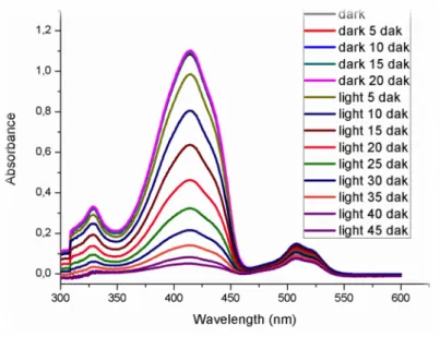
Figure 48. Singlet oxygen generation experiment in DCM solution. Decrease in Absorbance spectrum of trap molecule (DPBF) in the presence of 5.0 µM compound (33). ... 52
Figure 49. Absorbance decrease of DPBF at 414 nm with time in dichloromethane in the presence of BODIPY photosensitizer 33. ... 53
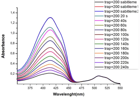
Figure 50. Singlet oxygen generation experiment in DCM solution. Decrease in Absorbance spectrum of trap molecule (DPBF) in the presence of 5.0 µM compound (37). ... 54
Figure 51. molecular orbitals of triplet and singlet excited states. ... 54
Figure 52. Singlet oxygen phosphorescence with sensitization from compound 33. 55 Figure 53. Photo-induced electron transfer example and working principle as sensor. ... 56
Figure 54. An example of Internal charge transfer with blue shift. ... 57
Figure 55. An example of Internal charge transfer with blue shift. ... 57
Figure 56. 1H-NMR of compound 31 ... 72 Figure 57. 13C-NMR of compound 31 ... 73 Figure 58. 1H-NMR of compound 32 ... 74 Figure 59. 13C-NMR of compound 32 ... 75 Figure 60. 1H-NMR of compound 33 ... 76 Figure 61. 13C-NMR of compound 33 ... 77 Figure 62. 1H-NMR of compound 34 ... 78 Figure 63. 13C-NMR of compound 34 ... 79 Figure 64. 1H-NMR of compound 35 ... 80 Figure 65. 13C-NMR of compound 35 ... 81 Figure 66. 1H-NMR of compound 36 ... 82 Figure 67. 13C-NMR of compound 36 ... 83 Figure 68. 1H-NMR of compound 37 ... 84 Figure 69. 13C-NMR of compound 37 ... 85 Figure 70. 1H-NMR of compound 38 ... 86 Figure 71. 1H-NMR of compound 39 ... 87 Figure 72. 1H-NMR of compound 40 ... 88 Figure 73. 1H-NMR of compound 40 ... 89 Figure 74. 1H-NMR of compound 41 ... 90
xii Figure 75. 1H-NMR of compound 43 ... 91 Figure 76. 1H-NMR of compound 44 ... 92 Figure 77. 13C-NMR of compound 44 ... 93 Figure 78. 1H-NMR of compound 45 ... 94 Figure 79. 13C-NMR of compound 45 ... 95 Figure 80. 1H-NMR of compound 46 ... 96 Figure 81. 13C-NMR of compound 46 ... 97 Figure 82. 1H-NMR of compound 47 ... 98 Figure 83. 13C-NMR of compound 47 ... 99 Figure 84. 1H-NMR of compound 48 ... 100 Figure 85. 13C-NMR of compound 48 ... 101 Figure 86. 1H-NMR of compound 49 ... 102
Figure 87. Mass spectrum of compound 30 ... 103
Figure 88. Mass spectrum of compound 31 ... 103
Figure 89. Mass spectrum of compound 32 ... 104
Figure 90. Mass spectrum of compound 33 ... 104
Figure 91. Mass spectrum of compound 38 ... 105
Figure 92. Mass spectrum of compound 42 ... 105
Figure 93. Mass spectrum of compound 41 ... 106
Figure 94. Mass spectrum of compound 43 ... 106
Figure 95. Mass spectrum of compound 45 ... 107
Figure 96. Mass spectrum of compound 46 ... 107
Figure 97. Mass spectrum of compound 47 ... 108
1
CHAPTER 1
1. INTRODUCTION
There are more than 100 diseases known as cancer and what these diseases have in common is their origin; all cancers are known to start due to abnormal growth of cells.1 A normal body consists of billions and trillions of cells that grow, multiply and die each and every second in order to repair injuries. However, in an abnormal body the growth and multiplication of the cells is not controlled and instead of dying these cells continue to grow as abnormal cells invading other tissues.2 Moreover, sometimes there is damage in the DNA (deoxyribonucleic acid) and normal cells either repair it or die but since cancer cells are not able to that there is a risk of inheriting such damages. Therefore, cancer is probably one of the most life threatening and challenging diseases the world has ever seen. The number of people carrying cancer is expected to increase in the upcoming years so, by then several treatments and diagnostic methods must be available in order to replace the nowadays-available methods (Chemotherapy and Radiotherapy) since they are very invasive and have several side effects. 3
As mentioned above, conventional cancer treatment methods such as Chemotherapy and Radiotherapy are very well known and mostly applied all over the world however, other methods such as; Stem cell transplant, Surgery, Immunotherapy and Hormone therapy are also known. The greatest challenge with most of them is the number of side effects they carry and the great discomfort they might cause to the patients while they are being used. That is why organic chemists and biochemists play an important role on the design and synthesis of molecules that are selective towards cancer cells in order to kill or diagnose them.4–6 Besides, Photodynamic Therapy is probably one of the best candidates for the selective treatment of
2
malignant cancers such as; gastrointestinal, head and neck, skin and gynecological cancers, premalignant cancers such as; actinic keratosis and non-malignant cancers such as; psoriasis, AMD-age related macular generation.7
In this cancer cell killing crusade, BODIPY (4,4-diflouro-4-bora-3a,4a-diaza-s-indacene) dyes have shown great promise due to their high fluorescence quantum yields and absorption coefficients, easy functionalization and synthesis and tunable absorption/fluorescence peaks. Because of the rich BODIPY chemistry, we are able to design and synthesize orthogonal BODIPY molecules and in chapter 4 we have shown three orthogonally mixed BODIPY molecule synthesis to be used on Photodynamic Therapy. The most important issue in Photodynamic Therapy is the sensitizer chosen because it must have low toxicity in the absorbance of light among other properties. Moreover, high triplet quantum yields are desired therefore the design is critical. 8–11
In Chapter 5 we will focus on the diagnostics of the singlet oxygen production for Photodynamic Therapy. Knowing whether the singlet oxygen is being produced at the presumed rates or not required several instruments however, it is now possible to determine that with a single experiment. Two designs are presented in this chapter with different working mechanisms; Photo-induced electron transfer (PET) and Photo-induced Internal Charge Transfer (ICT). So, depending on the system performed by each designed molecule the system can switch from fluorescent OFF to ON or vice versa or we can expect a color change of sort.
In chapter 4 we will introduce the orthogonal BODIPY molecules synthesized for the first time by our group and they are able to provide an Inter-system Crossing smoothly without incorporating heavy atoms. A similar working concept was introduced by our group where it was named doubly substituted tetra radical state (DS-TR) of S1 which was also shown to have a strong correlation with S1 – T1
Inter-system crossing to yield singlet oxygen (1O2).12 The detailed studies from literature
and the problem with heavy atoms will be explained in more details in the coming chapters of this thesis.
3
CHAPTER 2
2. BACKGROUND INFORMATION
2.1 Sensors
Any device that yields an output that can be measured by interaction of matter or energy is called sensor.13 Only until the beginning of the nineties that microscopic devices were considered sensors for instance, thermometer and pH electrodes. Due to the development of technology but specially nanotechnology a new page was open to properly designed molecules to be suitable candidates for sensing purposes. Therefore, nowadays we are able to design molecular sensors; they can signal a physical output upon interaction with an analyte. A molecular sensor basically is designed to have a selective interaction with the analyte which will form a measurable type of energy that can be detected with any conventional spectroscopic technique such as UV-visible, Fluorescence or electrochemical methods such as cyclic voltammetry. The vast use of optical sensors (UV-vis, Flourescence) lies in the fact that they are cheap and easy to use. For example, calorimetric sensor involves a color change due to a shift in absorbance when interacted with an analyte. Another example is the use of fluorescence spectroscopy, which senses analyte concentrations with a fluorescent signal transduction. Usually, this type of instruments work based on photophysical mechanisms that include Photo-induced electron transfer (PET), excimer or exciplex formation, Photo-induced charge transfer (PCT), aggregation-induced emission (AIE) and Förster resonance energy transfer (FRET). Some of these mechanisms shall be discussed in the upcoming chapters.14
While designing molecular sensors, three different approaches can be implemented, first binding site-signaling subunit, second displacement approach and chemodosimeter designs. In order for the first design to work, the molecular sensor should contain covalently bonded binding site(s) that are selective to an analyte and this binding site should be able to trigger an electronic modulation in the optical
4
signaling unit in terms of fluorescence emission change. As of the second approach, there shall be non-covalent interactions between the binding site and the signaling sites. There is a destruction of the molecular ensemble upon binding of analyte, which causes a displacement of the binding moiety yielding a detectable optical change. In the chemodosimeter designs there is a reaction on the molecular probe with specific analytes for instance a cation or anion that causes fluorescence alterations. This type of design usually results in an irreversible bond formation or breaking. 15
The design of Fluorescent molecular sensors became an attractive field after Tsiesn’s Ca2+ probe in 1980 and was followed by several other studies worldwide.
Applications such as real time imaging of biological systems/processes and medical diagnosis are very well know for fluorescent molecules. A molecular level understanding of processes in living organisms and their environment is extremely important since the detection of the rich array of analytes in these organisms is a great challenge but at the same time an opportunity for researchers to investigate biological systems in vivo and for biological disgnostic instrumentation development.16–18
2.2 Fluorescence
When a molecule absorbs light, depending on it energy electrons from singlet ground state level S0 are elevated to an excited state Sn. On its return, the electron
follows two different steps; one is the scattering of some of its energy, which is called “internal conversion” until S1 excited state then, from this state it is ready to
relax to the ground state S0 via four competitive processes.19
Depending on the type of relaxation, these processes are: flourscence, collosional quenching, inter-system crossing and phosphorescence. Fluorescence is probably the most important process where there is an emission of a photon with a radiative rate constant. In collosional quenching there is energy transfer from one excited molecule to another on its ground state. When an electron passes from S1 state to the T1 and then from T1 to So these processes are called intersystem crossing and
5
phosphorescence consecutively. These processes are shown in the Jablosnki Diagram below
Figure 1. Jablonski diagram.
Flourophore is a molecule/chromophore that emits light. Since the total energy absorbed by a chromophore is released in various manners such as a photon, the energy absorbed by a flourophore is always greater than the emitted energy. Meaning that, the emission spectrum maximum of the flourophore is shifted to longer wavelengths as compared to absorption spectrum. Such shifts are called “Stokes shift” in honor of the man who observed this phenomenon in 1852, sir George Stokes.20 This is illustrated below.
AB SO RB AN CE
)
FL O U RE SC EN CE ) INTERSYSTEM)) CROSSING) PH O SP H O RE SC EN CE)
S
0#S
1#S
n#T
1#S
0# IN TE RN AL ) )C O N VE RS IO N )6
Figure 2. Stokes shift representation.
Every flourophore has its own position of emission wavelength, intensity and lifetime, which are very important observables in the fluorescence spectrum characterization because the energy difference between ground state and singlet-excited states of different molecules differs. Fluorescence and absorption processes are faster than phosphorescence since absorption occurs in 10-15 s, fluorescence in
10-9 – 10-12 s whereas phosphorescence may last up to hours. The reason behind this is; for phosphorescence to occur primarily intersystem crossing must occur first. Fluorescence lifetime is the average time, which molecules remain in the excited state before they return to the ground state and this depends on the design of the flourophore itself.
Since fluorescence lifetime is an intrinsic property it is considered a state function therefore, it is independent from the method of measurement, initial conditions such as excitation wavelength while it is exposed to light. Moreover, the concentration and emission intensity do not affect the fluorescence lifetime. Nevertheless, factors like temperature, polarity and presence of fluorescence quenchers affect fluorescence lifetime. Knowing that fluorescence lifetime is basically first order kinetics decay, it can be calculated as follows:
T = 1/(k
f+k
nr)
7 Kf = rate constant of the radiative process Knr = rate constant of the nonradiative process
Flourescence intensity is directly proportional to the quantum yield (the number of photons radiatively emitted over number of photons absorbed by a molecule) and it can be calculated as:21
O = photons emitted/ photons absorbed = kf/(kf+knr)
The higher the quantum yields the brighter the fluorescence. It is possible to control fluorescence intensity by properly designing fluorescence switch on/off using PET, ICT and ET mechanisms.22,23
2.2.1 Photo-induced electron transfer (PeT)
Figure 3 - Schematic and molecular orbital representation of PeT working mechanism.
In photo-induced electron transfer system, there are three units: fluorophore (signaling unit), receptor for recognition of an analyte, and spacer between these two parts. There is no conjugation between fluorophore and receptor in a PeT system; however, they are close enough for electronic interaction by spacer.
As it is seen Figure 22, in a PeT system, firstly, an electron is excited from highest occupied molecular orbital (HOMO) of the fluorophore to its lowest unoccupied
SPACER Chromophore Receptor No flourescence SPACER Chromophore Receptor Flourescence hv hv
8
molecular orbital (LUMO) upon irradiation. If there is a receptor whose function is to accept or donate the electrons is linked to fluorophore without any conjugation and has its lone pair electrons occuring at separate orbital as it is seen in the figure above. When the energy of the receptor’s orbital is between the HOMO and LUMO of the fluorophore, an electron transfer occurs from HOMO of the receptor to the holes in the HOMO of the fluorophore which is created after the excitation process, which causes decrease in emission intensity and the quenching of the fluorophore. This phenomenon is known as photo-induced electron transfer (PeT). However, when there is an analyte such as a metal cation, it binds to the receptor and that decreases the energy of HOMO of the receptor due to stabilization, then PeT is prevented and quenching is finished.
There is another mechanism of PeT process which is called “oxidative PeT”. In this process, in the absence of an analyte, emission occurs normally. However, when there is an analyte such as metal cation, the energy of LUMO of the receptor decreases and takes places between HOMO and LUMO of the fluorophore. After the excitation of fluorophore, electron transfer occurs from LUMO of the excited molecule (fluorophore) to LUMO of the receptor instead of HOMO of the fluorophore during the relaxation process as it is seen in Figure 23. Compared to conventional PeT process, now firstly fluorophores are strongly fluorescent at the initial state ( there are also some exceptions, e.g., Nagano et al.)[ ] and upon binding, they becomes weakly or non-fluorescent due to the electron transfer between the LUMOs.24,25
9
There are many fluorescent chemosensors work via PeT mechanism. Acceptance moiety can change from crown ether to cryptand as it is seen in Figure 5. There has been reported many PeT sensors for recognition of specific metals such as zinc, sodium, magnesium, calcium ions. 26
Figure 5. Some examples of PeT based sensors. 2.2.2 Internal Charge Transfer (ICT)
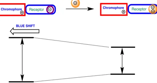
In internal charge transfer (ICT) system, there are two units: fluorophore and receptor. Unlike PeT process, there is no spacer in ICT, which means the fluorophore is directly attached to the receptor, in other words, there is a conjugation between the receptor and the π-electron system fluorophore moiety. Therefore, there is orbital overlapping in this conjugated system which causes the internal charge transfer. When an analyte is binded to the receptor, it causes excited state dipole and affects emission spectrum.
There are two types of stokes shift in the emission spectrum: blue shift and red shift depending on the interaction of analytes with receptor, which is an electron withdrawing or donating group as it is seen in Figure 25. If there is an electron donating group binding to the fluorophore, the electron donating ability of the electron donor group will decrease which causes the decrease in conjugation and blue-shift in the absorption spectrum of the fluorophore. To understand the changes in emission spectrum, one can use excited state of molecules. For example, there is a
N O O N O O O O N O O O O O 1 2
10
positive charge on amine groups at the excited state. Due to the interaction between the positively charged groups, there will be destabilization of excited state. Hence, energy gap between HOMO and LUMO increases.
Figure 6. Representation of ICT working mechanism for blue shift.
On the other hand, if there is a withdrawing group binding to the fluorophore such as carbonyl groups, the interaction between the cation and withdrawing group increases the electron witdrawing ability of the acceptor. This interaction between the cation and negatively charged acceptor group stabilize excited state, which results in decrease in energy gap of HOMO and LUMO and red-shifted absorption.
Chromophore Receptor Chromophore Receptor
11
Figure 7. Representation of ICT working mechanism for red shift
There are many examples of ICT based sensor in the literature. For instance, compound 22[ ] (figure 26) is a good example of ICT-based probe for Hg (II) ions. In this molecule, dithia-dioxa-aza macrocycle is the receptor part and in the literature it is known as Hg (II) selective ligand. Due to the interaction of positively charged groups that causes blocking of ICT process upon coordination of Hg (II) ions to the nitrogen donor atoms, blue shift is observed. In addition, when there is no Hg (II) ions in the environment, fluorescence is highly quenched because of ICT process from crown moiety to BODIPY core. On the other hand, with a Hg (II) ions, strong fluorescence intensity with a large blue shift will be observed.27–29
Chromophore Receptor Chromophore Receptor
12
Figure 8.. Examples of ICT sensors.
2.3 Photodynamic Therapy (PDT)
2.3.1 History of PDT
. The use of photoactive molecules started has been with us for too long for instance, back in time photoactive molecules were used for the treatment of vitiligo as the holy book of India (Atharava-veda) describes it however, it was first realized by Herodotes more than two hundred years ago. Oscar Raab first observed the toxic effects of acridine red molecule on different bacteria in the presence of light and after doing the control experiments he then concluded that a flourophore must be present in order to have light-induce toxicity. With this contribution to scientific literature, several groups nowadays are working on the crusade of finding cancer cure using light-induced molecules, which started with Tappeiner and Jesionek using eosin dye to treat skin cancer for the first time in history.
Following the same course of research, Teppeiner and Jodbauer in 1904 found out that oxygen is a critical component for photodynamic therapy nevertheless no mechanism was proposed until 1979 when electron spin resonance was used to monitor the generation of singlet oxygen. Meyer-Betz performed the first human trial of PDT on himself and he described apses and severe pain. Whereas, the first clinical applications were done at the Rosewell Park Cancer Institute back in 1978. In 1980, FDA approved photophrin (a mixture of haematoporphryn derivatives) as a
N B N O O S N S O O S N S F F 3 N B N F F N N 4
13
photosensitizer and from there on, several other molecules have been proposed to be used as PDT agents.30
2.3.2 Working mechanism
Singlet oxygen must be generated in order for PDT to work as desired. Three main components are required for PDT namely; Photosensitizer (flourophore), Light and Oxygen. First the flourophore must be excited from its ground state to the first excited state by appropriate photon energy. There is a relaxation on the viabrational states then there are two possibilities of relaxation; one is intersystem crossing and the other is relaxing to the ground state where we will have fluorescence. In PDT we require the relaxation through intersystem crossing because in this case there is a spin conversion and the energy can be later transferred to molecular oxygen in order to produce singlet oxygen. However, if we have the intersystem crossing and no oxygen present then a relaxation with phosphorescence is observed.30 This phenomenon is described on the Jablonski diagram for singlet oxygen production below:
Figure 9. Jablosnki diagram for singlet oxygen production.
Singlet oxygen can lead to formation of reactive oxidative species or oxidative damage of cells because it has a short lifetime of about 0.6 us and diffusion distance of 0.1 um. Therefore, only cellular structures on the vicinity of the photosensitizer
AB SO RB AN CE
)
FL O U RE SC EN CE ) PH O SP H O RE SC EN CE)
S
0#S
1#S
n#T
1#S
0# 1O
2) # 3O
2) Ene rgy)Tr ansf er)14
can be damaged and these reasons have been the moto for the synthesis of localized and more target oriented photosensitizers. 31
2.3.3 Oxidative damage of singlet oxygen
Two mechanisms rule the cytotoxicity mediated by the photosensitizer. When the photosensitizer reacts directly with the biomolecule and forms unstable radicals it is called type-I reaction. After the radicals are formed, they react with other substrates or molecular oxygen to for singlet oxygen. On the other hand, when energy is transferred directly to the photosensitizer and produces singlet oxygen it is called type-II reaction. The differentiation between the two mechanisms is difficult therefore it is usually assumed that they occur simultaneously.
After singlet oxygen is produced it immediately reacts with biomolecules in the vicinity provoking an oxidative stress since it is a powerful oxidant. Lipid peroxidation alters the membrane structure, integrity and its fluidity so lipid radicals are then formed causing another cytotoxic danger to the cells. Free amino and thiol groups are other targets of singlet oxygen and with this there is a disruption of the proper folding and functioning of the aminoacids. Another damage is caused to mithocondrial membrane permeability through oxidation of disulfide histidine amino acids and membrane proteins, which leads to more labile membrane, that permits the passage of mithocondrial constituents and ends up with cell death. Dioxyribose sugars and bases are easy targets for chemical reactions with singlet oxygen for instance thymine. In case of such modifications, DNA strands may break and after accumulation of such damages cell death is triggered.32–34
2.3.4 Requirements for photosensitizers
In order for a photosensitizer to be successfully used in PDT it must bear several properties for instance; photostability, easy synthesis, absorption coefficients ranging between 600 to 900 nm known as the therapeutic window. The reason for this is, the penetration depth of light is high at this range. Neither dark toxicity nor reactivity is required in the presence of light. An amphiphilic character is also required for safe cell penetration. In addition, high quantum triplet yields are required therefore an
15
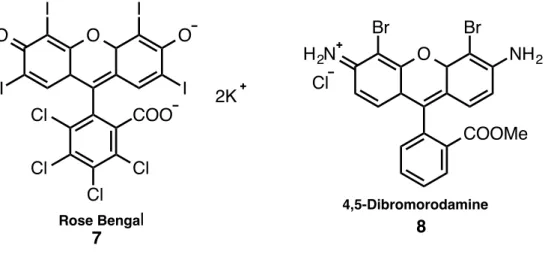
effective ISC and long triplet lifetimes are desired.35 Nevertheless, these are not easy requirements for example to decrease dark toxicity one should embed targeting moieties and depending on the photosensitizers it might be very difficult to do so. Although photofrin has low absorption coefficients and a rather complicated structure, it is the most widely used photosensitizer in medicine. As a consequence, scientists are have been looking for alternatives. Foscan was developed in order to diminish the high drug/light exposure and long-term photosensitivity presented by photofrin. Other photosensitizers such as rose Bengal, methylene blue, eosin B, phtalocyanine and BODIPY derivatives have been developed and employed over the years of research. 36,37
On the other hand, the efficiency of PDT is determined by the light source used. The penetration depth of blue light is quite limited but that of red light and near-IR can be more useful in tissue penetrations. Wavelengths outside the therapeutic window are not useful because for instance; if we use longer wavelengths, the energy will not be enough to penetrate the tissues and water and other molecules can absorb the incoming light causing no PDT reactions at all. Whereas when shorter wavelengths are used, the energy is so high that can damage tissues on its way to the required target. As result, light dose, light delivery and time of exposure are important factors for clinical efficacy.35,37,38
2.4 Photosensitizer families in literature
392.4.1 Phenothiazines
Methylene blue is one of the mostly used photosensitizer for PDT applications. It takes part of phenothiazolium family and with εmax of approximately 82,000 M−1 cm−1 it absorbs at 666 nm. Besides, it targets melanoma cells and a positive PDT action was observed in melanoma cell cultures. In clinical PDT treatments methylene blue is used for basal cell carcinoma and Kaposi’s sarcoma whereas in vitro it is used for testing adenocarcinoma, HeLa cervical tumor cells and bladder carcinoma. On the other hand, toluidine blue which asorbs at 596 nm and 630 nm with εmax 51,000 M−1 cm−1 of approximately is known to be undergoing phase two trials to treat
16 chronic periodontitis.40–42
Figure 10. Examples of phenothiazines sensitizers.43
2.4.2 Xanthenes
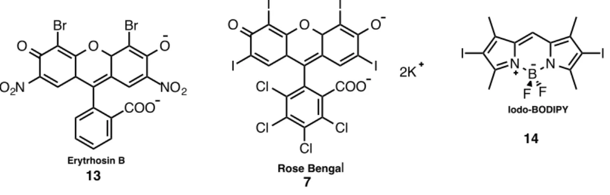
Water soluble rose Bengal is a xanthene photosensitizer and it absorbs at 549 nm and it has an εmax of approx.100,000 In addition, it is used in the treatment of cancinoma
and metastitatic melanoma. Another effective Xanthene sensitizer is 4,5-Dibromorhodamine with εmax ~ 100,000 M−1 cm−1 and absorbs light at 514 nm.
Moreover, it was investigated for PDT on graft-versus-host disease and it can destroy lymphocytes via apoptosis.44,45
Figure 11. Examples of xanthene sensitizers.
2.4.3 Phtalocyanines
As mentioned before in the previous chapters, the use of heavy metals enhances ISC and in the case of phtalocyanines, a metal complex formation is required. They absorb light in the range of 670 – 700 nm with εmax ~ 200,000 M−1 cm−1. For
instance, aluminum phthalocyanine tetrasulfonate is one typical example of this class S N N N Cl Methylene Blue 5 S N N H2N Cl Toluidine Blue 6 O Cl Cl Cl Cl COO I I O I I O 2K Rose Bengal 7 O COOMe Br Br NH2 H2N 4,5-Dibromorodamine 8 Cl
17
of sensitizers. Another example is Photosens with an absorption at 676 nm and in Russia it is used to treat stomach, oral, breast and lip cancer. Nevertheless, skin photoxicity may occur for many weeks as a side effect. Silicon phtalocyanine absorbs light at 675 nm and has completed phase I trials.46–49 In these trials it was able to treat actinic keratosis, skin cance, micosis e.t.c.
Figure 12. Example of phtalocyanine sensitizer. 2.4.4 Anthraquinones
Naturally occurring anthraquinone derivative “Hypericin” absorbs light at 590 nm with εmax ~ 44,000 M−1 cm−1 generates reactive oxygen species that target cancer cells. Although several clinical trials have been performed to treat squamous cell carcinoma and basal cell carcinoma, the results were unsatisfactory.50–54
Figure 13. Example of anthraquinone sensitizer.
N N N N O3S SO3 SO3 O3S M Phtalocyanine 9 O O OH OH OH OH HO HO Hypericin 10
18
2.4.5 Cyanines
Merocyanine 540 is a cyanine that targets leukemia and lymphoma cells. It was investigated for PDT in vitro to treat neuroblastoma and leukemia with considerable results. Besides, it absorbs at 556 nm with εmax ~ 110,000 M−1 cm−1 which is almost
within the therapeutic window.50,55,56
Figure 14. Example of cyanine sensitizer.
2.4.6 Curcuminoids
Curcumin absorbs at 420 nm with εmax ~ 55,000 M−1 cm−1. It can be isolated from
rhizomas of curcuma longa L and it is also a component of turmeric (a cooking spice). In PDT it was used as a disinfectant in oral surgery. This sensitizer upon PDT action destroys bacteria.
Figure 15. Example of curcuminoid sensitizer.
As listed below several non-porphyrin sensitizers have been proposed and FDA approved.39,50,57,58 O N N N S O O O O O O Merocyanine 11 O O OH O HO O Curcumin 12
19
2.5 Triplet Photosensitization in PDT
After intersystem crossing (S1 → Tn (n ≥ 1)) takes place, the triplet state of a
photosensitizer is populated by non-radiative electrons. Therefore, strong absorption is required in order to have a powerful triplet photosensitizer, as this is one of the most important requirements in PDT process. To trigger photophysical and photochemical processes long-lived triplet excited state is required because it acts as triplet energy donor. Triplet excited states play an important role not only in PDT applications but also photocatalytic organic reactions and triplet-triplet anhilation upconversion. Another use is in the phosphorescence imaging, sensing and electroluminescence.59–63
Pauli exclusion principle states that electrons have opposite spins while in their singlet excited state whereas in the triplet excited state they have parallel spins.64,65 As result, during the ISC a quantum mechanically forbidden process takes place because there must be a reversion of spins making triplet excited state less probably to observe. Much effort has been spent to increase the probability of triplet excited state existence within several years and several different results were published. For instance, Spin orbit coupling is one of the processes required because it favors ISC. During this process, there is a coupling between electron spin and orbital angular momentum and total angular momentum and energy are conserved.66–71
2.5.1 Incorporation of Heavy Atoms
Large atomic number decorated photosensitizers are known to be very effective in the induction of ISC because, when an electron moves around a largely positive nucleus it accelerates then the spin (µS) and angular (µL) momentum increase.
Therefore, the spin orbit coupling becomes more probable.
It is also possible to calculate the probability of getting SOC, as it is directly proportional to the fourth power of the nuclear charge (Z). As illustrated below:
20
Equation 1. Calculation of SOC probability.
Therefore, the atomic number of the photosensitizer increases, the higher the chances for ISC to occur. Heavy atoms have been the perfect candidates to increase the SOC in photosensitizers because the majority of the existing organic dyes have low triplet quantum yields. Although this incorporation has been successful in many cases, several drawbacks were later presented for instance; incorporation of Br, I, Ir, Rh, Pt, Ru and Os usually lead to dark toxicity. While heavy atoms such as transition metal complexes might seem safer than halogens, their absorption coefficients in the visible region are very small so they have a restricted use. Thus, organic photosensitizers such as Rose Bengal, eosin blue and BODIPY have been extensively employed in the research of PDT due to the easy incorporation of iodine and Bromine.72–77 Moreover, they have high absorption coefficients within the visible region, which is highly desired. Molecules are given below:
Figure 16. Halogenated sensitizers for PDT.
BODIPY chromophore is a highly remarkable sensitizer with high triplet quantum yields. In addition, they have high resistance to photobleaching, inert, reduced dark toxicity and insensitive to the environment. As result, they have been chosen as ideal candidates for future PDT clinical applications.
Negano et al. reported the first application of BODIPY molecule as a PDT agent in 2005 where 2,6-Diiodo-BODIPY was investigated for singlet oxygen generation on
!!!!!!!!!!!!!e
2!Z
4# !!!!!!!!!2a
om
2c
2n
3!
L.S!!
H
SO!=!
O COO Br Br O O NO2 O2N Erytrhosin B 13 O Cl Cl Cl Cl COO I I O I I O 2K Rose Bengal 7 N B N I I F F Iodo-BODIPY 1421
the heavy atom class. When low fluorescence quantum yields (about 2%) were observed it was considered a successful task since this indicated the high efficiency of ISC. Then, cancer cell studies in vitro were also performed and branded fruitful. Akkaya and coworkers also reported near-IR absorbing and water-soluble BODIPY derivatives such as di-styryl bromo-substituted BODIPY derivative. This molecules has a red-shifted absorption maximum and due to the oligoethylene glycol groups, the water solubility is increased. In the cancer cell experiments, cell membrane damage is observed via fluorescence microscopy which proves the cytotoxic effect of the sensitizer.50,78–82
Teg: Triethylene glycol
Figure 17. Di-styryl bromo-substituted BODIPY molecule.
2.5.2 Heavy Atom Free Photosensitization
As stated before, heavy atoms in most cases result in dark toxicity so since this in an undesired phenomenon, scientists have struggled with finding new heavy atom free photosensitizers. Within these attempts, various well known processes can be employed for example; designing chromophores with low lying n-π* transitions, the use of spin convertors and exciton coupling. In PDT, low levels of toxicity are required when compared to chemotherapy and other convectional methods even at cellular and organ levels. In this thesis, heavy atom free designed BODIPY molecules are presented and some information the other possible mechanisms are explained below. N B N Br Br F F TegO
TegO OTeg TegO OTeg
OTeg OTeg
OTeg TegO
22
2.5.3 Low-Lying n-π* Transition based chromophore design
In order for the Spin Orbit Coupling to take place the energy difference between singlet and triplet excited states has to be close enough. In some cases when the electronic configuration of the singlet and triplet excited states are similar, the energy difference may appear to be twice of the electron exchange integral (J). Nevertheless the energy gap between singlet and triplet excited states of the n-π* is much smaller. El-Sayed generalized that; organic molecules with an extended conjugation undergo π-π* transitions that decrease triplet quantum yields. According to him, S1 (n-π*) →
T1 (π-π*) is an allowed transition because the energy gap is small and the angular
momentum is conserved whereas S1 (π-π*) → T1 (π-π*) is a forbidden process.
According to the equation below, rate constant is directly proportional to the probability of getting SOC and inversely proportional to the energy gap between singlet and triplet excited states.
kISC ∝<T1|HSO|S1>2 / (ΔES1-T1)2
This proves that the smaller the energy gap, the higher the probability of o more effective ISC. One typical example of excited states with low-lying n-π* is benzophenone.
Figure 18. An example of excited states with low-lying n-p* orbitals.
Charge transfer mediated ISC was reported in a theoretical study by Dede et al. where quinolizinium flourophore was investigated and concluded that quinolizinium flourophore has an ISC at its deprotonated form and none at its protonated form. The reason for this presumed to be the aminophenyl moiety that transfers charge to the benzoquinolizinium ring and there is an n-π* type of charge transfer. When the quinolizinium is protonated on the other hand, π-π* was detected resulting in low triplet quantum yields which is in accordance with the El-Sayed’s rule.50,83–86
O
Benzophenone 16
23
Figure 19. An example of p-p* sensitizer. 2.5.4 Exciton Coupling
When two identical chromophores linked together but with no π-conjugation are excited, two delocalized ecited states for each chomophore are observed meaning that they are actually being excited separately. In this case, only when one of the singlet exciton states is close enough to the triplet excited state that ISC takes place.
Figure 20. Examples of sensitizers with exciton coupling states.
Flamigni et al. investigated an example of heavy atom free photosensitizer employing exciton coupling, where they synthesized dimeric BODIPY derivative and the absorption spectra of the dimer and monomer are different. The monomer typically shows ground state to singlet excited state excitation behavior with an absorption peak at 530 nm whereas; the dimeric form two different peaks are observed at 381 nm and 534 nm. This results in higher singlet oxygen generation rate due to the more effective ISC. 87–92
2.5.5 Spin Convertors
The selection rules on highly populated triplet excited states are very challenging since sensitizers should be synthesized taking them into consideration.
N N H+ N NH 17 NBN N B N F F F F N B N F F N B N F F 18 19
24
Inserting an energy acceptor that has an intrinsic character or also known as spin convertor to a sensitizer, may increase the probability for high triplet quantum yields. Moieties such as C60 are very prised because they can be easily coupled with
chromophores by a covalent bond and a more effective singlet oxygen production can be observed.
Figure 21. An example of sensitizer with spin convertor.
Zhao and coworkers used fullerene (C60) as a spin converter. In their work they
combined BODIPY sensitizer with fullerene moiety enhancing an intrinsic ISC property. On the other hand, it would not be possible to achieve such triplet quantum yields without the incorporation of fullerene and this is considered a good sensitizer because it has its absorption peak at the UV region. Besides, fullerene has an additional absorption peak at 700 nm as result it is suitable for intramolecular energy transfer processes and it acts like an antenna. The singlet excited state of the BODIPY molecule has 1.93 eV and the fullerene molecule has low lying singlet excited state at 1.72 eV therefore, upon excitation the electron passes from the high
N B N F F N N N 20
25
singlet excited state to the low lying singlet excited state. At this stage there is an intrinsic intersystem crossing which then results in easy triplet-to-triplet excited state transition.50,93–97
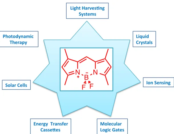
2.6 BODIPY
In 1968 Treibs and Kreuzer discovered BODIPY dyes and today they have a significantly popular area due to their wide applications. BODIPY has several uses in applications such as; solar cells, bimolecular labeling, molecular logic gates, drug delivery e.t.c. In addition, compared to other sensitizers, high molar extinction coefficients, high fluorescence quantum yields and depending on the design they may have high triplet quantum yields as well. The absorbance is within the therapeutic window and pH and solvent polarity do not affect them as much therefore, they tend to maintain their physical structure. Easy functionalization from 1 to 8 positions is probably one of the strongest suits of BODIPY because it can be modified for almost any function and solubility. Akkaya, Burgess, Negano, Rurack and Ziessel research groups have played an important role on investigating these modifications.36
Figure 22. BODIPY molecule.
2.6.1 BODIPY Applications
As mentioned before, several applications are depicted from literature as shown below. N B N F F 1 2 3 4 4' 5 6 7 8
26
Figure 23. Applications of BODIPY molecule.
Daub and Rurack first employed BODIPY as a chemosensor in 1997 and from that moment forward, various examples were presented in the literature. PET and ICT based sensors such as compound 21. It is a proton sensor because dimethyl amino group is a strong electron donor and when the molecule is excited weak fluorescence will be present whereas upon reaction with proton the PET is blocked and strong fluorescence is allowed. Some metal sensors such as compound 21 - 25 are also available. For instance, they sense cadmium and zinc metals. 98–107
Molecular)) Logic)Gates) Ion)Sensing) Liquid)) Crystals) Energy))Transfer) ))))))Casse:es) Photodynamic)) ))))))Therapy) Solar)Cells) Light)HarvesAng)) Systems)
N
B
N
F F
27
Figure 24. Examples of BODIPY PeT sensors.
In PDT, there is usually the need of heavy atoms to facilitate the ISC. In addition, water -soluble groups can be attached to in increase hydrophilicity for example compound 22 and 23. Akkaya et al. developed water soluble compound 20 to be used in PDT and Negano et al synthesized compound 21 with fluorescence quantum yields of around 0.02, which is consistent with the required because it has high triplet quantum yields.11,108–111 N B N N F F N B N F F HN N N N N B N F F N N N N N B N N B N F F F F N B N F F N N N 21 22 23 24 25
28
Figure 25. Water soluble BODIPY based molecules. N B N F F 28 N B N F F COOH N B N F F SO3Na SO3Na N N N COOH 27 26 I I N B N F F Br Br RO RO RO RO RO RO OR OR OR 15
29
CHAPTER 3
3. EXPERIMENTAL PROCEDURES
3.1
Methods and materials
All commercial chemicals were purchased from Merck, Sigma-Aldrich and ABCR and were used without any further purification. Merck TLC Silica gel 60 F254. was
used to monitor the reactions. Merck Silica Gel 60 (particle size: 0.040-0.063 mm, 230-400 mesh ASTM) was used for the column chromatography. All 1H NMR and
13C NMR spectra were recorded on Bruker DPX-400 in CDCl
3 and DMSO with
tetramethylsilane as internal standard. Chemical shifts were given in parts per million and the coupling constants (J) were in Hz. Mass spectra were recorded on Agilent Technologies 6224 TOF LC/MS. The absorption spectra were recorded on Varian Cary-100 spectrophotometer and Varian Cary 5000 UV-VIS-NIR absorption spectrophotometer. For fluorescence measurements Varian Eclipse Spectrofluometer was used. 1,3-Diphenylisobenzofuran (commercial) was used as a singlet oxygen trap in organic medium.
Figure 26. Symthesis of compound 30.
3.2
Synthesis of (30)
112To a 500 mL round-bottomed flask containing 250 mL argon-degassed dichloromethane, 2,4-dimethylpyrrole (1.05 mL, 10.26 mmol), 4-tert-butylbenzaldehyde (0.773 mL, 4.623 mmol) were added. Then, trifluoroacetic acid
O N B N F F BF3-OEt2 DIPEA, TFA, p-Chloroanil, DDQ, rt, 2-3 h 29 30 NH
30
(400 µL) was added to the reaction mixture and left to stirr overnight. Then, p-chloranil (1.36 g, 5.5 mmol) was added and mixed for 1 additional hour. After that, TEA (3.5 mL) was added and mixed for 1 additional hour and BF3.OEt2 (3.5 mL)
was added and the reaction mixture was left to stir at room temperature for 1h. When the starting material was consumed, water (100 mL) was added and the reaction mixture was extracted with DCM (3x100 mL), evaporated and dried over Na2SO4.
The product was purified by silica gel column chromatography using DCM:Hexane (1:1) as the eluant and the compound was obtained as purple redish solid (1.49 g, 85 %). 1H NMR (CD2Cl2, 250 MHz): δ = 7.42 (d, 2H), 7.11 (d, 2H), 5.91 (s, 2H), 2.42 (s, 6H), 1.30 (s, 6H), 1.27 (s, 9H). 13C NMR (CD2Cl2, 62.5MHz): δ=155.7, 153.2, 144.2, 143.4, 132.2, 128.2, 126.5, 121.8, 35.5, 31.8, 15.0, 14.8. ESI-HRMS (M-H+) calculated 380.2344, found 380.2297, Δ= 12.48 ppm
Figure 27. Symthesis of compound 31.
3.3
Synthesis of (31):
1 mL of DMF and 1 mL of POCl3 was stirred in an ice bath for 5 min under argon.
Then it was warmed to room temperature and waited for 30 minutes. To this mixture compound (30) (300 mg, 0.789 mmol) was added in dichloroethane (60 mL). The temperature was raised to 500C and stirred for 2 hours. The reaction was then cooled to room temperature and poured to an ice cold NaHCO3 solution (150 mL)This mixture was extracted with DCM (3x100 mL) and dried over Na2SO4. Solvent was evaporated in vacuo and purified by silica gel column chromatography using DCM:MeOH (98:2) as the eluent. Product 31 was obtained as an orange solid (289.9
N B N F F N B N F F O POCl3 DMF 31
31 mg, 90% yield). 1H NMR (400 MHz, CDCl3) δ 10.02 (s, 1H), 7.55 (dd, J = 6.5, 1.8 Hz, 2H), 7.19 (dd, J = 6.4, 1.8 Hz, 2H), 6.16 (s, 1H), 2.83 (s, 3H), 2.62 (s, 3H), 1.66 (s, 3H), 1.44 (s, 3H), 1.39 (s, 9H). 13C NMR (100 MHz, CDCl3) δ 185.87, 161.32, 156.27, 153.20, 147.35, 144.10, 142.96, 134.21, 131.06, 127.31, 126.25, 123.85, 34.86, 31.34, 15.02, 14.72, 13.00, 11.43. MS (TOF-ESI): m/z: Calcd: 408.2293 [M-H]+, Found: 408.2267 [M-H]+, ∆= 6.51 ppm.
Figure 28. Symthesis of compound 32
3.4
Synthesis of (32):
1 mL of DMF and 1 mL of POCl3 was stirred in an ice bath for 5 min under argon.
Then it was warmed to room temperature and waited for 30 minutes. To this mixture compound (31) (200 mg, 0.490 mmol) was added in dichloroethane (60 mL). The temperature was raised to 500C and stirred for 2 hours. The reaction was then cooled to room temperature and poured to an ice cold NaHCO3 solution (150 mL)This mixture was extracted with DCM (3x100 mL) and dried over Na2SO4. Solvent was
evaporated in vacuo and purified by silica gel column chromatography using DCM:MeOH (98:2) as the eluent. Product 32 was obtained as an orange solid (171 mg, 80% yield). 1H NMR (400 MHz, CDCl3) δ 10.07 (s, 2H), 7.61 (d, J = 8.3 Hz, 2H), 7.22 (d, J = 8.4 Hz, 2H), 2.89 (s, 6H), 1.73 (s, 6H), 1.41 (s, 9H). 13C NMR (100 MHz, CDCl3) δ 185.62, 160.51, 153.99, 148.42, 147.86, 132.03, 130.47, 128.02, 127.02, 126.88, 34.98, 31.32, 13.73, 11.95. MS (TOF-ESI): m/z: Calcd: 481.21045 [M-H]+, Found: 481.17248 [M-H]+, ∆= 74.13 ppm. N B N F F O N B N F F O POCl3 DMF O 32
32
Figure 29. Symthesis of compound 33.
3.5
Synthesis of (33):
To a 500 mL round-bottomed flask containing 250 mL argon-degassed dichloromethane, 2,4-dimethylpyrrole (2.10 mL, 20.5 mmol), 4-tert-butylbenzaldehyde (200 mg, 0.458 mmol) were added. Then, trifluoroacetic acid (500 µL) was added to the reaction mixture and left to stirr overnight. Then, p-chloranil (2.72 g, 11 mmol) was added and mixed for 1 additional hour. After that, TEA (5 mL) was added and mixed for 1 additional hour and BF3.OEt2 (5 mL) was
added and the reaction mixture was left to stir at room temperature for 1h. When the starting material was consumed, water (100 mL) was added and the reaction mixture was extracted with DCM (3x100 mL), evaporated and dried over Na2SO4. The
product was purified by silica gel column chromatography using DCM:Hexane (1:1) as the eluant and the compound was obtained as purple redish solid (160 mg, 40 %).
1H NMR (400 MHz, CDCl
3) δ 7.54 (d, J = 8.2 Hz, 2H), 7.21 (d, J = 8.2 Hz, 2H),
6.03 (s, 4H), 2.56 (s, 12H), 2.48 (s, 6H), 1.79 (s, 12H), 1.34 (s, 9H), 1.30 (s, 6H). 13C NMR (100 MHz, CDCl3) δ 156.00, 153.63, 153.45, 143.98, 142.31, 140.78, 132.91,
131.74, 131.04, 127.23, 126.89, 126.41, 121.34, 53.38, 34.84, 31.56, 31.26, 22.63, 14.61, 14.25, 14.08, 12.95, 12.39. ESI-HRMS (M-H+) calculated 869.46295, found
869.44414, Δ= 21.63 ppm N B N F F O O N B N F F N B N N B N F F F F BF3-OEt2 DIPEA, TFA, p-Chloroanil, DDQ, rt, 2-3 h H N 33
33
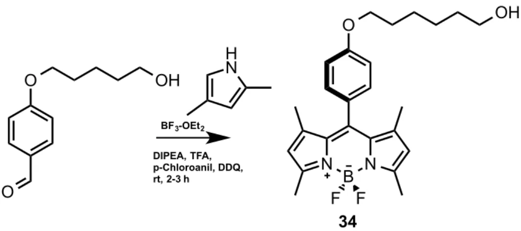
Figure 30. Symthesis of compound 34.
3.6
Synthesis of (34):
To a 500 mL round-bottomed flask containing 250 mL argon-degassed dichloromethane, 2,4-dimethylpyrrole (1.05 mL, 10.26 mmol), 4-((5-hydroxypentyl)oxy)benzaldehyde (300 mg, 1.44 mmol) were added. Then, trifluoroacetic acid (400 µL) was added to the reaction mixture and left to stirr overnight. Then, p-chloranil (1.36 g, 5.5 mmol) was added and mixed for 1 additional hour. After that, TEA (3.5 mL) was added and mixed for 1 additional hour and BF3.OEt2 (3.5 mL) was added and the reaction mixture was left to stir at room
temperature for 1h. When the starting material was consumed, water (100 mL) was added and the reaction mixture was extracted with DCM (3x100 mL), evaporated and dried over Na2SO4. The product was purified by silica gel column
chromatography using DCM:Hexane (1:1) as the eluant and the compound was obtained as purple redish solid (0.380 g, 60 %). 1H NMR (400 MHz, CDCl
3) δ 7.19 – 7.14 (m, 2H), 7.04 – 7.00 (m, 2H), 5.99 (s, 2H), 4.19 (t, J = 6.0 Hz, 2H), 3.91 (t, J = 5.9 Hz, 2H), 2.56 (s, 6H), 2.10 (dq, J = 12.1, 6.0 Hz, 2H), 1.44 (s, 6H). 13C NMR (100 MHz, CDCl3) δ 159.40, 155.27, 143.17, 141.81, 131.83, 130.41, 129.24, 127.18, 121.12, 115.06, 65.62, 60.20, 32.00, 14.58. ESI-HRMS (M-H+) calculated 438.24102 , found 438.241662, Δ= 6.0 ppm O O O N B N F F BF3-OEt2 DIPEA, TFA, p-Chloroanil, DDQ, rt, 2-3 h 34 H N OH OH
34
Figure 31. Symthesis of compound 35
3.7
Synthesis of (35):
1 mL of DMF and 1 mL of POCl3 was stirred in an ice bath for 5 min under argon.
Then it was warmed to room temperature and waited for 30 minutes. To this mixture compound (34) (200 mg, 0.469 mmol) was added in dichloroethane (60 mL). The temperature was raised to 500C and stirred for 2 hours. The reaction was then cooled to room temperature and poured to an ice cold NaHCO3 solution (150 mL)This mixture was extracted with DCM (3x100 mL) and dried over Na2SO4. Solvent was
evaporated in vacuo and purified by silica gel column chromatography using DCM:MeOH (98:2) as the eluent. Product 35 was obtained as (385 mg, 82% yield).
1H NMR (400 MHz, CDCl 3) δ 10.03 (s, 1H), 7.19 (d, J = 8.6 Hz, 2H), 7.07 (d, J = 8.6 Hz, 2H), 6.17 (s, 1H), 4.21 (t, J = 5.9 Hz, 2H), 3.81 (t, J = 6.3 Hz, 2H), 2.83 (s, 3H), 2.63 (s, 3H), 2.31 (dd, J = 12.0, 5.9 Hz, 2H), 1.73 (s, 3H), 1.50 (s, 3H). 13C NMR (100 MHz, CDCl3) δ 185.91, 161.45, 159.74, 156.39, 147.31, 143.66, 142.86, 134.47, 129.10, 126.37, 123.93, 115.42, 64.53, 41.39, 32.20, 15.11, 12.99, 11.81. MS (TOF-ESI): m/z: Calcd: 546.1621 [M-Br]-, Found: 546.16003 [M-Br]-, ∆= 3.79
ppm. O N B N F F O N B N F F O POCl3 DMF 35 OH OH
35
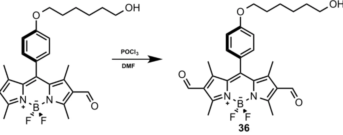
Figure 32. Synthesis of compound 36.
3.8
Synthesis of (36):
1 mL of DMF and 1 mL of POCl3 was stirred in an ice bath for 5 min under argon.
Then it was warmed to room temperature and waited for 30 minutes. To this mixture compound (35) (200 mg, 0.440 mmol) was added in dichloroethane (60 mL). The temperature was raised to 500C and stirred for 2 hours. The reaction was then cooled to room temperature and poured to an ice cold NaHCO3 solution (150 mL)This mixture was extracted with DCM (3x100 mL) and dried over Na2SO4. Solvent was
evaporated in vacuo and purified by silica gel column chromatography using DCM:MeOH (98:2) as the eluent. Product 36 was obtained as (174.6 mg, 80% yield). 1H NMR (400 MHz, CDCl 3) δ 10.08 (s, 2H), 7.23 – 7.19 (m, 2H), 7.14 – 7.10 (m, 2H), 4.24 (t, J = 5.9 Hz, 2H), 3.82 (t, J = 6.2 Hz, 2H), 2.90 (s, 6H), 2.33 (dd, J = 12.1, 6.1 Hz, 2H), 1.80 (s, 6H). 13C NMR (100 MHz, CDCl3) δ 185.63, 160.59, 160.21, 148.32, 147.46, 132.28, 128.86, 128.05, 125.67, 115.81, 64.63, 41.31, 32.15, 13.72, 12.33. O N B N F F O O N B N F F O POCl3 DMF O 36 OH OH