Research Article [Araştırma Makalesi]
Türk Biyokimya Dergisi [Turkish Journal of Biochemistry–Turk J Biochem] 2015; 40(1):24–30 doi: 10.5505/tjb.2015.99815
Atherogenic dyslipidemia, subclinical atherosclerosis,
non-alcoholic fatty liver disease and insulin resistance in
polycystic ovarian syndrome
[Polikistik over sendromunda insülin direnci, aterojenik dislipidemi, subklinik
aterosklerozis ve non-alkolik yağlı karaciğer hastalığı]
ABSTRACT
Objective: We aimed to explore the relationship between insulin resistance (IR) and small dense lipoprotein (sd-LDL) particles, carotid intima-media thickness (CIMT) and non-alcoholic fatty liver disease (NAFLD) in young normal weight PCOS cases.
Methods: This prospective, case-control study was designed in a University Hospital and 34 women with PCOS and 21 healthy controls were enrolled. Fasting plasma glucose, insulin, lipid (including sd-LDL particles) and hormone profiles, abdominal ultrasound and CIMT were evaluated.
Results: IR was present in 68% of PCOS group while in none of controls. High density lipoprotein (HDL), very low density lipoprotein (VLDL), triglycerides (TG), and sd-LDL were higher in patients with IR (p<0.05). A positive correlation of sd-LDL with IR, VLDL and TG was found. A significantly higher rate of NAFLD and CIMT was found in PCOS. Total-testosterone levels were weakly and positively correlated with CIMT (r=0.277, p=0.041). Conclusion: Insulin resistance and NAFLD are highly prevalent among young normal weight PCOS patients. When compared to controls levels of sd-LDL and CIMT are increased in PCOS. Insulin resistance is the key parameter for NAFLD and atherogenic dyslipidemia in PCOS. Hence, screening for NAFLD may be valuable for detection and prevention of liver disease. Higher levels of sd-LDL in insülin resistant PCOS cases necessiates treating PCOS for IR. Key Words: Polycystic ovarian syndrome, non-alcoholic fatty liver disease, carotid intima-media thickness, metabolic syndrome
Conflict of Interest: The authors have no conflict of interest. ÖZET
Amaç: Bu çalışmada, genç, normal kilolu PKOS hastalarında insülin direncinin ve küçük dan-siteli lipoprotein (sd-LDL) partiküllerinin, karotis intima-media kalınlığı ve non-alkolik yağlı karaciğer hastalığı ile ilişkisinin araştırılmasını hedefledik.
Metod: Otuzdört PKOS’lu hasta ile 21 sağlıklı kontrol hastasını içeren bu prospektif vaka-kontrollü çalışma bir üniversite hastanesinde yürütüldü. Hastaların açlık plazma glukoz, insülin düzeyleri, lipid (sd-LDL partiküllerini de içeren) ve hormon profilleri, abdominal ultrasonografi ve karotis intima-media kalınlığı değerlendirildi.
Bulgular: İnsülin direnci, PKOS’lu hastaların %68’inde mevcut iken kontrol grubunda hiç-bir hastada saptanmadı. Yüksek dansiteli lipoprotein (HDL), çok düşük dansiteli lipoprotein (VLDL), trigliserid (TG) ve sd-LDL insülin direnci olan hastalarda yüksekti (p<0.05). sd-LDL ile insülin direnci, VLDL ve TG arasında pozitif korelasyon saptandı. PKOS’da belirgin daha yüksek oranda non-alkolik yağlı karaciğer hastalığı ve artmış karotis intima-media kalınlığı izlendi. Total testosteron seviyeleri karotis intima-media kalınlığı ile zayıf pozitif korele idi (r=0.277, p=0.041).
Sonuç: İnsülin direnci ve non-alkolik yağlı karaciğer hastalığı normal ağırlıklı PKOS has-talarında yaygındır. Kontrollerle kıyaslandığında, sd-LDL ve karotis intima-media kalınlığı PKOS’da artmıştır. İnsülin direnci, non-alkolik yağlı karaciğer hastalığı ve aterojenik disli-pidemi için anahtar noktadır. Bu nedenle, karaciğer yönünden tarama karaciğer hastalığının tanınmasında ve önlenmesinde değerlidir. İnsülin dirençli PKOS vakalarında yüksek sd-LDL bu konuda tedaviyi gerekli kılmaktadır.
Anahtar Kelimeler: Polikistik over sendromu, non-alkolik yağlı karaciğer, karotis intima-me-dia kalınlığı, insülin direnci
Çıkar Çatışması: Yazarların çıkar çatışması yoktur. Gamze Sinem Çağlar1,
Mine Kiseli1,
Rabia Seker2,
Elif Didem Ozdemir1,
Demet Karadag3,
Selda Demirtas2
1University of Ufuk, Faculty of Medicine,
Department of Obstetrics and Gynecology, Ankara
2University of Ufuk, Faculty of Medicine,
Department of Biochemistry, Ankara
3University of Ufuk, Faculty of Medicine,
Department of Radiology, Ankara
Correspondence Address
[Yazışma Adresi]
Gamze Sinem Çağlar, MD.
Ufuk University Faculty of Medicine, Department of Obstetrics and Gynecology, Mevlana Bulvarı, No: 86-88, 06520 Balgat, Ankara,Türkiye Phone: +90 312 2044000
Fax: +90 312 2847786
E-mail: [email protected]
Registered: 09 February 2013; Accepted: 21 June 2014 [Kayıt Tarihi: 09 Şubat 2013; Kabul Tarihi: 21 Haziran 2014]
Yayın tarihi 24 Ocak 2015 © TurkJBiochem.com [Published online January 24, 2015]
Exclusion criteria were hyperprolactinemia, thyroid dys-function, adrenal dysdys-function, diabetes mellitus, pregnan-cy, alcohol consumption, history of chronic viral hepatitis, hemochromatosis, autoimmune liver disease, other chron-ic liver diseases, history of hepatotoxchron-ic, antihypertensive, lipid lowering or antiinflammatory agent usage. None of the patients had received any drugs known to interfere with hormone levels for at least 3 months before the study. All of the subjects were nonsmokers. Anthropometric measurements, including body weight, height and waist-to-hip ratio (WHR) were measured by the same observer. Waist circumference was measured in a hospital gown at the narrowest level between the costal margin and the iliac crest and hip circumference was measured at the widest level over buttocks. Body mass index (BMI) was calcu-lated by the formula: weight(kg)/height(m)². Hirsutism was determined by a modified Ferriman-Gallwey score >7 [10]. Systolic (SBP) and diastolic (DBP) blood pressure were measured twice in the right arm in relaxed sitting position. The average of two measurements were used. The features of metabolic syndrome were identified [11]. Blood samples were obtained on 2nd or 3rd days of
menstru-ation, after overnight fasting for at least 12 hours. In cases with oligoanovulation, blood was taken after progesterone withdrawal bleeding. Levels of glucose, insulin, hormone profile [follicle-stimulating hormone (FSH), luteinizing hormone (LH), estradiol (E2), prolactin (PRL), total and free testosterone (Total-T and Free-T, respectively), dehy-droepiandrosterone sulfate (DHEAS), 17-OHprogester-one (17-OHP) and thyroid-stimulating horm17-OHprogester-one (TSH)], and lipid profile [Total blood cholesterol (Total-C), HDL-C, LDL-HDL-C, VLDL-C and triglycerides (TG)] were deter-mined. Plasma glucose was determined with the glucose hexokinase method, lipid profile and serum alanine ami-notransferase (ALT), gamma-glutamyl transferase (GGT) levels were determined with the enzymatic method (Co-bas Integra 400 Plus, Roche Diagnostics, Mannheim Germany). VLDL-C was calculated with TG/5 formula (Fridewald Formula) [12]. Small dense lipoprotein par-ticles were measured by precipitation method [13]. After mixing with heparin-magnesium (containing 150 U/mL heparin-sodium salt and 90 mmol/L MgCl2), each serum
sample was incubated for 10 minutes at 37°C. The su-pernatant was removed after centrifuging at 15000 rpm (21885 g) for 15 min; LDL-cholesterol was measured by the automatic colorimetric method by Cobas Integra. Fasting insulin and glucose (FPG) levels were used for calculating homeostatic model assesment (HOMA-IR), (insulinxglycemia in µmol/L/22.5) and quantitative insu-lin sensitivity check index (QUICKI) (1/log insuinsu-lin+log glycemia in mg/dL). Insulin resistance was defined as HOMA-IR ≥2.1. HOMA-B scores, showing pancreatic ß cell function were assessed by the following formula: (fasting insulin in μU/ml) x 3.33 / (fasting glucose in mg/ dl - 3.5). Levels of FSH, LH, E2, PRL, DHEAS, Total-T, insulin and TSH were measured with
electrochemilumi-Introduction
Polycystic ovarian syndrome (PCOS) is a common en-docrine disorder affecting up to 10% of women in repro-ductive age [1]. Characteristic features are chronic oligo-anovulation, hyperandrogenemia and/or morphologically polycystic ovaries. Recently, PCOS has gained attention due to crucial metabolic aspects which are important for long-term sequelae of the condition.
In majority of women with PCOS, the components of metabolic syndrome are present and predispose to ath-erosclerotic cardiovascular disease and type-2 diabetes. Additionally, non alcoholic fatty liver disease (NAFLD) which is the hepatic manifestation of metabolic syndrome [2] is more common (41%) in women with PCOS than in controls and tend to occur in early ages [3]. Various histopathological degrees of progressive steatosis, lobular inflammation and fibrosis of the liver are known and se-vere form is non alcoholic steatohepatosis (NASH) which presents risk for advanced fibrosis, cirrhosis and hepato-cellular carcinoma [4].
In obese PCOS patients, elevated plasma TG and reduced high density lipoprotein-cholesterol (HDL-C) concentra-tions are seen [5]. Irrespective of obesity, low density lipo-protein cholesterol (LDL-C) levels are often modestly ele-vated in PCOS [6]. Among the subpopulations of particles of LDL-C, small and dense LDL particles (sd-LDL) are known to be more atherogenic than larger LDL species. When compared to body mass index (BMI) matched con-trol subjects the elevated concentrations and proportions of sd-LDL particles in patients with PCOS [6], increase the risk of coronary heart disease and type 2 diabetes [7]. Besides, in young PCOS women precocious atherosclero-sis has also been shown by increased intima-media thick-ness of the common carotid artery (CIMT) [8].
Atherogenic dyslipidemia, excess weight and IR all play dominant roles in developing metabolic problems in PCOS. However, screening strategies for metabolic comorbidities in PCOS are still lacking. Therefore, this study is designed to explore the relationship between IR and sd-LDL particles, CIMT and NAFLD in young nor-mal weight PCOS cases. In addition, the predictive fac-tors of NAFLD in PCOS are evaluated.
Materials and Methods
The patients admitted an Out-patient Clinic to Obstetrics and Gynecology Department of University were enrolled in the study. Thirty four women with PCOS constituted the study group. The diagnosis of PCOS was made as proposed at the Rotterdam Consensus Meeting [9]. The controls (n=21) were healthy volunteers without any fea-tures of clinical or biochemical hyperandrogenism who had regular menstrual cycles. The study was approved by the Institutional Ethics Committee and informed consent was obtained from all patients and controls.
assessment of the distance between the lumen-intima and intima-adventitia interfaces in longitudinal frames acquired during arterial diastole [15]. The mean of mea-surements of CIMT made at the greatest thickness on both sides was used for statistical analyses. Ultrasono-graphic measurements were performed by the same ex-perienced radiologist.
Statistical analysis
Data analysis was performed by using SPSS for Win-dows, version 11.5 (SPSS Inc., Chicago, IL, United States). Whether the distributions of continuous vari-ables were normal or not was determined by Shapiro Wilk test. Descriptive statistics for continuous variables were shown as mean ± SD and median (min-max), the categoric variables were shown as case number and (%). The median differences between groups were analyzed by nescence assays (ELECYS 2010 HITACHI, Roche
Di-agnostic, Germany). Levels of 17-OHP and Free-T were measured by radioimmunoassay. The intra- and interassay coefficients of variation (CV) were <1.9% and <4%, re-spectively, for all assays performed.
Fatty liver was diagnosed by abdominal ultrasound using accepted criteria which includes a bright hepatic echo pat-tern, increased attenuation of the ultrasound beam, and loss of intrahepatic architectural details [14]. Ultrasonographic diagnosis of fatty liver in the absence of alcohol intake or use of any medication was defined as NAFLD [14]. The measurements of CIMT was conducted using a high resolution ultrasound machine (Logic Q7, General Elec-tric, USA) in all cases. The posterior carotid wall at 1 cm below the bifurcation of the common carotid artery was imaged in B-mode and CIMT was estimated by visual
Table 1. Demographic, hormone and insulin resistance parameters of PCOS and control groups
Parameters Control group PCOS Group p
n=21 n=34 FSH† (mIU/mL) Mean±Sd 5.3±1.3 5.5±1.6 0.655 LH‡ (mIU/mL) Mean±Sd 6.5±2.4 8.6±6.3 0.897 E2§ (pg/mL) Median (min-max) 59(14-230) 33(5-146) 0.120 Total-T¥ (ng/dL) Median (min-max) 0.20(0.10-0.60) 0.30(0.03-1.30) 0.216 Free-Tæ (pg/dL) Mean±Sd 2.2±0.85 2.2±0.93 0.100 17-OH Pþ (ng/dL) Mean±Sd 1.6±0.41 1.5±0.53 0.728 dHEASœ (µg/dL) Mean±Sd 265±111 262±135 0.494 TSH¢ (µIU/mL) Median (min-max) 1.4(0.4-4.0) 1.9(0.5-4.3) 0.310 PRL# (ng/mL) Mean±Sd 14.0±5.8 12.6±5.7 0.390
Fasting insulin (µIU/mL)
Median (min-max) 7.9 (2.5-11.5) 9.3 (3.9-57.4) 0.052 FPGø (mg/dL) Mean±Sd 85.8±7.7 88.3±6.2 0.188 Homa-IR□ Median (min-max) 1.8 (0.5-2.4) 2.0 (0.8-14.0) 0.025* QUICKIÆ Mean±Sd 0.36±0.03 0.34±0.03 0.024* HOMA-B฿ Mean±Sd 139.3±78.5 170.5±102.4 0.239
†follicle-stimulating hormone; ‡luteinizing hormone; §estradiol; ¥total testosterone; æfree testesterone; þ17
OH-progesterone; œdehydroepiandrosterone sulfate; ¢thyroid-stimulating hormone; #prolactin; øfasting plasma glucose; □homeostatic model assesment-insulin resistance; Æquantitative insulin sensitivity check index; ฿homeostatic model
er in insulin resistant cases (22.7±1.4 vs 21.6±1.5 kg/m2,
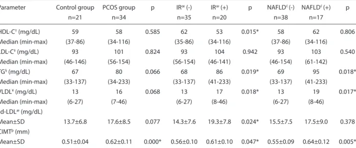
p=0.011; 0.77±0.06 vs 0.71±0.05, p=0.000; respectively). There was no significant difference in parameters of lipid profile (Total-C, LDL-C and HDL-C, VLDL, TG) be-tween cases with PCOS and controls (p>0.05, Table 2). Although not statistically significant, sd-LDL were higher in PCOS patients (p=0.077, Table 2). Levels of VLDL and sdLDL were parallel to each other. Among the lipid parameters HDL, VLDL, TG, and sd-LDL were signifi-cantly higher in insulin resistant cases (p<0.05, Table 2). According to the Spearman’s Rank Correlation analyses neither the demographic characteristics (age, BMI, WHR) nor hormone profiles were correlated with sd-LDL levels. However, fasting insulin levels, HOMA-B and insulin resistance parameters were correlated with sd-LDL lev-els (Table 3). Moreover, a positive correlation of sd-LDL with VLDL, and TG was also found (Table 3). Two cases had PCOS and metabolic syndrome.
Carotid intima-media thickness was significantly higher in PCOS patients (p>0.05, Table 2). In addition, CIMT was significantly higher in insulin resistant PCOS cases when compared with non-insulin resistant participants (0.61 vs 0.56 mm, p=0.047). The Spearman’s Rank Cor-relation analyses of CIMT showed no corCor-relation with demographic characteristics (age, BMI, WHR), lipid pro-files including sd-LDL levels or insulin resistance param-eters (p>0.05). A positive but weak correlation was found between total-T and CIMT (r=0.277, p=0.041) (Table 3). The results of the liver function tests (ALT, GGT) are giv-en in Table 4. Whgiv-en patigiv-ents with or without insulin resis-tance were compared with respect to their liver function tests, GGT levels were found to be significantly higher in insulin resistant cases (13.8±5.8 vs 10.9±3.4, p=0.040). A statistically significantly higher rate of NAFLD was found in PCOS cases when compared with controls Mann Whitney U test. For nominal variables Pearson’s
Chi-Square or Fisher’s Exact test were used. Strength of association between continuous variables were calculated by Spearman’s Rank correlation test. Multiple Logistic Regression Backward method was used to determine the independent predictors which mostly affect NAFLD. Any variable whose univariable test had a p value <0.25 was accepted as a candidate for the multivariable model along with all variables of known clinical importance. Odds ra-tio (OR) and 95% confidence intervals (CI) for each inde-pendent variable were also calculated. A p value less than 0.05 was considered statistically significant.
Results
The mean age and BMI did not differ between patients and controls (26.0±2.5 vs 26.1±2.8, p=0.981 and 22.0±1.1 vs 22.1±1.9, p=0.816; respectively). No significant differ-ence was found in PCOS and controls regarding the WHR [median (min-max) 0.74(0.61-0.97) in PCOS and 0.73 (0.62-0.87) in controls]. Oligomenorrhea and anovulation was present in 82% of patients with PCOS. Hirsutism and acne was observed in 44.1% and 38.2% of PCOS patients, respectively. The mean SBP and DBP were similar be-tween groups (SBP: 105±10 vs 100±12 mmHg and DPB: 65±8 vs 60±9 mmHg; p>0.05). The results of hormone profiles of PCOS patients and controls are given in Table 1. Fasting glucose, insulin levels and HOMA-B were sim-ilar between groups whereas insulin resistance calculated by QUICKI and HOMA-IR was significantly higher in patients with PCOS (Table 1). Insulin resistance defined as HOMA-IR≥2.1 was present in 20 cases with PCOS (68%). None of the controls had insulin resistance. When all the demographic characteristics of the participants were compared in cases with (n=20) and without insulin resistance (n=35), BMI and WHR were significantly
high-Table 2. Lipid profile, sd-LDL and CIMT in PCOS, controls, insulin resistance and NAFLD groups
Parameter Control group PCOS group p IRœ (-) IRœ (+) p NAFLd¢ (-) NAFLd¢ (+) p
n=21 n=34 n=35 n=20 n=38 n=17 HdL-C†(mg/dL) 59 58 0.585 62 53 0.015* 58 62 0.806 Median (min-max) (37-86) (34-116) (35-86) (34-116) (37-86) (34-116) LdL-C‡(mg/dL) 93 101 0.824 93 104 0.942 93 103 0.540 Median (min-max) (46-146) (56-154) (56-154) (46-141) (46-154) (61-142) TG§(mg/dL) 67 80 0.066 68 86 0.019* 69 95 0.018* Median (min-max) (33-137) (34-233) (33-137) (41-233) (33-137) (41-233) VLdL¥(mg/dL) 13 16 0.068 13 17 0.018* 13 19 0.017* Median (min-max) (6-27) (7-46) (6-27) (8-46) (6-27) (8-46) sd-LdLæ (mg/dL) Mean±Sd 13.7±6.8 17.6±8.5 0.077 14.3±7.6 19.3±7.8 0.024* 15.5±7.5 17.5±9.0 0.378 CIMTþ (mm) Mean±Sd 0.51±0.04 0.62±0.11 0.000* 0.56±0.10 0.61±0.10 0.047* 0.55±0.09 0.64±0.12 0.005*
†high density lipoprotein cholesterol; ‡low denstity lipoprotein cholesterol; §triglycerides; ¥very low density lipoprotein cholesterol; æsmall dense low denstity
in inflammation, hepatic cell destruction, necrosis and fibrosis. Therefore, increased risk of liver disease in es-pecially young patients with PCOS, needs to be clarified. In the general population, prevalence of NAFLD ranges from 3% to 24% [17]. In women with PCOS, increased prevalence (15-55%) of NAFLD based on abnormal ami-notransferase levels and/or ultrasonographic evidence of hepatic steatosis, were shown by a few studies [3,18,19]. Our results were parallel to the literature documenting 44% NAFLD in young normal weight PCOS patients depending upon ultrasonographic diagnosis. Even if the histologic diagnosis is superior, it is not an applicable procedure for all patients. Therefore, ultrasonography is a practical tool for early detection of NAFLD. Diagno-sis and intervention at this stage, may slow, or even stop progression to NASH. A previous study by Gambarin-Gelwans and colleagues [19] found a 40% prevalence of NAFLD in lean (BMI<25) PCOS cases, similar to ours. In our study BMI and insulin resistance parameters did not differ between patients with or without NAFLD, but pa-tients with insulin resistance had two-fold more NAFLD when compared with cases without insulin resistance. In addition to insulin resistance, abnormal lipid profile was found to be associated with NAFLD in PCOS [20]. The finding of significantly higher levels of TG and VLDL in PCOS patients with NAFLD strengthens this aspect. The regression analysis performed in our study showed that ALT and VLDL levels were best predictors for NAFLD. In PCOS patients, other than well documented dyslip-idemia, increased levels of proatherogenic lipid altera-tions such as small dense lipoprotein particles have been (44.1% vs 9.5%, respectively, p=0.006, Table 4). Neither
the demographic (BMI, WHR, age) nor the hormone pa-rameters differed between cases with or without NAFLD (p>0.05). When patients with or without insulin resistance were compared, NAFLD was diagnosed in 45% of the in-sulin resistant cases whereas this rate was 22% in patients without insulin resistance (p=0.087) (Table 5). Regardless of having PCOS, cases with NAFLD were found to have significantly higher levels of VLDL and TG when com-pared with patients without NAFLD (Table 2). In addi-tion, CIMT was significantly higher in cases with NAFLD (Table 2). The predictive value of the parameters on the risk for subsequent NAFLD development was examined by multivariable analysis using the variables that might be associated with NAFLD. The final model in logistic re-gression analysis of the statistically significant continous variables showed that ALT and VLDL levels are predic-tive for NAFLD [VLDL OR(95%CI):1.14(1.011-1.298), p=0.033; ALT OR(95%CI): 1.06(1.006-1.122), p=0.029].
Discussion
In this study, insulin resistance was found in 68% of young, normal weight PCOS cases, which is consistent with previous data (65-70% prevalence) [16]. BMI and WHR were higher in insulin resistant PCOS cases. Insu-lin resistance is the determinant factor in accumulation of triglycerides in the liver and oxidative stress plays role Table 3. The results of Spearman’s Rank Correlation analyses
for sd-LDL and CIMT
sd-LdL฿ CIMT^ r p r p Total-T† 0.103 0.453 0.277 0.041* Free-T‡ 0.097 0.480 -0.038 0.786 Fasting insulin 0.379 0.004* 0.184 0.179 FPG§ 0.018 0.897 0.147 0.283 HOMA-Bæ 0.373 0.005* 0.152 0.269 Homa-IRþ 0.302 0.025* 0.172 0.210 QUICKIœ -0.357 0.008* -0.142 0.302 Total-C¢ 0.020 0.886 0.052 0.705 HdL-C# -0.208 0.127 0.084 0.543 LdL-Cø 0.079 0.566 -0.038 0.785 VLdL□ 0.436 0.001* -0.061 0.660 TGÆ 0.438 0.001* -0.059 0.669 sd-LdL฿ -0.110 0.424 CIMT^ -0.110 0.424
†total testosterone; ‡free testesterone; §fasting plasma glucose; æhomeostatic
model assesment-pancreatic ß cell function; þhomeostatic model
assesment-insulin resistance; œquantitative insulin sensitivity check index; ¢Total
cholesterol; #high density lipoprotein cholesterol; ølow denstity lipoprotein
cholesterol; □very low density lipoprotein cholesterol; Ætriglycerides; ฿small dense low denstity lipoprotein; ^carotid intima media thicknesses;
*statistically significant.
Table 4. Liver function tests, NAFLD and metabolic
syndrome in PCOS and control groups
Parameters Control group PCOS group p
n=21 n=34 ALT† (U/L) Median (min-max) 12(5-19) 15(8-83) 0.110 GGT‡ (U/L) Median (min-max) 9.4(6.5-18) 12(6-28) 0.610 NAFLd§ n(%) 2(9.5) 15(44.1) 0.007* Metabolic syndrome n(%) 0(0) 2(5.8)
-†alanin aminotransferaz; ‡gamma-glutamyl transferase; §nonalcoholic fatty
liver disease (NAFLd); *statistically significant.
Table 5. Liver Disease in cases with and without insulin resistance
Liver ultrasonography NAFLd‡ (+) NAFLd‡ (-) %
n=17 n=38
IR† (-) 8 27 %23 (8/35)
IR† (+) 9 11 %45 (9/20)
the data, and performed the analysis and interpretation of data. M.K. and E.D.O. contributed to the acquisition of data. Manuscript preparation was performed by G.S.C., M.K and R.P. D.K. is the radiologist performing the ca-rotid intima–media thickness measurements and abdomi-nal ultrosound in all cases. S.D performed the biochemi-cal analyses of the serum samples. Drafting the article or revising it critically for important intellectual content was performed by all authors. All authors have approved the final version of the manuscript.
Conflict of Interest
There are no conflicts of interest among the authors.
References
[1] Ehrmann DA. Polycystic ovary syndrome. N Engl J Med 2005; 352(12):1223-36.
[2] Neuschwander-Tetri BA. Fatty liver and the metabolic syndrome. Curr Opin Gastroenterol 2007; 23(2):193-8.
[3] Cerda C, Pérez-Ayuso RM, Riquelme A, Soza A, Villaseca P, et al. Nonalcoholic fatty liver disease in women with polycystic ovary syndrome. J Hepatol 2007; 47(3):412-7.
[4] McCullough AJ. The clinical features, diagnosis and natural his-tory of nonalcoholic fatty liver disease. Clin Liver Dis 2004; 8(3):521-33.
[5] Conway GS, Agrawal R, Betteridge DJ, Jacobs HS. Risk factors for coronary artery disease in lean and obese women with the poly-cystic ovary syndrome. Clin Endocrinol (Oxf) 1992; 37(2):119-25. [6] Pirwany IR, Fleming R, Greer IA, Packard CJ, Sattar N. Lipids
and lipoprotein subfractions in women with PCOS: relationship to metabolic and endocrine parameters. Clin Endocrinol (Oxf) 2001; 54(4):447-53.
[7] Austin MA, Breslow JL, Hennekens CH, Buring JE, Willett WC, et al. Low-density lipoprotein subclass patterns and risk of myocar-dial infarction. JAMA 1988; 260(13):1917-21.
[8] Talbott EO, Guzick DS, Sutton-Tyrrell K, McHugh-Pemu KP, Zborowski JV, et al. Evidence for association between polycystic ovary syndrome and premature carotid atherosclerosis in middle-aged women. Arterioscler Thromb Vasc Biol 2000; 20(11):2414-21. [9] The Rotterdam ESHRE/ASRM-Sponsored PCOS Consensus
Workshop Group. Revised 2003 consensus on diagnostic criteria and long-term health risks related to polycystic ovary syndrome. Fertil Steril 2004; 82(suppl 3):1193-7.
[10] Ferriman D, Gallway JD. Clinical assessment of body hair growth in women. J Clin Endocrinol Metab 1961; 21:1440-7.
[11] Grundy SM, Cleeman JI, Daniels SR, Donato KA, Eckel RH, et al. Diagnosis and management of the metabolic syndrome: an Ameri-can Heart Association/National Heart, Lung, and Blood Institute Scientific Statement. Circulation 2005; 112(17):2735-52. [12] Onwubuya EI, Anisiuba BC, Osuji CU, Ahaneku JE. Changes in
lipids and lipoprotein indices in relation to the severity of hyper-tension in newly diagnosed hypertensive nigerians. ISRN Cardiol 2012; 2012:972341.
[13] Hirano T, Ito Y, Saegusa H, Yoshino G. A novel and simple method for quantification of small, dense LDL. J Lipid Res 2003; 44(11):2193-201.
[14] Neuschwander-Tetri BA1, Caldwell SH. Nonalcoholic steatohepa-titis: summary of an AASLD Single Topic Conference. Hepatology 2003; 37(5):1202-19.
[15] Pignoli P, Tremoli E, Poli A, Oreste P, Paoletti R. Intimal plus me-dial thickness of the arterial wall: a direct measurement with ultra-sound imaging. Circulation 1986; 74(6):1399-406.
reported [6,21]. One third of women with PCOS who have a normal lipid pattern, may have atherogenic lipo-protein abnormalities [21]. Confirming these previous data, we found higher levels of sd-LDL in PCOS patients. Moreover, in the insulin resistant PCOS, significantly lower levels of HDL-C and higher levels of TG, VLDL and sd-LDL were found. The correlation between insu-lin resistance parameters and sd-LDL is in accordance with the report of Rizzo et al [22]. Additionally, positive correlation between TG and VLDL levels and sd-LDL in our study, supports the previously documented associa-tion with plasma TG concentraassocia-tions [6]. Insulin resistance in accordance with hypertriglyceridemia seems to be the crucial factor for increased cardiovascular risk in PCOS. Recently, a meta-analysis was conducted to determine whether CIMT is higher in women with PCOS compared to women without PCOS [23]. The results of the analysis showed that the mean difference in CIMT among women with PCOS compared with controls was 0.072 mm (95% CI: 0.040-0.105, p<0.0001) [23]. Regarding these results, the authors suggest that women with PCOS are at a greater risk for premature atherosclerosis [23]. In our study, sig-nificantly higher CIMT was found in women with PCOS when compared to controls. Neither the demographic fea-tures nor the hormone or lipid levels were found to be cor-related with CIMT. However, total testosterone was posi-tively correlated with CIMT. Similarly, Coksuer et al [24] also showed a positive correlation of CIMT with testos-terone levels in obese young women with PCOS. Larger studies with PCOS population are required to draw strong conclusions, but until then androgen excess seems to be the determining factor for increased CIMT in women with PCOS [25]. The lack of association between insulin re-sistance parameters and CIMT in our study supports the previous suggestion that insulin resistance per se may not be adequate for endothelial dysfunction in young, lean patients with PCOS [26]. Older age, duration of insulin resistance and obesity may be confounding factors for en-dothelial deterioration in PCOS.
In conclusion, screening for insulin resistance and dyslip-idemia should be the first step in young PCOS patients. When detected, insulin resistance and hypertriglyceridemia in PCOS should to be treated to prevent long term conse-quences of the disease. Screening for NAFLD may be valu-able for early detection and prevention of liver disease in insulin resistant patients. And these patients should be un-der strict control as they age. Larger population based long term observational studies are required to better understand if treatment of comorbidities associated with PCOS de-crease the later cardiovascular risk for these patients.
Acknowledgements
The authors wish to thank all patients for their participa-tion in this study, and all personnel at the obstetrics and gynecology department for their enthusiastic contribu-tion. This study has no financial support. G.S.C. collected
forms of atherogenic dyslipidemia in ovulatory versus anovula-tory polycystic ovary syndrome phenotype. Hum Reprod 2009; 24(9):2286-92.
[23] Meyer ML, Malek AM, Wild RA, Korytkowski MT, Talbott EO. Carotid artery intima-media thickness in polycystic ovary syn-drome: a systematic review and meta-analysis. Hum Reprod Up-date 2012; 18(2):112-26.
[24] Coksuer H, Koplay M, Oghan F, Haliloglu B, Keskin N. Evalua-tion of carotid wall thickness and vertebro-basilar system insuffi-ciency in patients with obese polycystic ovary syndrome. J Obstet Gynaecol Res 2011; 37(8):997-1003.
[25] Luque-Ramírez M, Mendieta-Azcona C, Alvarez-Blasco F, Esco-bar-Morreale HF. Androgen excess is associated with the increased carotid intima-media thickness observed in young women with polycystic ovary syndrome. Hum Reprod 2007; 22(12):3197-203. [26] Arikan S, Akay H, Bahceci M, Tuzcu A, Gokalp D. The evalua-tion of endothelial funcevalua-tion with flow-mediated dilataevalua-tion and ca-rotid intima media thickness in young nonobese polycystic ovary syndrome patients; existence of insulin resistance alone may not represent an adequate condition for deterioration of endothelial function. Fertil Steril 2009; 91(2):450-5.
[16] DeUgarte CM, Bartolucci AA, Azziz R. Prevalence of insulin re-sistance in the polycystic ovary syndrome using the homeostasis model assessment. Fertil Steril 2005; 83(5):1454-60.
[17] Clark JM. The epidemiology of nonalcoholic fatty liver disease in adults. J Clin Gastroenterol 2006; 40 Suppl 1:5-10.
[18] Setji TL, Holland ND, Sanders LL, Pereira KC, Diehl AM, et al. Nonalcoholic steatohepatitis and nonalcoholic Fatty liver disease in young women with polycystic ovary syndrome. J Clin Endo-crinol Metab 2006; 91(5):1741-7.
[19] Gambarin-Gelwan M, Kinkhabwala SV, Schiano TD, Bodian C, Yeh HC, et al. Prevalence of nonalcoholic fatty liver disease in women with polycystic ovary syndrome. Clin Gastroenterol Hepa-tol 2007; 5(4):496-501.
[20] Vassilatou E, Lafoyianni S, Vryonidou A, Ioannidis D, Kosma L, et al. Increased androgen bioavailability is associated with non-alco-holic fatty liver disease in women with polycystic ovary syndrome. Hum Reprod 2010; 25(1):212-20.
[21] Berneis K, Rizzo M, Hersberger M, Rini GB, Di Fede G, et al. Ath-erogenic forms of dyslipidaemia in women with polycystic ovary syndrome. Int J Clin Pract 2009; 63(1):56-62.