Research Article
Nihal Karaka
ş*, Mehmet Evren Okur, Nurşah Öztunç, Derya Çiçek Polat and
Ay
şe Esra Karadağ
Laurocerasus officinalis Roem. fruit extract
induces cell death through caspase
mediated apoptosis in gastric cancer cell lines
Laurocerasus officinalis Roem. meyve ekstrelerinin
mide kanseri hücrelerinde kaspaz arac
ılı apoptoz
yoluyla sitotoksik etkilerinin incelenmesi
https://doi.org/10.1515/tjb-2020-0378
Received February 23, 2020; accepted October 22, 2020; published online January 8, 2021
Abstract
Objectives: Laurocerasus of
ficinalis Roem. fruits are
tra-ditionally used for several health problems. Although there
are some studies about its antiproliferative effects on
dif-ferent cancer cells, no study was reported about its
potential therapeutic efficacy against gastric cancers
which is the most malignant disease in the digestive system
with high morbidity and mortality.
Methods: This study was aimed to evaluate L. of
ficinalis fruit
extract phytochemical contents as well as to compare
anticancer effects on gastric cancer cells. The antioxidant
activities were determined by ABTS and DPPH assays.
Anti-cancer effects were measured by cell viability assays, then
apoptotic proteins were analyzed by western blotting and
flow
cytometry.
Results: Laurocerasus of
ficinalis fruit methanol extract
showed moderate antioxidant activity by ABTS
•and DPPH
•assays. Signi
ficant cytotoxic activities and caspase mediated
apoptosis were detected in the extract treated MKN-45 and AGS
gastric cancer cells respectively while sparing healthy cells.
Conclusion: Our results showed that the L. of
ficinalis
Roem. extract has signi
ficant anticancer efficacy on gastric
cancer cell lines; therefore, it can be further studied to
determine its potential therapeutic components.
Keywords: antioxidant activity; apoptosis; gastric cancer;
Laurocerasus of
ficinalis; Rosaceae.
Öz
Giri
ş: Laurocerasus officinalis Roem. meyveleri halk arasında
çe
şitli sağlık problemlerine karşı kullanılmaktadır. Farklı
kan-ser hücreleri üzerindeki antiproliferatif etkileri ile ilgili baz
ı
çal
ışmalar olmakla birlikte, sindirim sistemindeki ciddi bir
rahats
ızlık olan, yüksek morbidite ve mortaliteye sahip mide
kanserlerine kar
şı herhangi bir çalışma bildirilmemiştir.
Yöntem: Bu çal
ışma L. officinalis meyve ekstrelerinin
fitokimyasal analizi ile beraber karşılaştırmalı olarak mide
kanseri hücrelerine kar
şı sitotoksik etkilerinin
incelenme-sini amaçlam
ıştır. Antioksidan aktivite ABTS ve DPPH
yöntemleriyle ara
ştırılmıştır. Antikanser etkinliği ise hücre
canl
ılığı yöntemi ile araştırılmış ve sonrasında apoptotik
*Corresponding author: Nihal Karakaş, Department of Medical Biology, School of Medicine, Istanbul Medipol University, Beykoz, Istanbul, Turkey; and Research Institute for Health Sciences and Technologies (SABITA), Istanbul Medipol University, Istanbul, Turkey, Phone:+90 216 681 51 00, E-mail: [email protected]. https://orcid.org/0000-0002-9096-1512
Mehmet Evren Okur, Department of Pharmacology, Faculty of Pharmacy, University of Health Sciences, Istanbul, Turkey, E-mail: [email protected]
Nurşah Öztunç, Research Institute for Health Sciences and
Technologies (SABITA), Istanbul Medipol University, Istanbul, Turkey Derya Çiçek Polat, Department of Pharmaceutical Botany, Faculty of Pharmacy, Ankara University, Ankara, Turkey,
E-mail: [email protected]
Ayşe Esra Karadağ, Department of Pharmacognosy, School of Pharmacy, Istanbul Medipol University, Istanbul, Turkey; and Graduate School of Health Sciences, Anadolu University, Eskişehir, Turkey, E-mail: [email protected]
Open Access. © 2021 Nihal Karakaş et al., published by De Gruyter. This work is licensed under the Creative Commons Attribution 4.0 International License.
proteinleri western blot ve
flow sitometre yöntemleri ile
ölçülmüştür.
Sonuçlar: L. of
ficinalis meyve metanol ekstresi, ABTS ve
DPPH testlerine göre orta düzeyde antioksidan aktivite
göstermi
ştir. Sağlıklı hücrelerde sitotoksik etki
gözlemlen-mezken, ekstre uygulanm
ış MKN-45 ve AGS mide kanseri
hücrelerinde önemli sitotoksik aktiviteler ve kaspaz arac
ılı
apoptoz saptanm
ıştır.
Tart
ışma: Sonuçlar, L. officinalis Roem ekstresinin mide
kanseri hücre hatlar
ı üzerinde önemli antikanser
etkinli-ğine sahip olduğunu göstermiştir; bu nedenle, potansiyel
terapötik bile
şenlerini belirlemek için ileri çalışmalar
gerçekle
ştirilebilecektir.
Anahtar kelimeler: Laurocerasus of
ficinalis; Rosaceae;
mide kanseri; apoptoz; antioksidan aktivite.
Introduction
Laurocerasus of
ficinalis Roem. (synonym: Prunus
lauroce-rasus L.) is a food plant of the Rosaceae family. It grows
naturally in Turkey and is known as
“laz kirazı, karayemiş
or ta
flan”. Cherry laurel is native of Central and Western
Asia, Anatolia, and Southern Europe and cultivates in
temperate regions and is mostly used as ornamental plant
[1]. Its fruits are usually consumed as jam, marmalade, fruit
juice, tea, and in canned or pickled styles [2]. Fruits and
seeds are used in the treatment of kidney stones, stomach
ulcers, bronchitis, strengthening the bones, the acid-base
balance of blood (seeds), eczemas and hemorrhoids, and as
a diuretic, antispasmodic, antitussive (fruits) as a folk
medicine in Turkey [3]. The major bioactive components
are 2-O-
β-d-glucopyranosyl-2-hydroxyphenyl-acetic acid,
(
+)-catechin and
kaempferol-3-O-β-d-xylopyranosyl-(1→2)-O-
β-d-glucopyranoside have been noticed in the cherry
laurel leaves extract [4]. L. officinalis fruit stems nutritional
and pharmaceutical value from its vanillic, caffeic,
chloro-genic, and benzoic acid with fructose, glucose [5], mannitol,
ascorbic acid, antocyanins [6] and tannin content [7].
Although many drugs have been developed for the
treatment of cancers, there are concerns about the
the-rapeutic effects and safety of these drugs. The major
problem of chemotherapeutic drugs used as a standard
treatment in various types of cancers is the toxicity [8].
However, products from plants have been proven to be
effective and safe in the treatment of cancers. Therefore,
cancer drug discoveries are also directed to plant derived
products obtained from natural plants [9]. These products
act as anti-cancer agents by interfering with the initiation,
development, and progression of cancer through the
modulation of various mechanisms including cellular
proliferation, differentiation, apoptosis, angiogenesis,
and metastasis [10]. Studies have greatly agreed on the
protective effect of fruits and vegetables in reducing the
risk of stomach cancer, because fruits and vegetables
contain the antioxidant substances [11
–13].
Laurocerasus of
ficinalis is used for stomach ulcer
treatment as a folk medicine in Turkey and therefore its
contents make it a potential therapeutic candidate for
dif-ferent gastrointestinal problems.
Gastric cancer is a malignant disease and ranks fifth for
cancer incidence and second for cancer deaths [14]. The
underlying mechanisms of gastric cancer are mostly linked
to Helicobacter Pylori (H. Pylori) infection and abundance
of the H. Pylori makes its mortality pattern variative for
different ethnics. Eradication of H. Pylori provides a good
prognosis of the disease [15
–17].
Unfortunately, the infection is hard to clear by the host
and results in a chronic inflammatory state with continued
oxidative stress within the tissue. Reactive oxygen/nitrogen
species are released by the effected immune and epithelial
cells; then damage the surrounding tissue and lead to
gastric carcinogenesis [18]. Therefore, H. Pylori behavior
and host response determine the progress of the disease,
and alternative treatment options are needed to overcome
chemotherapy resistance developed by the bacterium [19].
There have been many anti-cancer studied on
L. of
ficinalis and showed a selective cytotoxic effect in lung,
colon, prostate, liver, and cervical cancer cell lines [20
–22].
But no studies have been found with anti-cancer activities
against gastric cancer L. of
ficinalis. In this study, we
focu-sed on its potential anti-cancer effects against gastric
cancer since it can be eaten and in direct contact with the
stomach. Besides cell viability assays, we showed
apop-totic cell death by western blotting and Annexin V/PI
stainings upon treatment with the extract.
In the present study, L. officinalis fruits total phenolic
content and
flavonoid contents were determined,
after-ward; their in vitro antioxidant and anti-cancer activities
against gastric cancer showed that it can be further studied
and characterized for therapeutic bene
fits.
Materials and methods
Materials
The standard chemicals were purchased from Sigma Chemical Co. (St. Louis, USA) and the HPLC-grade solvents were purchased from Merck. Methanol was purchased from Sigma-Aldrich, Germany. All other reagents and solvents used were of analytical grade.
Preparation of samples
Laurocerasus officinalis fruits were collected from Ankara University, Ankara, Turkey (Date: 18.07.2017), and identified by Derya Çiçek Polat. L. officinalis fruit, which is dark purple, were pureed and extracted with methanol on a magnetic stirrer (Heidolph MR3001, Germany) (250 g sample, 400 mL× 3 days) followed by filtration [1]. The extract was distilled using an evaporator (Heidolph WB2000, Germany).
Total phenolic and flavonoid contents
Folin Ciocalteu technique was used to detect total phenolics of the fruit extract. The mixture was prepared with fruit extract (5 mL), Folin-Ciocalteau’s reagent (0.25 mL), and Na2CO3(0.2 mL) and kept for
15 min at 45°C. The absorbance reading of samples was performed at 765 nm. A calibration curve (R2=0.981) was used for calculating the
total phenolic content (TPC) of the extract and the outcome was sho-wed as mg gallic acid equivalent (gae)/100 g extract [1, 23, 24]. The basis of this method is based on the redox reaction of phenolic com-pounds in the basic medium, reducing the Folin-Ciocalteu reagent and converting the phenolic compounds into oxidized form. The color intensity of the resulting complex is directly proportional to the con-centration of phenolic substances [25].
The aluminum chloride colorimetric method was performed to detect the total flavonoid content of L. officinalis fruit extract. 50 μL of the extract was mixed with methanol (up to 1 mL) and added 4 mL water, then 5% NaNO2 solution; 10% AlCl3 solution was added.
Afterward, NaOH (1 mol/L) was added and water was used to adjust to 10 mL (total volume). After waiting, the absorbance of the mix was read at 510 nm. The content offlavonoid was detected by the calib-ration curve (R2=0.9978) [26] and the outcome was displayed as mg quercetin equivalent (qe)/100 g extract. All experiments were done in triplicate. The basis of this method is that Aluminum chloride forms acid stable complexes with the C-4 keto groups and either the C-3 or C-5 hydroxyl group offlavonoids. Moreover, aluminum chloride is also complex with ortho-dihydroxyl groups of A or B-rings offlavonoids. The results are usually given as equivalent to standardflavonoids quercetin [27].
DPPH scavenging assay
To determine the L. officinalis fruit extract antioxidant ability, the DPPH (2,2-diphenyl-1-picrylhydrazyl) radicals were utilized according to the spectrophotometric protocol [1, 28]. The absorbance was read at 517 nm using a spectrophotometer. The radical scavenging activity was calculated according to the following equation:
DPPH· RSA % = ([Absorbance control– Absorbance test sample]/ [Absorbance control])× 100
All experiments were done in triplicate. Ascorbic acid was ser-ved as a positive control. IC50rates were detected from a calibration
curve [23].
ABTS scavenging assay
2,2′-azino-bis-3-ethylbenzthiazoline-6-sulfonic acid (ABTS) radical scavenging activity of L. officinalis was determined according to Re et al. [29]. Stock ABTS· solution was composed by reacting aqueous of
ABTS· with potassium persulfate solution. The mixed solution was incubated for 12–16 h in the dark at room temperature. The absorbance of reaction mixtures was measured at 734 nm. Three independent experiments were performed. An analog of vitamin E, Trolox was used as the positive control [1, 26]. The results were compared with Trolox and expressed as IC50as follows:
% ABTS inhibition = ([Absorbance control– Absorbance test sample]/[Absorbance control])× 100
LC-MS analysis
For Liquid Chromatography Mass Spectroscopy (LC-MS) analysis the extract was dissolved in methanol and filtered. The extract was analyzed using LC-MS (1200LC, Agilent). Ionization was achieved in negative mode with ESI. For the chromatropic separation, LC-MS was run on 4.6 × 250 mm, i.d. 5 µm particle size, octadecyl silica gel analytical C18 column, and its temperature was maintained at 40°C. The elution gradient consisted of mobile phases were A: Acetoni-trile:Water (10:90, v/v) and B: AcetoniAcetoni-trile:Water (90:10 v/v). The gradient established in the time frame 0–40 min, B% 15–100. The solventflow rate is maintained at 0.7 mL/min. The injection volume is 20μL. The range of 100–1000 amu was scanned and recorded for MS analysis [30].
Cell culture
AGS-human gastric adenocarcinoma (ATCC, #CRL-1739) cell line and Human Primary Dermal Fibroblasts (HDFa) (ATCC, # PCS-201-012) were purchased from ATCC (U.S.). The human gastric adenocarcinoma cell line, MKN-45 (DSMZ, #ACC409) was obtained from DSMZ (Germany). AGS and MKN-45 cells were grown in RPMI and DMEM (Gibco) medium respectively in the presence of 10% fetal bovine serum (Gibco) with the addition of 1% antibiotics (penicillin/streptomycin) in 5% CO2incubator
(at 37°C). The subculture of cells was performed every 4–5 days as the cells reach the confluency. The cells were harvested from the flask with Trypsin/EDTA 0.25% (Gibco) and for cell viability assays, 5× 10³ cells/well were seeded into 96 black well plates (Corning).
Cell viability assays
Extracts were dissolved in growth medium and filtered using 0.20μm PESfilters (Sarstedt) to prepare stock solutions, and for serial dilutions as afinal concentration to normalize measurements. After seeding into 96 well plates, for 24 h, cells were incubated in 5% CO2(37°C)
incu-bator. Then, 1–10 mg/mL of L. officinalis extract was added as tripli-cates. After 48 h of treatment, Cell Titer Glo reagent (Promega) added into each well according to the manufacturer’s guide, and viable cells were determined by reading luminescence signal under SpectraMax i3x Multi-Mode Detection Platform.
Western blotting
Cells were seeded at a density of 2× 10⁵cells/well into six well plates for
western blot sample collection. Then incubated at 37°C in 5% CO2for
24 h. The culture medium was discarded, and cells were treated with 0 mg/mL (control) or 5 mg/mL of L. officinalis extract (Control wells were treated with an equal amount of extract solvent; dH2O). After 48 h of
treatment, protein lysates were obtained using Ripa lysis buffer (Thermo Fischer Scientific; #89900) from each well. Protein samples were equally loaded (25μg/well) and run on SDS-PAGE. Then, Bio-Rad semi-dry western blotting protocol was applied. Then the membrane was incu-bated with blocking buffer (5% BSA or 5% skim milk accordingly) for 1 h at room temperature. Afterward, the membrane was probed with anti-bodies against PARP, caspase-3 andβ-actin (anti-PARP (CST; #9542), anti-cleaved caspase 3 (CST; #9161), (CST #4970), anti-rabbit (CST; #7074) and HRP conjugated anti-mouse (GenDEPOT; #W3903) antibo-dies were used). Thefirst antibody incubation was performed at 4 °C for overnight and after washing with TBST three times, membranes were then probed with HRP conjugated secondary antibodies for 2 h at room temperature. The membranes were washed and incubated with a 1:1 ratio of Clarity Western ECL Substrate (Bio-Rad), then analyzed for protein bands by ChemiDoc-MP (Bio-Rad).
Annexin V/Propidium Iodide apoptosis stainings
After seeding into 100× 20 mm culture dishes, cells were incubated at 37°C in 5% CO2for 24 h. Then the culture medium was discarded, and
cells were treated with 0 mg/mL (control) and 5 mg/mL, of L. officinalis extract. To detect early apoptotic cells, an earlier time point was deter-mined than the viability assays. Therefore, after 36 h of treatment with L. officinalis extract, Annexin V-FITC/Propidium Iodide (PI) early apoptosis double staining protocol was applied according to manu-facturer’s instructions (CST #6592 Annexin V-FITC Early Apoptosis Kit). Then, early apoptotic, late apoptotic, necrotic, and live cell percentages were determined throughflow cytometry analysis.
Statistical analysis
All statistical comparisons of antioxidant activity studies were per-formed by one-way ANOVA followed by Dunnett’s tests. p<0.05 was considered statistically significant. For in vitro cell based assays, statistical comparisons were performed by unpaired Student’s t-test assuming equal variance. Differences were considered as statistically significant at 0.003<p*≤0.005 and 0.0005<p**≤0.003; p***≤0.0003 and 0.01<p#≤0.05. Data were expressed as means ± S.E.M.
Results
Phenolic and flavonoid contents
Total phenolics were calculated by using the Folin-Ciocalteu
method. Total flavonoids in the fruit extract were measured
using the aluminum chloride colorimetric method.
L. officinalis fruit total phenolic and flavonoid content were
detected, and the results which are similar to our previous
study [1] are given in Table 1.
In vitro antioxidant assays
The free radical scavenging activity of the extract was
determined using ABTS and DPPH experiments and the
outcomes are assembled in Table 2, as previously reported
[1]. The antioxidant activity of L. of
ficinalis was investigated
by similar and different methods. When the results were
compared, similar results were observed in similar studies.
It is known that the collection of the plant, its drying, the
area it was grown, and the extraction method even caused
a change in the antioxidant capacity. Therefore, some
minor differences can be observed between previous
stu-dies [6, 31
–34].
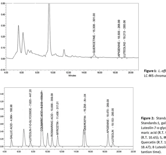
Qualitative chromatographic analysis by
LC-MS
The flavonoid compositions of the methanol extract were
detected by LC/MS. Standards were detected according to
mass analyses and qualitative analyses using
reversed-phase chromatography. The flavonoid components of
L. of
ficinalis fruit were characterized as luteolin, quercetin,
and apigenin (Figure 1, Figure 2) (Table 3).
Cell viability assays
Treatment with
L. officinalis fruit extract led to
signi
ficantly decreased cell viability in MKN45 and AGS
human gastric cancer cell lines
Cytotoxic effects of the L. of
ficinalis on MKN45 (human
gastric cancer cell line), AGS (gastric adenocarcinoma cell
line), and primary human
fibroblast cells by measuring
their metabolically active state. According to our
findings,
cell viability signi
ficantly decreased when treated with
Table: Total flavonoid and phenolic content of L. officinalis fruit extract.
Total flavonoid content mg QE/ g extract
Total phenolic content mg GAE/ g extract L. officinalis fruit extract . ± . mg qe/ g . ± . mg gae/ g
Table: ABTS and DPPH scavenging activities of L. officinalis extract.
L. officinalis extract References IC ± SD, mg/mL
ABTS . ± . . (trolox) DPPH . ± . . (ascorbic acid)
5
–10 mg/mL extract in both MKN-45 and AGS cancer cell
lines. Treatment of gastric cancer cell lines with 5 mg/mL
L. officinalis extract showed 65.6% (p=0.0031) and 25.2%
(0.0038) signi
ficantly decreased cell viability in AGS and
MKN-45 cells, respectively. Besides that, 10 mg/mL
extract of L. of
ficinalis led to highly decreased viability
of cells with 1.1% in MKN45 and 27.4% in AGS cell lines
(p
≤0.0003) Conversely, treatment of primary human
fibroblast cells with the same concentrations (5–10 mg/mL)
of L. officinalis extracts showed statistically non-significant
cytotoxicity (Figure 3A). Accordingly, L. of
ficinalis extract
showed 3.770 mg/mL; 5.606 mg/mL and 29.42 mg/mL IC
50values for MKN-45; AGS and primary human
fibroblast cells
respectively (Figure 3B).
Detection of apoptosis
L. officinalis extract induced apoptosis in both AGS and
MKN-45 cell lines through caspase-3 and PARP
cleavages
Apoptosis was analyzed in 5 mg/mL extract treated
MKN-45 and AGS gastric cancer cell lines. Compared to
control treatments (0 mg/mL), cleaved PARP and cleaved
caspase-3 presence were detected in L. of
ficinalis extract
treated AGS and MKN-45 gastric cancer cell lines
(Figure 4A). According to Image J analysis of AGS cell line,
cleaved PARP intensity increased approximately four
folds (p
≤0.0001), and cleaved caspase three intensity
increased
approximately
eight
folds
(p=0.0025)
(Figure 4B). In L. of
ficinalis extract treated MKN-45 cell
Figure 2: Standard chromatogram of LC-MS. Standards:1, gallic acid (R.T. 4.96); 2, Luteolin-7-o-glycoside (R.T. 7.62); 3, Cou-maric acid (R.T. 9.21); 4, Rosmarinic acid (R.T. 10.65); 5, Myrcetin (R.T. 11.43); 6, Quercetin (R.T. 15.25); 7, Apigenine (R.T. 18.47); 8 Luteolin (R.T. 19.15) (R.T.=Re-tantion time).
Table: L. officinalis methanol extract flavonoid compounds. Compounds RT Base peak (m/z)
Quercetine . .
Apigenine . .
Luteolin . .
Figure 1: L. officinalis fruit methanol extract LC-MS chromatogram.
line; cleaved PARP intensity increased approximately
one and a half folds (p=0.0029) and cleaved caspase-3
intensity increased approximately
five folds (p=0.0010)
(Figure 4C).
These data demonstrated the cleavage of apoptotic
proteins and biochemical verification of cell death induced
by L. officinalis extract in both AGS and MKN-45 gastric
cancer cell lines. Additionally, caspase cleavage showed
that L. of
ficinalis extract induced cell death occurs in a
caspase dependent manner.
Apoptotic cell populations were determined
in
L. officinalis fruit extract treated AGS cell
line
We analyzed the apoptotic phase of cell populations in
L. officinalis extract treated AGS gastric adenocarcinoma cell
line. Different from cell viability assays, AGS cells were either
treated with distilled water and 5 mg/mL of L. of
ficinalis just
for 36 h to track early apoptotic cells as well as late apoptotic
cells using Annexin V-FITC Early Apoptosis Detection Kit
(CST, U.S.). According to staining, L. of
ficinalis extract
trea-ted AGS cells showed a shift from alive to death cell state,
which is significantly determined as apoptotic cells either
from early or late stages (Figure 5A, B). The quantitative
analysis was performed and when compared to control cells,
viable cells decreased from 89.65 to 66.22%, and early
apoptotic cells increased from 1.55 to 19.77%, while late
apoptotic cells increased from 4.78 to 11.78%. Necrotic cell
increase in population was not found statistically signi
ficant
(from 4.02 to 2.23%) (Figure 5C).
Discussion
In this study, we evaluated the potential use of L. of
ficinalis
fruit methanol extract against gastric cancer cell lines as it
is one of the most malignant cancers studied and
tradi-tionally used in Turkey for the treatment of different types
of human illness, especially for gastrointestinal problems
[6, 33, 35, 36]. Firstly, L. of
ficinalis fruits TPC and TFC were
determined and they have been investigated with respect
to their in vitro antioxidant and anti-cancer activities
against gastric cancer.
In our study, we found the total phenolic compounds
and total flavonoids which are responsible for antioxidant
activity were also rich in fruits.
The antioxidant activities of fruits were measured
using the ABTS and DPPH methods which are most
Figure 3: Cytotoxic effects of L. officinalis extracts on MKN45 and AGS gastric cancer cell lines and primary human fibroblast cells. Cell viability of a) AGS and MKN-45 cell line decreased in a concentration dependent manner upon 48 h treatment with 1–10 mg/mL L. officinalis extracts. Decrease in viability offibroblast cells were found statistically not significant. Cell viability assays were performed as three independent experiments. Data are expressed as±SEM. Differences were considered as statistically significant at 0.003<p*≤0.005 and 0.0005<p**≤0.003; and p***≤0.0003. b) Normalize of dose vs. response graphs and IC50values of AGS; MKN45 cell lines and primary humanfibroblast cells.
Accordingly, L. officinalis extract showed IC50values of 3.770 mg/mL in MKN45 cell line; 5.606 mg/mL in AGS cell line; and 29.42 mg/mL in
common for antioxidant activity. These methods are also
used to estimate the antioxidant activity because of the
relatively short time required for analysis [34]. Many
antioxidant studies have been conducted on L. of
ficinalis
and reported to be related to the total amount of
flavonoids
and phenolic compounds. Compared with these other
Figure 4: Western blot analysis of cell death in L. Officinalis fruit extract treated AGS and MKN45 gastric cancer cell lines. a) AGS and MKN45 gastric cancer cell lines were treated with distilled water (0 mg/mL) (left band) and 5 mg/mL of L. Officinalis (right band) for 48 h to detect apoptosis. In comparison with control bands (0 mg/mL), cleaved PARP and cleaved caspase three presence were detected in 5 mg/mL treatments. According to Image J analysis of b) AGS and c) MKN-45 gastric adenocarci-noma cell lines; cleaved PARP and cleaved caspase-3 bands were detected as compa-red to control treatments. Data are expres-sed as±SEM. Differences were considered as statistically significant at
0.0005<p**≤0.003; and p***≤0.0003. Experiments were performed as triplicates and band densities were normalized to actin controls using image J analysis.
Figure 5: Flow cytometry analysis of apoptosis in 5 mg/mL L. officinalis treated AGS gastric adenocarcinoma cell line. AGS cells were either treated with a) distilled water (left) or b) 5 mg/mL of L. officinalis (right) for 36 h to detect early apoptotic cells using Annexin V-FITC Early Apoptosis Detection Kit (CST, U.S.). Pan-nels P2-Q1; P2-Q2; and P2-Q3 indicate necrotic cells; late apoptotic cells, and early apoptotic cells respectively. Each staining was performed as triplicates. Data are expressed as±SEM and diffe-rences were considered as statistically significant at 0.01<p#≤0.05.
studies, it has been shown to have a high antioxidant effect
because of
flavonoids and phenolic compounds like other
studies [6, 31
–34, 37].
Oxidative stress causes many diseases such as gastric
diseases [38, 39]. Many studies have shown that plant
components especially
flavonoids can be effective and
protective against oxidative damage [40, 41]. The
flavonoid
compositions of the fruit extract were determined by LC/MS
analyses. The flavonoid components of the extract were
characterized as luteolin, quercetin, and apigenin.
Quer-cetin has been high antioxidant activity, which is a member
of the
flavonoid’s family. It is the most effective scavenger
of ROS [42]. Some studies have shown that quercetin is
effective against gastric cancer [43, 44]. And also previous
studies have shown that luteolin and apigenin also have
effective activity for gastric cancer [45–48].
In the present study, the cytotoxic activity of the
extract can be thought to be due to these flavonoid group
compounds. In previous cytotoxic activity studies, it can be
realized that the cytotoxicity may cause by flavonoids
[49, 50]. Previous studies have shown that L. of
ficinalis
fruits have a selective cytotoxic effect in lung, colon,
prostate, liver, and cervical cancer cell lines and also in
these studies indicated that the effect was caused by the
phenolic compounds and
flavonoids having antioxidant
effects [20
–22].
Our results revealed that ranging from 5 to 10 mg/mL
concentrations of the extracts induce highly signi
ficant
cell death in AGS and MKN-45 cell lines whilst preserving
human
fibroblasts healthier. We also investigated the
background of cytotoxicity of the extract in terms of cell
death pattern. Regarding LC/MS
findings, luteolin,
quer-cetin and apigenin contents of L. Of
ficinalis methanol
extract, might lead to apoptotic cell death in gastric
can-cer cells when treated. Protein cleavages (PARP and
cas-pase 3) of the apoptotic cascade showed that the
apoptosis caused by the extracts follows a caspase
dependent manner. Furthermore, when the apoptotic cell
population was analyzed via Annnexin V/PI stainings, we
detected a statistically signi
ficant shift to apoptotic cell
copopulation from live cell state (including both early and
late apoptosis stages).
These findings indicate that as well as antioxidant
activities, L. of
ficinalis fruit extracts have anti-cancer
effects against gastric cancer and when further studied,
the active components can be an alternative or adjuvant
to standard chemical drugs used in the clinics. Since
many of the active phytochemicals are toxic to normal
cells, cancer selective behavior of L. officinalis fruit extract
for certain concentrations can be promising for
transla-tional approaches and L. of
ficinalis can be potentially
used against several cancers upon investigation. For this
reason, the results of this study have a novelty to further
study in detail and suggest to examine L. of
ficinalis fruit
methanol extract contents as potential anti-cancer
the-rapeutic candidates.
Research funding: None declared.
Author contributions: All authors have accepted
responsibility for the entire content of this manuscript and
approved its submission.
Competing interests: The authors declare no con
flict of
interest.
References
1. Ayla S, Okur ME, Günal MY, Özdemir EM, Çiçek Polat D, Yoltaş A, et al. Wound healing effects of methanol extract of Laurocerasus officinalis Roem. Biotech Histochem 2019;94:180–8.
2. İslam A. ‘Kiraz’ cherry laurel (Prunus laurocerasus). N Z J Crop Hortic Sci 2002;30:301–2.
3. Yeşilada E, Sezik E, Honda G, Takaishi Y, Takeda Y, Tanaka T. Traditional medicine in Turkey IX: folk medicine in north-west Anatolia. J Ethnopharmacol 1999;64:195–210.
4. Küpeli Akkol E, Kırmızıbekmez H, Küçükboyacı N, Gören AC, Yesilada E. Isolation of active constituents from cherry laurel (Laurocerasus officinalis Roem.) leaves through bioassay-guided procedures. J Ethnopharmacol 2012;139:527–32.
5. Var M, Ayaz A. Changes in sugar composition in cherry laurel (CV oxygemmis) fruit during development and ripening. Pakistan J Bot 2004;36:389–94.
6. Kolayli S, Küçük M, Duran C, Candan F, Dinçer B. Chemical and antioxidant properties of Laurocerasus officinalis Roem.(cherry laurel) fruit grown in the Black Sea region. J Agric Food Chem 2003;51:7489–94.
7. Colak A, Özen A, Dincer B, Güner S, Ayaz FA. Diphenolases from two cultivars of cherry laurel (Laurocerasus officinalis Roem.) fruits at an early stage of maturation. Food Chem 2005;90:801–7. 8. Livshits Z, Rao RB, Smith SW. An approach to
chemotherapy-associated toxicity. Emerg Med Clin North Am 2014;32:167–203. 9. Demain AL, Vaishnav P. Natural products for cancer
chemotherapy. Microb Biotechnol 2011;4:687–99. 10. Rajesh E, Sankari L, Malathi L, Krupaa J. Naturally occurring
products in cancer therapy. J Pharm BioAllied Sci 2015;7:183. 11. Serafini M, Bellocco R, Wolk A, Ekström AM. Total antioxidant
potential of fruit and vegetables and risk of gastric cancer. Gastroenterology 2002;123:985–91.
12. Metere A, Giacomelli L. Absorption, metabolism and protective role of fruits and vegetables polyphenols against gastric cancer. Eur Rev Med Pharmacol Sci 2017;21:5820–58.
13. Borek C. Dietary antioxidants and human cancer. Integr Canc Ther 2004;3:333–41.
14. Venerito M, Link A, Rokkas T, Malfertheiner P. Gastric
cancer– clinical and epidemiological aspects. Helicobacter 2016; 21:39–44.
15. Suzuki H, Mori H. Gastric cancer after helicobacter pylori eradication. Jpn J Canc Chemother 2018;45:1123–7.
16. Guggenheim DE, Shah MA. Gastric cancer epidemiology and risk factors. J Surg Oncol 2013;107:230–6.
17. Ang TL, Fock KM. Clinical epidemiology of gastric cancer. Singap Med J 2014;55:621–8.
18. Butcher LD, den Hartog G, Ernst PB, Crowe SE. Oxidative stress resulting from Helicobacter pylori infection contributes to gastric carcinogenesis. CMGH 2017;3:316–22.
19. Díaz P, Valderrama MV, Bravo J, Quest AFG. Helicobacter pylori and gastric cancer: adaptive cellular mechanisms involved in disease progression. Front Microbiol 2018;9:1–10.
20. Aydin A, Erenler R, Yılmaz B, Tekin Ş. Antiproliferative effect of Cherry laurel. J Turk Chem Soc Sect A Chem 2016;3:217. 21. Demir S, Turanİ, Demir F, Ayazoglu Demir E, Aliyazicioglu Y.
Cytotoxic effect of Laurocerasus officinalis extract on human cancer cell lines. Marmara Pharm J 2017;21:121–6.
22. Çakir B, Gülserenİ. Investigations on apoptotic activities of cherry laurel extracts in HCT-116 human colon carcinoma cells. Indian J Pharm Educ Res 2019;53:S264–72.
23. Okur ME, AylaŞ, Çiçek-Polat D, Günal MY, Yoltaş A, Biçeroğlu Ö. Novel insight into wound healing properties of methanol extract of Capparis ovata Desf. var. palaestina Zohary fruits. J Pharm Pharmacol 2018;70:1–13.
24. Spanos GA, Wrolstad RE. Influence of processing and storage on the phenolic composition of Thompson seedless grape juice. J Agric Food Chem 1990;38:1565–71.
25. Singleton VL, Orthofer R, Lamuela-Raventós RM. [14] Analysis of total phenols and other oxidation substrates and antioxidants by means of folin-ciocalteu reagent. Methods Enzymol 1999;299: 152–78.
26. Okur ME, Polat DC, Ozbek H, Yilmaz S, Yoltas A, Arslan R. Evaluation of the antidiabetic property of Capparis Ovata Desf. Var. Paleastina Zoh. Extracts using in vivo and in vitro approaches. Endocr Metab Immune Disord - Drug Targets 2018; 18:489–501.
27. Mihai CM, Mărghitaş L, Bobiş O, Dezmirean D, Tămaş M. Estimation offlavonoid content in propolis by two different colorimetric methods. Sci Pap Anim Sci Biotechnol 2010;43: 407–10.
28. Blois MS. Antioxidant determinations by the use of a stable free radical [10]. Nature 1958;181:1199–200.
29. Re R, Pellegrini N, Proteggente A, Pannala A, Yang M, Rice-Evans C. Antioxidant activity applying an improved ABTS radical cation decolorization assay. Free Radic Biol Med 1999;26:1231–7. 30. Gulsoy-Toplan G, Goger F, Yildiz-Peko A, Gibbons S, Sariyar G,
Mat A. Chemical constituents of the different parts of Colchicum micranthum and C. chalcedonicum and their cytotoxic activities. Nat Prod Commun 2018;13:535–8.
31. Orhan IE, Akkol EK. Estimation of neuroprotective effects of Laurocerasus officinalis Roem. (cherry laurel) by in vitro methods. Food Res Int 2011;44:818–22.
32. Liyana-Pathirana CM, Shahidi F, Alasalvar C. Antioxidant activity of cherry laurel fruit (Laurocerasus officinalis Roem.) and its concentrated juice. Food Chem 2006;99:121–8.
33. Karabegović IT, Stojičević SS, Veličković DT, Todorović ZB, Nikolić NČ, Lazić ML. The effect of different extraction techniques on the composition and antioxidant activity of cherry laurel
(Prunus laurocerasus) leaf and fruit extracts. Ind Crop Prod 2014; 54:142–8.
34. Cai Y, Luo Q, Sun M, Corke H. Antioxidant activity and phenolic compounds of 112 traditional Chinese medicinal plants associated with anticancer. Life Sci 2004;74:2157–84. 35. Celep E, Aydin A, Yesilada E. A comparative study on the in vitro
antioxidant potentials of three edible fruits: cornelian cherry, Japanese persimmon and cherry laurel. Food Chem Toxicol 2012; 50:3329–35.
36. Erdemoglu N, Küpeli E, Yeşilada E. Anti-inflammatory and antinociceptive activity assessment of plants used as remedy in Turkish folk medicine. J Ethnopharmacol 2003;89:123–9. 37. Karahalil F,Şahin H. Phenolic composition and antioxidant
capacity of cherry laurel (Laurocerasus officinalis Roem.) sampled from Trabzon region, Turkey. Afr J Biotechnol 2011;10:16293–9. 38. Tandon R, Khanna RD, Dorababu M, Goel R. Oxidative stress and
antioxidants status in peptic ulcer and gastric carcinoma. Indian J Physiol Pharmacol 2004;48:115–8.
39. Bhattacharyya A, Chattopadhyay R, Mitra S, Crowe SE. Oxidative stress: an essential factor in the pathogenesis of gastrointestinal mucosal diseases. Physiol Rev 2014;94:329–54.
40. Butera D, Tesoriere L, Di Gaudio F, Bongiorno A, Allegra M, Pintaudi AM, et al. Antioxidant activities of Sicilian prickly pear (Opuntiaficus indica) fruit extracts and reducing properties of its betalains: betanin and indicaxanthin. J Agric Food Chem 2002;50: 6895–901.
41. Lee J-C, Kim H-R, Kim J, Jang Y-S. Antioxidant property of an ethanol extract of the stem of Opuntiaficus-indica var. Saboten. J Agric Food Chem 2002;50:6490–6.
42. Boots AW, Haenen GRMM, Bast A. Health effects of quercetin: from antioxidant to nutraceutical. Eur J Pharmacol 2008;585: 325–37.
43. Borska S, Chmielewska M, Wysocka T, Drag-Zalesinska M, Zabel M, Dziegiel P, et al. In vitro effect of quercetin on human gastric carcinoma: targeting cancer cells death and MDR. Food Chem Toxicol 2012;50:3375–83.
44. Wang K, Liu R, Li J, Mao J, Lei Y, Wu J, et al. Quercetin induces protective autophagy in gastric cancer cells: involvement of Akt-mTOR- and hypoxia-induced factor 1α-mediated signaling. Autophagy 2011;7:966–78.
45. Wu B, Zhang Q, Shen W, Zhu J. Anti-proliferative and
chemosensitizing effects of luteolin on human gastric cancer AGS cell line. Mol Cell Biochem 2008;313:125–32.
46. Wu H, Huang M, Liu Y, Shu Y, Liu P. Luteolin induces apoptosis by up-regulating miR-34a in human gastric cancer cells. Technol Canc Res Treat 2015;14:747–55.
47. Shukla S, Gupta S. Apigenin: a promising molecule for cancer prevention. Pharm Res 2010;27:962–78.
48. Kuo C-H, Weng B-C, Wu C-C, Yang SF, Wu DC, Wang YC. Apigenin has anti-atrophic gastritis and anti-gastric cancer progression effects in Helicobacter pylori-infected Mongolian gerbils. J Ethnopharmacol 2014;151:1031–9.
49. Hirobe C, Qiao Z-S, Takeya K, Itokawa H. Cytotoxicflavonoids from Vitex agnus-castus. Phytochemistry 1997;46:521–4. 50. Seelinger G, Merfort I, Wölfle U, Schempp CM. Anti-carcinogenic