S’^: / .^· "i í^·; ! ^"ïl / :!!^.Л‘ .'■ *г‘ W4*^^‘v w v ; > ^ * s i - / «•■ w·'^·', * г х r ¿ v ^ / ; ; -v .4 ^ E C ü L A r l T^TST'vvt -%r ¿ 9 · ' Ci i л^"^"';“·: ; ігѵ i r;¿- J ï ^ . í.·^. .4 . ; ..^. :Y AN* / /(‘ / O y , J í?> ?
гсі
■ H i t é
ФЭ
81993
MOLECULAR CLONING and CHARACTERIZATION OF THE
COMMON lb SUBTYPE OF HCV FROM TURKEY
A THESIS SUBMITTED TO
THE DEPARTMENT OF MOLECULAR BIOLOGY AND GENETICS
AND
THE INSTITUTE OF ENGINEERING AND SCIENCE
OF BILKENT UNIVERSITY
IN PARTIAL FULFILLMENT OF THE REQUIREMENTS
FOR THE DEGREE OF MASTER OF SCIENCE
By
ASLI ÓZTAN
α ρ . İO İ • Н4б -0 3 i. ‘foQa
π 9052
I certify that I read this thesis and that in my opinion it is fully adequate, in scope and in quality, as thesis for the degree of Master of Science.
I certify that I read this thesis and that in my opinion it is fully adequate, in scope and in quality, as thesis for the degree of Master of Science.
P^rof. D r Hikmet Akkiz
I certify that I read this thesis and that in my opinion it is fully adequate, in scope and in quality, as thesis for the degree of Master of Science.
Assist. Prof. I
5
ik ju luApproved for Institute of Engineering and Science.
Prof.Dr. Mehmet Baray, Director of Institute of Engineering and Science
ABSTRACT
MOLECULAR CLONING and CHARACTERIZATION OF THE COMMON lb SUBTYPE OF HCV FROM TURKEY
Ash Oztan
MSc. in Molecular Biology and Genetics Supervisor: Prof. Mehmet Öztürk
July 1999
Hepatitis C Virus is a major cause o f acute and chronic hepatitis worldwide. 80-90% o f Hepatitis C Virus infections become chronic and 75% o f these cases lead to liver disease, including cirrhosis, liver failure and hepatocellular carcinoma. Hepatitis C Virus was first identified by molecular cloning o f the viral genome in 1989. Hepatitis C Virus is an enveloped virus containing a positive stranded RNA genome with a size o f around 9.5 kilobases. In terms o f genomic organization, it was accepted as a member o f Flaviviridae family as a new genus named Hepaciviruses. The single-stranded RNA genome encodes a single open reading frame, which is transcribed into a single polypeptide o f 3010 or 3030 amino acids and cleaved into viral proteins Core, E l, E2/p7, NS2, NS3, NS4A, NS4B, NS5A, NS5B by host and viral proteases. Sequencing, serotyping and RFLP studies indicate that Hepatitis C Virus genome is highly variable. There are six distinct genotypes and at least 74 subtypes with different distributions between the geographic areas. Variability is not equally distributed throughout the genome. 5' UTR, some parts o f the 3' UTR and capsid protein are the most conserved regions. The predominant genotype in the Turkish population was found to be lb by sequencing o f the 5-UTR. In this study, the entire sequence encompassing the complete coding region and partial non coding regions o f the genotype lb obtained from a HCV-infected Turkish patient was cloned to investigate its evolutionary relationship with other genotypes and to study its overall genome organization,. In order to characterize the viral genome, viral RNA was extracted from the serum, cDNA was synthesized, the HCV genome was amplified by PCR in 7 overlapping fragments, PCR fragments were cloned into bacterial vectors and cloned inserts were sequenced by automated sequencing methodology. The partial sequence data covering 70% o f the cloned HCV genome indicate that the Turkish lb genotype displays high homology to other lb genotypes, but differs from others by distinct amino acid changes. To our knowledge, this is the first report about the HCV genome structure from Turkey. The HCV subgenomic fragments obtained here will serve to further molecular and immunologic studies on this dominant form o f HCV found in Turkish patients.
ÖZET
TÜRKİYE DE BASKIN OLARAK GÖRÜNEN HEPATİT C VİRÜSÜ Ib ALT TİPİNİN MOLEKÜLER KLONLANMASI VE KARAKTERİZ AS YONU
Aslı Öztan
Moleküler Biyoloji ve Genetik Yüksek Lisans Tez Yöneticisi: Prof. Mehmet Öztük
Temmuz 1999
Hepatit C Virüsü dünyada akut ve kronik hepatitin en önemli nedenidir. Hepatit C Virüsü enfeksiyonlarinin yüzde 80 ile yüzde 90 gibi bir oranı kronikleşir ve buna bağlı olarak siroz, karaciğer yetmezliği ve karaciğer kanseri gibi hastalıklara sebep olur. Hepatit C Virüsü ilk defa 1989 yılında örtüşen bir çok kopya DNA'nın klonlanması ile bulunmuştur. Bu zarflı, pozitif tek iplikli RNA virüsü 9.5 kilobazlık bir genoma sahiptir. Genom organizasyonuna göre HCV Flaviviridae virüs ailesinin altında Hepasivirusler diye adlandırılan yeni bir generada sınıflandırılmıştır. Virüsün tek iplikli RNA genomu 3010 ile 3030 amino asitten oluşan tek bir polypeptid sentezleyen açık okuma çerçevesi içerir ve viral ya da hücre proteazları tarafından Core, E l, E2/p7, NS2, NS3, NS4A, NS4B, NS5A ve NS5B virüs proteinlerine parçalanır. Dizi analizi, serotipleme ve RFLP metodları ile virüs genomunun çok sayıda değişime uğradığı anlaşılmıştır. Şimdiye kadar farklı coğrafi alanlara yayılmış 6 değişik genotip ile 74 civarında altip bulunmuştur. Virüs genomunun dizisinde gözlemlenen hızlı değişim genomun tüm bölgelerine eşit şekilde dağılmamıştır. 5'- UTR, 3'-UTR’ın bir kısmı ve kapsid proteini en çok korunan bölgelerdir. Türk popülasyonunda en çok rastlanan genotip 5' UTR dizi analizinin yapılması ile Ib olarak saptanmıştır. Bu çalışmada da ilk Türk HCV izolatının karakterize edilmesi ve virüsün evriminin incelenmesi amacı ile bir Türk hastadan izole edilen Hepatit C Virüsü genomu klonlanmış ve dizi analizi yapılmıştır. İzlenen yöntemler virüs RNA sının hastanın serumundan izole edilmesi, kopya DNA nm sentezlenmesi, genomun örtüşen 7 tane polimeraz zincir reaksiyonu ile çoğaltılması, bu fragmentların bakteri plasmidlerine klonlanması ve klonlanan bölgelerin otomatik dizi analizi yöntemi ile analiz edilmesidir. Klonlamış HCV genomunun %70 lik dizi verisine göre, Türk Ib genotipinin diğer Ib genotipleriyle homolojisi yüksek olmakla birlikte, bazi amino asit farklılıkları da belirlenmiştir. Elimizdeki verilere göre, bu çalışma Türkiye’den HCV genomu yapısıyla ilgili ilk rapordur. Burada elde edilen HCV alt genom parçaları ileride Türkiye’de baskın olan bu alt tür ile ilgili moleküler ve immünolojik çalışmalarda kullanılabilecektir.
ACKNOWLEDGEMENTS
1 would like to express my greatest gratitude to my thesis advisor Prof. Mehmet Öztürk, who had understood my passion in virology and gave me a chance to be a part o f the virology project. It was a great opportunity for me to share his scientific vision during this study.
1 would like to thank to my undergraduate advisor Prof Ashhan Tolun, for giving me the "first" chance and for her undoubtfUll trust and encouragement throughout the years; Uğur Yavuzer, for all the discussions that enhanced my scientific way o f thinking, for her trust in me more than I had in myself during the nightmares and above all for seeing rather than looking; Cengiz Yakicier for being a person as he is; Işık Yuluğ for sharing her scientific experiences, all the support during these two years and her endless tolerance to me; Rengiil Çetin Atalay, who is not that much old at all, for all the chats in the lab and her kind personal interest.
"Diin baştan başa onlarla açıklandı, yarın onlarla kuruldu baştanbaşa. (A.Timuçin)” Gülayşe, the blue color in my picture, things wouldn't be the same without your smile, encouragement and unquestionable belief Thank you not only for all the sleepless nights, the difficult days and pains that you were with me but for all the successes and happiness that we could share with each other. Bleda, the whisperer o f my conscience,
"Birbirinden en ayrı gemilerde bile bizim için yo l aynı olurdu., nehirden yukarı- çünkü bizi ayni kaynak bekliyor (R.M.Rilke)", for being a better thorn in the game than me. Tolga Emre, my favorite puzzle, you are the magenta in the picture, sometimes breaking things down, but surely one o f the irreplaceable .
Hilal, the mother goddess Kybele, surviving against the most difficult experience o f her legend throughout the centuries. "Onlar sava.fçılardır, acıyı ve .sevinci direncin dupduru suyuyla yoğururlar. (A.Timuçin)". Thank you for each A, T, G and C in this thesis and every second, minute and hour you had spend for me both professional and personal.
My special thanks go to Esra, the one and only virology group member, for sharing your deep knowledge on HCV and for being the guide o f me in the virology world; Tuba for all the units o f the enzymes she shared with me and her patience on answering my endless questions; Ayça, the ultimate support and ocean blue on my palette, for sharing the great joy o f being on "çimenler" or "manzara" and all the great memories in Boğaziçi; Suha, the one whom I had sheltered not to scream all the way out; Tunç, the soul mate in a parallel universe, for being a part o f my unconscious land and taking Aruoba and the dark side o f the moon with you; Ersan, the "kirk yılda bir gibisin", for being the safest ground.
Finally, I would like to thank to my family Akin and Güler Öztan, for bringing me up as I am. They gave me the chance to explore my own limits and never loose their faith in me to date.
TABLE OF CONTENTS
SIGNATURE PAGE... II ABSTRACT... Ill ÖZET... IV ACKNOWLEDGEMENTS... V TABLE OF CONTENTS... VI LIST OF FIGURES... VIII LIST OF TABLES...IX ABBREVATIONS...X1. INTRODUCTION... 1
LI VIRAL GENOME ORGANIZATION...6
l . U UNTRANSIA TED REGIONS... 6
1.1.2 STRUCTURAL PROTEINS... 8
1.1.3 NON-STRUCTUML PROTEINS... 12
1.2 AIM OF THE STUDY AND STRATEGY... 20
2. MATERIALS AND METHODS... 22
2.1 MATERIALS... 22
2.1.1 Chemicals and Enzymes...22
2.1.2 Equipment...\23
2.1.3 Plastic Disposables... 23
2.1.4 Bacterial Strain... 24
2.1.5 Solutions and Media... 24
2.2 M ETHODS...26
2.2.1 Patient... 26
2.2.2 Viral RNA Extraction and cDNA synthesis... 26
2.2.3 Oligonucleotide Synthesis... ... 26
2.2.4 Polymerase Chain Reaction... 28
2.2.5 Agarose Gel Electrophoresis o f DNA Fragments... 30
2.2.6 Electrophoresis Markers... 30
2.2.7 Cloning ofPCR Products... 31
2.2.8 Transformation ofE.C oli... 33
2.2.9 Small Scale Preparation o f Plasmid DNA (Mini-Preperation)... 33
2.2.10 Restriction Enzyme Digestion o f DNA... 34
2. 2.11 Medium and Maximum Scale Isolation (Midi-Preperation)...34
2.2.12 Quantification o f DNA... 34
2.2.13 A utomated Sequencing o f DNA Fragments...35 vi
3. RESULTS...36
3.1 Introduction...36
3.2 Strategyfor Amplificationofthe Viral Genome... 37
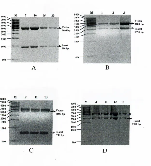
3.3 Strategyfor Cloningofthe PCR Products...·...42
3.4 Sequencingofthe Cloned Fragm ents... 46
3.4.1 ZEUS... 47 3.4.2 CHARON...47 3.4.3 NS2... 48 3.4.4 NS3... 48 3.4.5 NS4... 49 3.4.6 NS5A... 49 3.4.7 NS5B... 50 4. DISCUSSION... 60 5. PERSPECTIVE...62 6. REFERENCES... 64 7. APPENDIX... 72
F igurel.l Incidence o f Hepatitis C Virus cases world wide 2 Figure 1.2 Schematic illustration o f HCV genome and polyprotein. 4 Figure 1.3 Schematic illustration o f HCV polyprotein processing. 5 Figure 1.4 M odel o f secondary and tertiary stru ctu re o f the HCV IRES. 7
Figure 2.1 DNA size markers 30
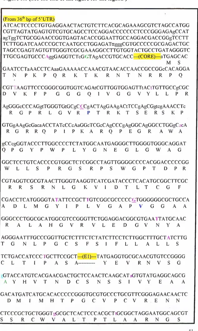
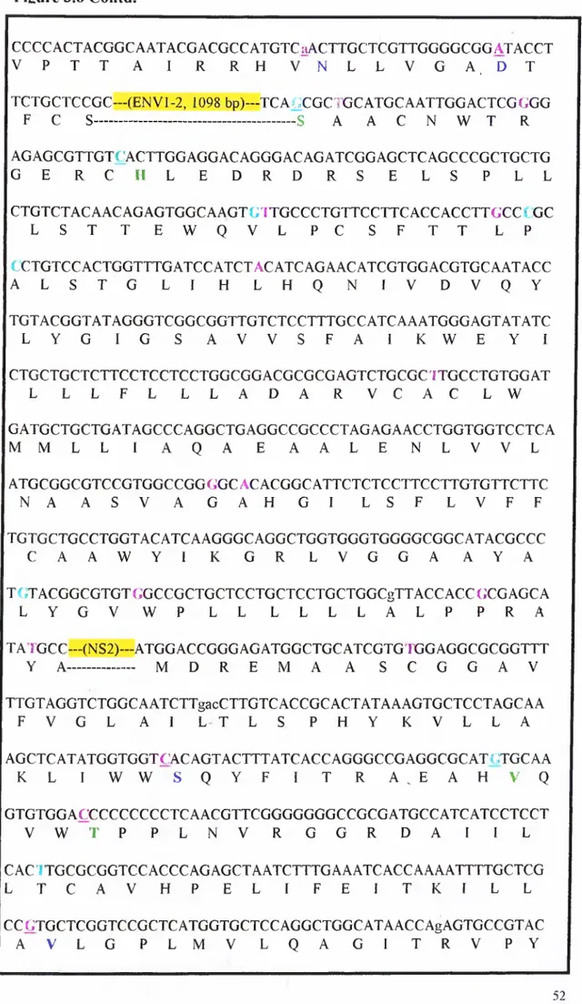
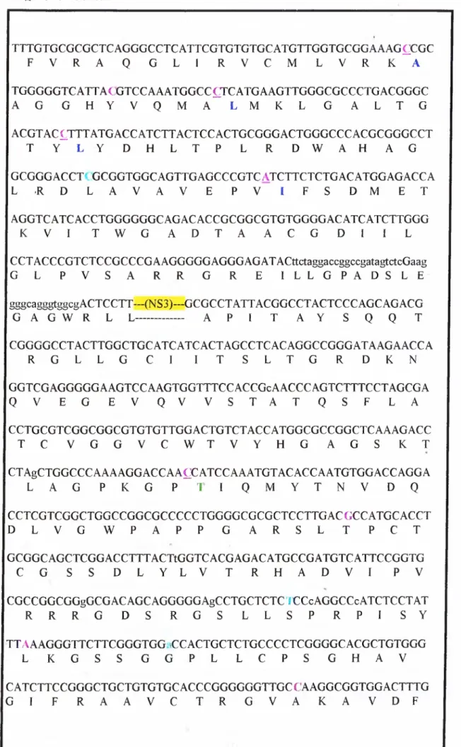
Figure 2.2 A.) pGEM®-T Easy Vector Map and B.) Multiple Cloning Site 32 Figure 3.1 Amplification strategy o f HCV genome 38 Figure 3.2 Agarose gel electrophoresis o f PCR products.(I) 40 Figure 3.3 Agarose gel electrophoresis o f PCR products.(II) 4 1 Figure 3.4 Agarose gel electrophoresis results o f cloning experiments (I). 44 Figure 3.5 Agarose gel electrophoresis results o f cloning experiments (II) 45 Figure 3.6 RNA and amino acid sequences o f the Turkish isolate o f the
HCV subtype lb. 51
LIST OF FIGURES
LIST OF TABLES
Table 2.1 PCR Primers 27
Table 2.2 Sequencing Primers 27
Table 2.3 PCR Conditions for Fragments Smaller Than 1 kb. 29 Table 2.4 PCR Conditions for Fragments Longer Than 1 kb. 29 Table 2.5 Cloning conditions o f PCR products 35 Table 3.1 Isolates o f Submitted HCV lb Complete Genomes 37 Table 3.2 Summary o f codon differences between the HCV lb Turkish
isolate and other characterized HCV lb genomes. 59
ABBREVATIONS
A Ala Arg Asn Asp Asx bp C cDNA C-Terminus Cys ddHzO DNA dNTP ds EDTA EtBr G Gin Glu Glx Gly HBV HCC HCV His HIV HVR IFN -a He IPTG IRES ISDR kb LB Leu Lys Met NANBH NS N-Terminus Adenine (A) Alanine (R) Arginine (N) Asparagine (D) Aspartic Acid (B) Asparagine base pairs CytosineComplementary Deoxyribonucleic Acid Carboxyl-terminus
(C) Cysteine
deionized distilled water Deoxyribonucleic Acid deoxynucleotide triphosphate double strand
diaminoethane tetra-acetic acid Ethidium Bromide
Guanine (Q) Glutamine (E) Glutamic Acid (Z) Glutamic Acid (G) Glycine Hepatitis B Virus Hepatocellular Carcinoma Hepatitis C Virus (H) Histidine
Human Immunodeficiency Virus Hyper Variable Region
Interferon Alfa (I) Isoleucine
Isopropylthio-b-D-galactoside Internal Ribosome Entry Site
Interferon Sensitivity Determining Region kilobase(s)
Luria-Bertoni Media (L) Leucine
(K) Lysine (M) Methionine
Non-A Non-B Hepatitis Non-structural Protein Amino-Terminus
NTP Nucleoside triphosphate
PCR Polymerase Chain Reaction
Phe (F) Phenylalanine
Pro (P) Prolin
1. INTRODUCTION
Hepatitis C Vims is a major cause o f non-A, non-B hepatitis worldwide. In 80-90% o f the cases, the HCV infections become chronic hepatitis which may lead to cirrhosis, liver failure and hepatocellular carcinoma (Caselmann
et al.,
1996). HCV infection is typically mild in its early stages and rarely recognized until it has caused significant damage to the liver. The cycle o f disease from infection to significant liver damage can take 20 years or more. Known risk factors o f the HCV are mainly transfusions o f blood and blood products, intravenous dmg usage, parental or sexual transmission and needle-stick accidents (Okamotoei al.,
1991a). Since 1990, after the introduction o f blood tests for detection o f antibodies against HCV proteins, the posttransfusion incidence o f HCV has decreased in the developed countries. The risk o f transfusion related hepatitis is in the range o f 1 in 100,000 units transftised in these countries. In developing countries lack o f detection tests for blood donors, multiple usage o f syringes, low socioeconomic levels and lack o f public education leads to a drastic increase in new HCV cases. In Turkey mandatory detection o f antibodies against HCV in the blood donors' sera started in 1997. The data based on tests made on blood donors in Turkey showed that the 1,5% o f the population is anti-HCV positive (Figure 1.1; Thomaset al.,
1994). Persistent HCV infection is common in transplant recipients. It was reported that HCV is the cause o f chronic hepatitis in approximately 10% o f all transplant recipients. Antiviral therapy with interferon-alpha is effective in only a minority o f transplant patients, and since allograftsm
# M O V .^1
%
W
or
ld
w
id
e
F
fe
v
a
le
ii
c
e
o
í
H
e
p
a
ti
ti
s
0
u re l· !: I n c id e n c e o f H e p a ti ti s C V ir u s c a se s w o rl d w id efrom HCV infected donors are quite efficient in transmitting the virus, great attention is paid to the appropriate use o f organs from HCV-positive donors. At present, these organs should be particularly targeted for patients in emergency need o f lifesaving heart, liver, or lung transplants (Fishman
et al.,
1996).Hepatitis C virus was first identified in 1989 by molecular cloning o f the viral genome in several overlapping cDNA fragments (Choo
ei al.,
1989). The virus genome is approximately 9.5 kb positive single stranded RNA molecule (Figure 1.2). RNA encodes a single polypeptide which is cleaved into structural and non-structural proteins by viral and host proteases (Figure 1.3). Genome replication takes place in the cytoplasm through an RNA intermediate. In terms o f the presence o f a lipid envelope, sensitivity to organic solvents, single stranded positive RNA genome, a long open reading frame, organization o f the polyprotein and cleavage o f polyprotein into several viral proteins, HCV was classified as a member o f the Flaviviridae and has been placed in a new monotypic genus in this family, named hepaciviruses The nucleotide sequence is highly variable, the most divergent isolates sharing only 60% nucleotide sequence homology. Variability rate o f the genome was estimated to be 1.44X10'^ base substitution/site/year. According to this data, theoretically all o f the nucleotides may change during 700 years o f infection in chimpanzees. However variability/stability o f the genome varies by region within the genome. HVRl and some regions o f the 3 ’-UTR were found to be the most variable sites between various HCV strains (Simmonds P., 1998). Until today isolates from all over the world have been sequenced and a classification system was proposed due to the differences in the 5 ’-UTR o f the different HCV groups and a phylogenetic tree was constructed where3 9 k b 5 · 3 ' p 2 2 g p $ l> g p T D p 2 1
9
7
0
P 2 7 p 5 6 P & 6 U T R C E l E 2 /N S 1 2 A ? 2 B L JO
i
1- =1 -I -· O -I L- -f-· CjO •T? I? H-· O I? CO 1. D -H· O D L. -1^ iO <l· •l· o !i- O. "O c o o >l· N S 3 a> o ■1— 'aJ a- V I <l· - I-· O L· Cl 4 A 4 B 5 C o O o o W CO 5 A L. O -H· O O 0 il· W L. 01£
'o CL 5 B <v Vli L. 0^£
IT· 'oC
l.
Ct
H
u re 1 .2 : S c h e m a ti c i ll u st ra ti o n o f H C V g e n o m e a n d p o ly p ro te in .0
1
1 21
1
3 4 5 1 t > ■' ? i1 9 k b 1_ _1
p 2 S1
fl p S SE
E
V 7 0 ■ ■B
B
ii
S'/ " 3 ’P
o
ly
p
r
o
te
in
S
y
n
th
e
sis
a
E
ii
,E
2
tB
N
S
|i
a
N
S
3
K S 5 2 \ . f l N S S B i p 7H
o
st
S
ig
n
a
l
P
e
p
ti
d
a
se
^
N
S
2
/3
C
le
a
v
a
g
e
iz
zj
)
N
S
3
C
is
-C
le
a
v
a
g
e
r—
S
N
S
3
T
r
a
n
s-C
le
a
v
a
g
e
F ig u r e 1 .3 : S c h e m a ti c i ll u st ra ti o n o f H C V p o ly p ro te in p ro c e ss in g .HCV isolates were grouped into 6 equally divergent genotypes and each into several subtypes. Main types are numbered 1 to 6 and subtypes have been lettered a, b and c in order o f discovery (Simmonds
el al.,
1993). Besides these clearly divergent isolates, there are different variants o f the virus within the same individual reflecting the high variability rate o f the viral genome. The quasispecies are not different enough to be grouped as a separate type or subtype but diverged enough to escape from the host’s immune system. Some genotypes o f HCV such as la, lb, 2a and 2b have shown a broad worldwide distribution, whereas others have been seen in specific geographical areas. Genotype 1 b seems to be the most common variant in Japan, 1 a is commonly seen in Europe and United States, and type 4 is the predominant genotype in Middle East and Central Africa. Turkey is between the regions o f Southern Europe and Middle East, but interestingly the predominant genotype o f Turkish population was found to be lb at a frequency higher than Japan (Yildizet al.,
unpublished data).1.1 VIRAL GENOME ORGANIZATION
l . I . l UNriMNSLATED REGIONS
l .I .l .I 5 ’-UJR
Complete 5’ UTR spans the 3 4 1 nucleotide region at the 5’ site o f the genome (Lee
ei al.,
1992) but shorter sequences have been detected and reported (Okamotoet al.,
1990, Chooet al.,
1991, Katoet al.,
1990). 5’-UTR is themost conserved region in the genome and used in genotyping studies because o f the differences specific to the various types.
F ig u re 1.4: M odel o f secondary and tertiary stru c tu re o f the HCV IRES pro p o sed by H onda
et al.,
1996.A large conserved stem-loop structure (Figure 1.4) which acts as the Internal Ribosome Entrance Site (1RES; Wang
etal.,
1993) is present at the proximal part. HCV viral genome translation is cap-independent and the 1RES is involved in the regulation o f replication and translation (Hondaet a l,
1999). 1RES provides a structure which is able to direct ribosomes to the AUG codon at position 342 for translation initiation. p57 polypyrimidine tract binding protein (PTB; Aliet al.,
1995) and 2 subunits o f eIF3 were found to bind to the HCV 1RES (Burattiet al.,
1998).3 ’ end o f the HCV genome has a tripartite structure with a conventional 3 ’ end sequence, a poly(U) tract in some isolates and a highly conserved 98 nucleotide sequence which is called 3'X tail. Poly(U) tract appears to be highly heterogenous between different isolates and even within the same infected liver. In contrast 3 ’X tail is highly conserved between two most divergent genotypes la and lb except two additional U residues at the end in lb isolates (Tanaka
el al.,
1996,'Yamadaei al.,
1996). It was reported that this region folds into a stem-loop structure and polypyrimidine tract binding protein binds to 3'X tail just like 5’- UTR (Itoet al.,
1998). These finding indicate that 3 ’-UTR region might also be important in replication like 5'UTR.1.1.2
STRUCTUML PROTEINS
1.1.2.1 CORE
L I . 1.2 3 ’-UTR
The first structural protein is the HCV core protein (p22). It does not have any N-glycosylation sites. Amino acid sequence was found to be well conserved between the different HCV isolates (Bukh
et al.
1994), which may reflect the importance o f the function o f this protein for the survival o f the virus. Also because o f this property core protein can be used for efficient detection o f anti HCV antibodies (Doomet al
1994).The core protein is cleaved from the polyprotein by the cellular signal peptidase which recognize a specific sequence in the peptide chain. The protein then undergoes a complex process o f proteolytic maturation and two main different products have been described: C l 73 (residues from 1 to 173) and C l 91
when the expression o f C l 73 takes place in the absence o f C 191, it is translocated to the nucleus (Santolini
el al
1994).HCV core protein is thought to be a multifunctional protein. It was shown that core protein binds to cellular membranes, RNA molecules and 60S ribosomal subunit (Santolini
et al.,
1994). There is also a domain for binding to HCV El protein (Loet al.,
1996). There are some data about the multimerization and dimerization ability o f the core protein (Santoliniet al.
1994). It can be detected in viral particles in patients serum by immunoelectron microscopy (Kaitoet al.,
1994) and related to the capsid protein C o f flaviviruses (Santoliniet al
1994). All these data indicate that the core protein is the nucleocapsid protein o f the virus and it is involved in the viral assembly. Besides having a role in viral encapsulation, it was shown to have a trans-acting regulatory role. It possesses a nuclear localization signal and as described previously, some forms o f core protein were detected in the nuclei. HCV core protein is involved in the transcriptional repression o f p53 (Rayet al.
1997) and repression p21WM'l/Cipl/Sidl promoter activities (Rayet al
1998), and also trans activates c-myc oncogene (Rayet al.
1996). Yeast two hybrid library screening studies have identified an interaction with the cytoplasmic tail o f Lymphotoxin-P receptor (Matsumotoet al.
1997, Chenet al
1997) which is involved in host defense mechanism. Lymphotoxin-P receptors are the members o f tumor necrosis factor receptors (TNFR). After the identification o f Lymphotoxin-P receptor, it was shown that core protein also binds to the cytoplasmic domain o f TNFR 1 which is another member o f the TNFR family. This binding enhances TNF-induced apoptosis (Zhuet al.,
1998). In a recent study a transgenic mice model was used to show that the core protein induces hepatocellular carcinoma (Moriyaet al
1998).1.1.2.2 ENVELOPE PROTEINS
Other structural proteins o f HCV are E l and E2 glycoproteins. Molecular mass o f E l varies from 28 to 35 kDa depending on the strain and conditions o f expression (Ralston
et
a /.,1993), Between residues 350-390 a stretch o f hydrophobic amino acids is present, which is thought to act as a transmembrane anchor (Heinzet al.,
1992). In an immunoprécipitation experiment, the E l protein was found to be precipitated by an anti-core antibody in the presence but not in the absence o f the core protein, indicating that the E 1 protein can interact with the core protein. This interaction is independent o f whether the E l and the core genes are linked incis
or separated in different DNA constructs for expression. Deletion-mapping studies indicate that the carboxy-terminal sequences o f both the core and the E l proteins are important for their interaction. Since a small portion o f E l sequence is exposed to the cytosolic side o f the endoplasmic reticulum, the interaction between the core and the E l proteins most likely takes place in the endoplasmic reticulum membrane (Loet al.,
1996). Other envelope protein E2, gp72 is more heavily glycosylated than E l. It is encoded by E2/p7 and has a protein backbone o f 32 kDa (Bradleyet al.,
1992). Full length gp72 is not secreted but remains membrane-associated and there is an ER retention signal in the C terminal 29 amino acids (Cocquerelet al.,
1998). The C terminal position o f the E2 protein is not absolutely clear at present. The location o f E2 was predicted to be between amino acids 384-729 (Takamizawaet al,
1991). It was reported that (Hijikataet al.,
1991) the full E2 region does not extend aa 740 and the N terminal position o f NS2 lies about amino acid 810 (Matsuuraet al.,
1994).Therefore residues encoding the predicted structural protein p7 could not be detected and its expression status is not known. Comparison o f the available E2/p7 sequences revealed the presence o f hypervariable regions (Kafo
el a i,
1992) at the N terminal o f the E2 protein. HVRl is one o f these regions and it is located at the downstream o f the cleavage site between E l and E2/p7, covering 30 N terminal residues o f the E2/p7 protein (Weineret al.,
1991). It lacks a conserved secondary structure and resembles the V3 loop o f HIV g p l2 0 (Weineret al.,
1992). Specific antibody reactions were detected against peptides corresponding to linear epitopes in V R l, indicating that the N terminal part o f E2 region encodes antigenically distant variants, subjected to immune selection (Weineret al.,
1992; Lesniewskiet al.,
1993). The observed hypervariability may result from sequential mutations leading to escape mutants (Katoet al.,
1993) and variability rate is related with the immune pressure. This hypothesis was substantiated by the lack o f variability in the HVRl in an agammaglobulinémie patient over a period o f 2.5 years (Kumaret al.,
1994). Escape from the host immune system by means o f mutations in HVRl might be involved in the mechanism o f persistent infection by HCV, which results in chronic hepatitis and hepatocellular carcinoma.It was observed that E l and E2 form heterodimers which indicates that envelope proteins are involved in virus morphogenesis at budding. Characterization o f HCV glycoprotein complex formation indicates that a majority o f these proteins are misfolded aggregates (Deleersnyder
et al.,\991).
Analysis o f HCV glycoprotein assembly in viral and nonviral expression systems showed similar results, which indicates that tendency toward aggregation is not due to the expression systems used but it is an intrinsic property o f HCV glycoproteins. Itwas observed that formation o f stable E1-E2 complexes is slow because o f the slow folding o f these proteins. In the absence o f E2, E l does not fold properly which suggests that E2 plays a chaperon-like role in the folding o f E l (Michalak
et
al.,
1997). In addition, it was shown that ER chaperons like BiP, calnexin and calreticulin interact with E l and E2 (Choukhiet a i,
1998). In a relatively recent study (Pileriet a i,
1998) it was found that E2 binds human CD81, a tetraspanin expressed on various cell types including hepatocytes and B lymphocytes. Binding o f E2 was mapped to the major extracellular loop o f CD81. This observation indicates that E2 is involved in virus-host interaction and extremely important in understanding viral interactions, cell culture studies and foture vaccination studies.1.1.3
NON-STRUCTUML PROTEINS
Non-structural proteins o f the HCV are located at the C-terminus o f the genome. They are involved in the viral life cycle in the host cell, rather than being a structural component o f the mature virion. Cleavages in the nonstructural protein region are accomplished by two viral protease activities located within the NS2-NS3 region. Cleavage at the NS2-NS3 Junction occurs through an intramolecular (cis) mechanism in a zinc-dependent reaction (Hijikata
et
a/., 1993, Grakouiet
a/., 1993a). In contrast, cleavages mediated by the NS3 serine protease release NS3, NS4A, NS4B, NS5A and NS5B proteins (Grakoui A. c /a /., 1993b).HCV NS-2 is a transmembrane protein with a C-terminus located into the lumen o f the endoplasmic reticulum and N-terminus located in the cytosol. There is a proteolytic function mapped to the C-terminus o f the protein and it is involved in the autocatalytic cleavage o f NS2/NS3 region in combination with serine protease NS3. Proteolysis at the NS2-NS3 junction was proposed to be catalyzed by a zinc protease. This hypothesis was based on observations that the NS2-NS3 proteolytic activity in
\n-vitro
transcription/translation assays was inhibited by metal chelators such as EDTA and phenanthroline, and stimulated by exogenous zinc, but there is no direct structural evidence to support it (Pieroniet al.,
1997). An alternative to the metalloprotease hypothesis involves cysteine-protease- mediated cleavage at the NS2-NS3 site. In a recent study a relation between NS2- 3 autoproteolysis and phosphorylation o f NS5A was reported. This was supported by the loss o f phosphorylated form o f NS5A upon partial or complete deletion o f the NS2 region from the HCV polyprotein. Also deletion o f the amino acid residues between 810 and 907 o f NS2 protein was shown to abolish the phosphorylated form o f NS5 A and resulted in an impairment o f the autocleavage at the junction o f NS2/NS3. Site-directed mutagenesis experiments that disrupt the NS2-3 autoprotease activity and studies that show the expression o f NS2-3 precursor is suiFicient to restore phosphorylation in trans are the other supports o f this suggestion. Based on these data, apart form the its protealytic role, NS2 protein was postulated to be involved in phosphorylation o f the NS5A and become on o f the most important proteins in HCV life cycle (Liuet a i,
1999).1.1.3 .1 NON-STRUCTURAL PROTEIN 2 (NS2)
I. L 3.2 NON-STRUCrURAL PRO TEIN 3 (NS3)
HCV NS3 protein is a 70-kDa multifunctional enzyme with three known catalytic activities in two different domains. The serine protease function was mapped to the N-terminal o f the protein, whereas nucleoside triphosphatase (NTPase) (Preugschat
el al.,
1996) and RNA helicase functions (Kadareel ciL,
1997) reside in the remaining C-terminal region. The HCV NS3 helicase can unwind dsRNA as well as dsDNA and RNA-DNA heteroduplexes in the 3'-to-5' direction by using NTP or dNTP as the energy source. The helicase and NTPase activities are inhibited in the presence o f monovalent cations and NTPase activity is enhanced by the presence o f polynucleotides (Taiel al.,
1996). Despite the high degree o f sequence variability in the genome o f HCV, serine protease domain o f NS3 protein is highly conserved and carries common serine protease domains, like His-1083, Asp-1107 and Ser-1165 residues that form the enzyme catalytic triad at the active site o f the protein. Temporal hierarchy in NS3 mediated protein cleavage was shown by several transient protein expression studies in cultured mammalian cells and in vitro transcription/translation assays (Faillael al.,
1995; Hahmel al.,
1995; Lin and Rice, 1995). The cleavage between NS3 and NS4A is the first event in the cascade and occur in cis to produce a'm ature noncovalent NS3-NS4A complex. Then cleavage at the following junctions occur in trans. The difference between the cis- and trans-cleavage sites is the presence o f threonine as the PI residue in the NS3/NS4A Junction, whereas cysteine in the other junctions (Grakouiel al.,
1993c). Binding o f NS4A and presence o f a single Zn'^ atom per molecule are the necessary factors for the NS3 proteinase function. Binding o fNS4A enhances the trans-cleavage o f the protease by stabilizing the protein. There are several interaction studies between the cellular proteins and HCV NS3 protein. It was observed that NS3 is co-localized with wild type p53 but not with mutant p53 in the nucleus, whereas when expressed alone it is localized both in the cytoplasm and in the nucleus to the same extend (Ishido
el al.,
1997). NS3 suppresses actionomycine D-induced apoptosis (Fujitael al.,
1996) through the decreased expression o f p53 (Ishidoel al.,
1997), but interestingly expression o f p2|W.u i^ a downstream element o f p53 induced apoptosis, is not effected (Fujitael
al.,
1996). NS3 was also shown to be involved in signal transduction pathway by inhibiting and sequestering Protein Kinase A (PKA) activity in a cyclic AMP (cAMP)- independent manner (Borowskiel al.,
1997)./■ 1.3.3 NON-SmUCTURAL PROTEIN 4 (NS4)
The HCV NS4 protein is cleaved into 2 regions NS4A and NS4B, by transacting NS3 serine protease. NS4A, as stated before, is the structural component o f NS3 protease. A 14 amino acid hydrophobic region o f NS4A has been identified as being sufficient for this protease cofactor function o f the protein (Butkiewicz
el al.,
1996). The P strand in the central domain o f the NS4A intercalates into the N-terminal part o f the NS3 proteinase and becomes an integral part o f the protein. The N-terminal 30 residues o f the NS3 proteinase form P strand with the neighboring molecules in the absence o f NS4A (Kimel al.,
1996, Love at al., 1996). It was also observed that NS4A forms nonionic detergent-stable complex with the NS4B-NS5A protein complex through the central domain which may indicate that four nonstructural proteins might functionas a multisubunit protein complex. This protein complex might be involved in the replication o f HCV genome in combination with viral RNA-dependent RNA polymerase and some possible cellular cofactors (Lin
et al.,
1997). It was also shown that NS4A is involved in the phosphorylation o f the central region o f NS5A (Asabeet al.,
1997)./■
1.3.4 NON-STRUCTURAL PROTEIN5 iNS5)
The HCV NS5 region encodes two proteins, NS5A and NS5B. NS5A is a phosphoprotein, which exists in differentially phosphorylated forms o f 56 k-Da and 58 k-Da (Kaneko
e( a l,
1994). When only the NS5A region o f the HCV polyprotein is expressed in cultured cells, expression o f the 56-kDa product is higher than the 58-kDa product, whereas production o f 58-kDa protein is increased in the presence o f NS4A. Both forms o f the HCV NS5A protein are located in the perinuclear membrane fraction (Tanjiel al.,
1995). In a recent· study, it was shown that NS4A-dependent hyperphosphorylation o f NS5a is genotype specific. In the presence o f NS4A it was observed that HCV-2a NS5A is phosphorylated but not hyperphosphorylated and it was not associated with NS4A, whereas H C V -lb NS5A is hyperphosphorylated and associated with NS4A. These results suggest that different phosphoiylation status o f HCV NS5A region might reflect different virological features o f these two genotypes (Hirotaet al.,
1999). It was reported that NS5A protein, without its 146 amino-terminal amino acids and fused to the DNA-binding domain o f GAL4, strongly activates transcription in yeast and human hepatoma cells (Katoel al.,
1997). Another important characteristic o f this protein is the presence o f interferon sensitivitydetermining region (ISDR). Like other viral transactivator proteins known to repress interferon-induced gene expression, the ISDR in NS5A overlaps one o f the acidic amino acid regions, putative domains conferring this activity. Presently, the only therapeutic approach to hepatitis caused by HCV is long-term treatment with a-interferon, alone or in combination with ribavirin (Main
et ciL,
1995). Patient age, duration o f infection, presence o f cirrhosis before the treatment and genotype are the factors that influence the response to the treatment. Both la and lb exhibit a high level o f resistance to interferon. This resistance was correlated with mutations between amino acid positions 237-276 o f NS5A protein (Fukumaet al.,
1998). ISDR was thought to be involved in IFN mechanism via PKR pathway. IFN-induced cellular antiviral response is mediated by Mx proteins, the 2 ’-5 ’ oligoadenylate synthetase, RNAse L and PKR. Upon induction by IFN, these proteins block viral gene expression at multiple levels. PKR is known to phosphorylate the a subunit o f the eukaryotic initiation factor 1 (eIF-2a) and leads to cessation o f protein synthesis. Many viruses have evolved mechanisms to block PKR activity, such as the PKR kinase inhibitor activity o f HIV tat protein. It was found that NS5A represses PKR activity through a direct interaction with the protein kinase catalytic domain upon binding to PKR (Galeet al.,
1998). Recently it was reported that NS5A protein represses transcription o f the cell cycle regulatory gene p21W AFl, while it activates the human profiferating cell nuclear antigen gene in murine fibroblasts and human hepatoma cells. Furthermore, introduction o f NS5A into murine fibroblasts (NIH3T3) promoted anchorage- independent growth and tumor formation in nude mice. According to these data, NS5A might exhibit a role in cell growth regulation in addition to the other activities (Ghoshet al.,
1999).The amino acid sequence studies on NS5B have shown that there is a homology with the other known RNA-dependent RNA polymerase sequences (Miller and Purcell, 1990). A Gly-Asp-Asp sequence m otif is characteristic o f RNA-dependent RNA polymerases o f positive strand RNA viruses and it is present between the residues 2737 and 2739 o f the NS5B region o f the HCV genome. According to this sequence data, NS5B was identified as the region that encodes viral polymerase and later this activity was demonstrated in vitro (Chung and Kaplan, 1992). In a cell-free system, the RNA-dependent RNA polymerase activity o f the protein has been demonstrated and, by using the baculovirus expression system the functional and biochemical characteristics o f the NS5B protein were identified (Behrens
et ciL,
1996). Another result o f this study was about the unspecificity o f the polymerase activity. RNA synthesis was observed with unrelated input RNA molecules which suggests that the NS5B protein is necessary but not sufficient for the replication o f the HCV genome. Some other viral or cellular proteins are needed for the polymerase specificity. The localization o f the NS5B protein was found to be at the perinuclear region in association with the nuclear membrane and the endoplasmic reticulum or Golgi complex (Hwanget
al.,
1997). This finding suggests that HCV replication might take place in the membrane complex and this is consistent with the replication region o f other RNA viruses. It has been suggested that, in some isolates, a secondary structure o f the genomic RN A exists at the region encoding the C terminal part o f the NS4 region. This structure formation might serve as an IRES which might be located upstream o f a common inframe ATG codon and accelerate the replication rate o f the viral genomic RNA (Okamotoet al.,
1992).As a summary, chronic hepatitis C virus (HCV) infection occurs in about 3 percent o f the world population and is a major cause o f liver disease. HCV infection is also associated with cryoglobulinemia, a B lymphocyte proliferative disorder (Rice
et al.,
1999). High incidence o f the virus, direct involvement with several liver and some immunologic diseases, lack o f a successful therapy or vaccine lead to an increasing interest in molecular biology studies on HCV. As an RNA virus, the mutation rate o f the virus is high and the differences between the caused pathologies, immunity levels and the resistance o f some o f the variants to IFN therapy show that characterization o f the variants is one o f the most important steps in HCV studies.1.2 AIM OF THE STUDY AND STRATEGY
As a rapidly evolving RNA virus, HCV contains several base changes throughout the genome. As summarized previously this variability is not evenly distributed between the regions. Based on the classification system proposed by Simmonds
ei al.
in 1994, 6 main genotypes and several subtypes were defined. The characterization o f a predominant genotype in a selected population is necessary due to the differences between the pathologies caused , immune responses created and responses to the IFN treatment between the genotypes. The analysis o f the sequence data will provide information about the molecular biology o f the virus, its persistence, treatment, evolution and possible vaccination studies. The recent data based on tests made on blood donors in Turkey showed that 1,5% o f the population is anti-HCV positive. To date several clinical data about HCV status in Turkey were published but the molecular data o f the HCV virus type in Turkish population was not stated.In this study we aimed to characterize a HCV genome isolated representing the predominant genotype o f Turkish population. Based on the data obtained from a previous study o f genotyping o f the HCV in the Turkish population, a patient with lb subtype was selected. The viral genome was divided into 7 overlapping PCR fragments each corresponding to one or two viral protein and amplified. PCR products were cloned into bacterial vectors and sequenced. Sequencing data were compared with known lb genomes and differences were discussed. The strategy o f the project is summarized below.
RNA
а :
щ
Ш
cDNA synthesis
A inplifícatioii o f th e v irai genom e in 7 overfapping fragm ents
C loning o f P Ç R pro d u cts
Selection o f colonies#
Sequencing ;of thefragm ents:·
Sequence d a ta processing a n d conclusion
2. MATERIALS AND METHODS
2.1 MATERIALS
2.1.1 Chemicals and Enzymes
All enzymes or reagents were purchased from the sources listed below:
Restriction Enzymes Ribonucléase A RNAse Inhibitor Cloned
Pfu
Polymerase dNTPsIPTG X-Gal
Gel Extraction Kit Plasmid Purification Kit PCR Purification Kit 100 bp plus DNA Marker 1 kb DNA Marker
First Strand cDNA Synthesis Kit Expand High Fidelity PCR System pGEM-T Easy Vector System Ampicilin
MB1 Fermentas and Appligene Sigma Promega Stratagene MBI Fermentas Sigma Sigma Qiagen Qiagen Qiagen MBI Fermentas MBI Fermentas MBI Fermentas Boehringer Mannheim Promega Sigma
All other chemicals were purchased from Sigma, Difco or Carlo-Erba. Oligodeoxynucleotides were synthesized on a Oligo lOOOM DNA Synthesizer (Beckman) in the Molecular Biology and Genetics Department, Bilkent University and lontek company, Bursa, Turkey.
2.1.2 Equipment
The list o f equipment used and their manufacturers is given below.
Automatic Pipettes P2 PIO P20 P200 PI 000
GeneAmp PCR System 9600 and 2400 Centrifuges Biofuge (pico) Avanti J-25 I GS-15 R Gel Tanks Horizontal Minicell pH M eter Spectrophotometer DU 640 Power Supply PAC 300 UV Transilluminator Gilson Gilson Rainin, Pipet-plus Rainin, Pipet-plus Rainin, Pipet-plus Perkin Elmer Heraus Instruments Beckman Beckman
E-C Apparatus Corporation E-C Apparatus Corporation Beckman
BioRad
BioRad GelDoc 2000
2.1.3 Plastic Disposables
The list o f plastic disposables used and their manufacturers is given below:
Microcentrifuge Tubes 0.2 ul 1.5 ul 2.0 ul Costar Petri Dishes Syringes LP Fefarma 23
Filter Test Tubes 15 ml 50 ml Tips Filter Tips Costar
Costar and Nunc
Costar and USA/Scientific Plastics Greiner
2.1.4 Bacterial Strain
The bacterial strain used in this study is
E.coli
FIB 101. The genotype o f the strain is:thi-1, hsdS20 (rB-, mB ), supE44, recAJS, ara-14, leuB6, proA2,
lacYl, galK2, rpsL20 (strr), xyl-S, mtl-l
Bacteria were grown in liquid culture in Luria-Bertoni (LB) medium. For selective medium ampicillin was used in this study. For long term studies,
E.coli
strains were stored in glycerol in 1/1 ratio, at -70°C.2.1.5 Solutions and Media
2.1.5.1 General Solutions
EDTA. 0.5 M (stock solution), pFl 8.0
CaCb; IM (stock solution), 50mM (working solution)
IX TAE: 40 mM Tris-Acetate, ImM EDTA (pH 8.0)
IX T B E : 45mM Tris-borate, ImM EDTA (pH 8.0)
EtBr; 10 mg/ml water (stock solution) .
30 ng/ml (working solution)
1X Gel Loading Buffer: 0.25% bromophenol blue, 0.25% xylene cyanol, 50% glycerol, ImM EDTA
X-Gal 50 mg/ml, disssolved in N,N -dimethyl-formamide, stored at -20°C.
2.
1.5.2 Sohitiom for KNA Extraction
Guanidium Thiocyanate
N-Laurylsarosine
Sodium Acetate
4M, prepared with RNase free ddH20 and stored at 4°C.
0.5%, prepared with RNase free ddH20 and stored at 4°C.
25mM (pH 7.0), prepared with RNase free ddH20 and stored at 4°C.
2 .
1.5.3 SoltUions for Plasmid DNA Isolation:
Solution I Solution II: Solution HI: Phenol/Chloroform: 50 mM Glucose, 25 mM Tris-Cl, pH 8.0, 10 M EDTA 0.2 N NaOH, 1% (wt/vol) SDS 3 M Potassium Acetate, pH 4.8 1/1 ratio o f phenol and chloroform.
2 .
1.5.4 Microbiological Media and AnUbiotic
LB: 1% g bacto-tryptone, 0.5% yeast extract, 1% NaCl. . LB-Agar: 1% g bacto-tryptone, 0.5% yeast extract, 1% NaCl,
1.5% Agar.
Glycerol Stock Solution: 65% glycerol, 0.1 M MgS0 4, 0.025 M Tris-Cl (pH 8.0).
Ampicillin: 100 mg/ml solution in ddH20, sterilized by filtration and stored at -20°C (stock solution). 100 ug/ml (working solution)
2.2.1 Patient
A female, 59 years old, Turkish patient from the Gastroenterology Department o f Çukurova University in Turkey, who was found to be anti-HCV antibody positive with second-generation ELISA test was chosen for complete genome sequencing o f Hepatitis C Virus. HCV RNA detection with PCR amplification was positive and according to the sequencing results o f the 5-U TR, the genotype o f the sample was previously identified as subtype lb.
2.2.2 Viral RNA Extraction and cDNA synthesis
Total RNA was extracted from patient's sera by single step guanidium thicyanate method (Ausbel
et al.,
1991) with minor modifications (Wilsonet al.,
1995).
RNA was extracted from 300 ul o f serum with the freshly made lysis buffer containing guanidine-HSCN and mercaptoethanol in the presence o f an RNA carrier; RNA was then recovered by isopropanol precipitation and resuspended in 10 ul D E P C -ddH 20 and denatured at 90 °C. First strand cDNA . was synthesized by First Strand cDNA Synthesis kit (Cataloge# K1612, MBI Fermentas). cDNA synthesis was performed on 10 ul resuspended RNA in a 20 ul reaction volume by using 0.2 ug o f random primer, 40 U o f M-MuLV Reverse Transcriptase, 20 U o f Ribonucléase Inhibitor (Promega), and ImM (each) deoxyribonucleotide at 37 °C for 1 hr.
2.2.2 Oligonucleotide Synthesis
2.2 METHODS
Two sets o f primers were synthesized for this study. First set for amplification reactions and second set for the sequencing reactions. Sequences o f the primers used in this study were given in Table 2.1 and Table 2.2.
Oligonucleotides used in PCR were synthesized by Lutfiye Mesci (Bilkent University, Department o f Molecular Biology and Genetics), at Oligo lOOOM
DNA Synthesizer by utilizing cyanoethyl-phosphoramidite chemistry. After synthesis oligonucleotides were cleaved and deprotected by UltraFast Cleavage and Deprotection Kit (Beckman) and dried by the SpeedVac (Beckman). The concentrations o f the oligonucleotides were determined by using oligodeoxynucleotide quantification option o f the spectrophotometer. Oligonucleotides for the internal regions used in sequencing reactions were synthesized by lONTEK Çursa, Turkey).
T able 2.1: PCR Primers
Primer Fragment Region Site Primer Sequence
FI ,F Zeus 5'ÜTR+Core 36 ATCACTCCCCTGTGAGGAAC
CoreR 964 (G/A)GAGCA(G/A)TCGTTCGTGACAT El F Charon .... ... 850 CCCGGTTGCTCTTTCTCTATC E2R 2791 ATGC(A/G)GCCATCTCCCGGTC NS2F r ...NS2... 273ÎT(C/T)CT(A/G)(C/T)TG(G/T)C(G/A)TTACCACC NS2R 3449 GT(T/C)TG(T/C)TG(G/A/T)G(A/C)GTAGGCCGT NS3F NS3 NS3... 3354 CCGAAGGGGGA(A/G)GGAGAT NS3R 5326 GCACCCA(G/A)GTGCT(A/C/G)GT(G/A)ACGAC NS4F t NS4 NS4A+NS4B 5283 ATGCATGTCGGC(T/C)GACCT NS4R 6329 TG(G/A)AGCCA(G/A)GTCTTGAAGTC
N SS A F r”nS5A ...NS5A... 6142 TATGTGCCTGAGAGCGACG
NS5AR 7695 (A/G)CG(C/T)AGCAAAGAGTTGCTCA
N S S B F r ' NS5B NS5B 7543 AGCGACGGGTC(T/C)TGGTCTAC
NS5BR 9397 CCTGGAGTG(G/T)TT(A/G)GCTCCC
T able 2.2: Sequencing Primers
P rim er Fragm ent > Region Site Sequence
E2FseqI Charon Envelope 2 159]' TGGCACATCACAGGACTG
NS3Fseql NS3 NS3 3771 CGGCGGGGCGACAGCAGGG
NS4Rseql 1...NS4" ■ ' NS4A + NS4B 5875 CACCTTCCCAAGGCCTAT
NSSAfseql NS5A NS5A 6627 GTGGGGGATTTCCACTACG
NSSArseql [ NS5A NS5A 7294 TCTTCCTCCGTGGAGGT
NSSBfseql NS5B NS5B 8014 TGGCAAAAAATGAGGTTTTCTG
NSSBRseql
1 .
NS5B 8888" TAGGGCTTTCTCAAGTTGCT
T7 Vector Forward Primer, - TAATACGACTCACTATAGGG
M13 Vector
jvj.ujrijpjc
Reverse Primer, Multiple Cloning Site
- CAGGAAACAGCTÀTGAC
HCV genome was amplified in 7 overlapping PCR fragments. Primer sets for each fragment were designed from the most conserved regions in the desired area. Alignments o f the regions that the primers were designed from were given in Appendix A.
Due to the insufficient amount o f template DNA, two PCR reactions with the same primer sets were set up for each fragment. Fragments smaller than 1 kb were amplified with Cloned
Pfu
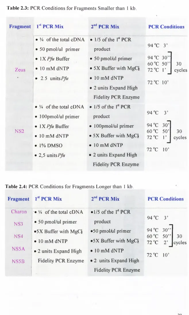
DNA Polymerase (Catalog# 600153, Stratagene) in the first round in order to decrease the mutation rate and in the second round Expand High Fidelity PCR System (Catalog# 1732650, Boehringer Mannheim) was used to obtain PCR products with A nucleotide overhangs. Fragments larger than 1 kb were amplified with Expand High Fidelity System in both PCR cycles. Optimized PCR reaction conditions for fragments smaller than 1 kb and larger than 1 kb are given in Table 2.3 and 2.4 respectively. Amplifications o f all fragments were performed as hot start PCR.Table 2.3: PCR Conditions for Fragments Smaller than 1 kb.
Fragment r ‘ PCR M ix 2"''PCR Mix
Zeus
NS2
• 14 o f the total cDNA I · 50 pm ol/ul prim er > I X / > Buffer ; · 10 mM dNTP ! · 2,5 u n i t s / ^ r
• 14 o f the total cDNA • lOOpmol/ul prim er • IX P ^ /B u ffe r • lO m M d N T P • 1% DMSO • 2,5 units/y?/ • 1/5 o f the f PCR product • 50 pmolAil prim er • 5X Buffer with M gCi • 10 mM dNTP
• 2 units Expand High Fidelity PC R Enzyme • 1/5 o f the
f
PCRproduct
• 1 OOpmol/ul prim er • 5X Buffer with M gCi • 10 mM dNTP
• 2 units Expand High Fidelity PCR Enzyme PCR Conditions 94 “C 3 ’ 94 "C 3 0 ” 6 0 “C 5 0 ” 7 2 ^ 1 ’ 72 °C 10’ 30 cycles 94 “C 3 ’ 94 °C 3 0 ’ 6 0 °C 50’ 72 “C r 72 "C 10’ 30 cycles
Table 2.4: PCR Conditions for Fragments Longer than 1 kb. Fragment 1** PCR Mix
Charon ) · 14 o f the total cDN A NS3 ! · 50 pmol/ul prim er
; ^5X Buffer with M gCi NS4
' · 10 m M dNTP
i
NS5A
1
i · 2 units Expand High NS5B i Fidelity PCR Enzyme 2"** PCR Mix • 1/5 o f the f PCR product •50 pmolAil prim er •5X Buffer with MgCi • 10 mM dNTP
• 2 units Expand High Fidelity PCR Enzyme PCR Conditions 94 “C 3 ’ 94 "C 3 0 ” 60 ”C 50” 72
X T
72 T 10’ 30 cycles 292.2.5 Agarose Gel Electrophoresis o f DNA Fragments
DNA fragments were separated by gel electrophoresis using agarose o f 1% w/v in IX TAE or TBE buffer (Maniatis
et al.,
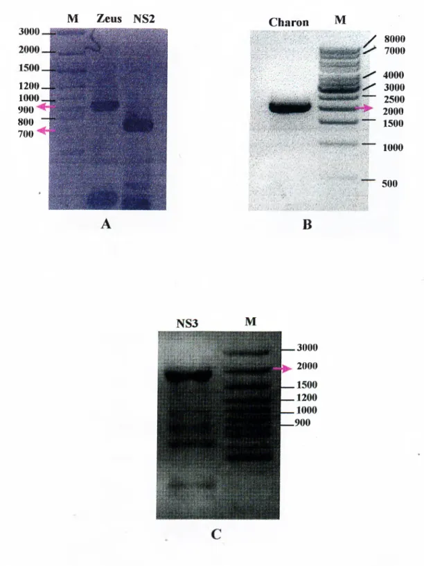
1982). 0.2-0.4 ug EtBr/ml o f gel was added to visualize the DNA bands. DNA samples were loaded into the wells with IX loading buffer. Electrophoresis was performed by running the gel for 1 hour at 100 V. The DNA bands were visualized by illuminating with UV light at 302nm and the gels were photographed.The size and concentration o f the PCR fragments were checked in the agarose gel. Bands with the correct size were isolated from the agarose gel slices by using QIAquick Gel Extraction Kit (Catalog#: 28106, Qiagen Inc.), according to the manufacturers instructions.
2.2.6 Electrophoresis Markers
The length o f DNA fragments were estimated by comparing them to known molecular weight standards, which had been run on the same gel. DNA markers used in this study were 1 kb DNA Ladder (Catalog# SM0311, MB I Fermentas) and 100 bp DNA Ladder Plus (Catalog# SM0321S, M Bl Fermentas. Sizes o f the fragments are given in Figure 2.1.
A
B
Figure 2.1: DNA size markers. Panel A; 100 bp DNA Ladder Plus and Panel B; Ikb DNA Ladder.
Primers for PCR reactions do not contain any linker sites. In order to clone the amplified fragments, pGEM®-T Easy Vector System (Cataloge
#
TM042, Promega) was used. The vector map is given in Figure 2.2. pGEM®-T Easy Vector is a linearized vector with T overhangs in the multiple cloning site.BsiZl,
/¿■coRJ,Notl
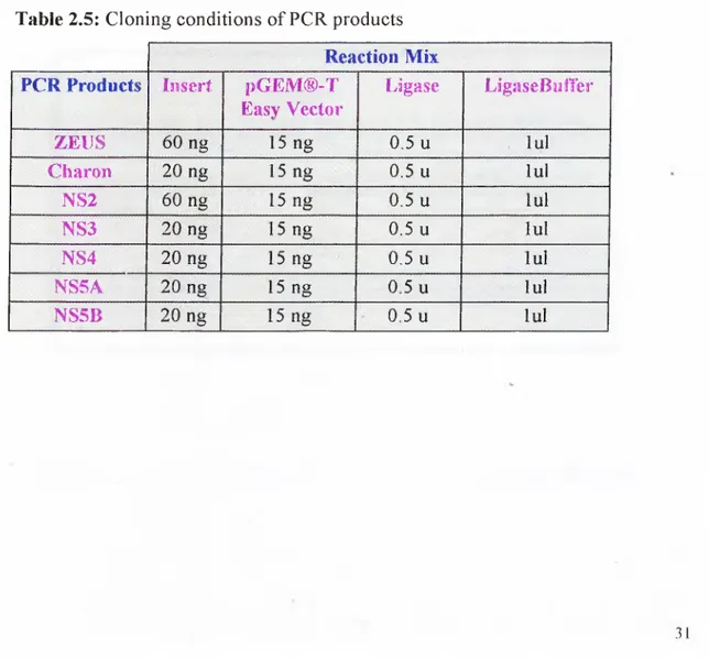
enzyme sites are present at the two sides o f the cloning site and they are used to check the presence o f an insert with a single restriction enzyme reaction. T7 and M13 primer sequences, that are present at the two ends o f the cloning site, were used for sequencing o f the insert. Multiple cloning site o f the vector is in the middle o f the LacZ gene. Presence o f an insert interrupts gene expression and colonies containing insert appears white whereas colonies containing religated vectors appear as blue, in the presence o f X-Gal and IPTG. Cloning reactions were set up in 10 ul total volume and optimized conditions are given in Table 2.5.2.2.7 Cloning ofP C R Products
Table 2.5: Cloning conditions o f PCR products Reaction Mix PCR Products In sert , i)G EM ® -T
Easy Vector L ig ase . LigaseBulTer • Z E llSv 60 ng 15 ng 0.5 u lul C h aro n 20 ng 15 ng 0.5 u lul NS2 60 ng 15 ng 0.5 u lul : 'NS3 ^ 20 ng 15 ng 0 .5 u lu l : NS4 ; 20 ng 15 ng 0.5 u lul n s s a; 20 ng 15 ng 0 5 u lul NS5B 20 ng 15 ng 0.5 u lu l .31
■| üiiïsri; 54
m
26 37 43 43 49 52 ^4 70 ;?? 77 QSm
97 1G5 119 127 141 T7 TîüTtîiCiiptor· SlürlS '.,. t r í í M ÍA G Í^ DÏCAC lAlAei if3r?,î>3A A f i 0 a i^ÛCCü flC01O <5CAÎ<3 CÎCCC (32^CC3CCAlCj 3 ',..A C A ,n ATÔOr «SAOf« ATATO CCiSOT Î.AACC C0<3«30 TÔOAO CGîAC 0C<300 S^ÔTAC
--- ...1 . . , . . . . ™ ! ™ ™ ,/1.1! ;l iíW¿l
T7 Pionieiti'?
Sohl
AVn»IiK ï3Ô C c o œ o < 3A A H Ç Û A V m , . _ , A AJCAO W3Î<> M i ?C ec<i<?cC<Î^CÏ 0CAÇC? TCiMO
CGCGG GCGCG C7ÎAAGCfA r - ^ ^ ^ V s ' mí T G
..I |¿actj ¿c<í^ i
ATGAC TTAAG COOCO GCGGACGTCC AGCTG
....JL.■...JЦ i L _ j v^i Éi?0Hl > i»teM Sch'l
SF6 Tfífjníe’ipjlon S“/3i5
CATAJ QQGAGfiGCT CCGAACGCGT IGGAf OCATAGGTTQ A G W TCTAT AGTGTCACCT AAAT . . . 3' GTATA CCCT CTCGA GGGT7 GCGCA ACCTA COTAT CGAAC TCAÎA AGATA TCACA G^GGA TITA. . . 5'
i .Î I ,1 ± I
iX'ch I Í5CI
ikJtX'i
.»‘♦irrS STÇ rjcrnctli^fB
Transformation o f plasmid DNA into
E.coli
was achieved by using calcium chloride method. The following procedure is based on Ausubelei al.
(1991).Preparation o f Competent Cells: 200 ul o f HBlO l overnight grown culture was inoculated into 5 ml LB medium and cells were grown at 37°C, shaking at 200 rpm to an optical density at 590 nm (OD390) o f 0.4. 1.5 ml o f growing cells were centrifuged at 13,000 rpm for 1 min and gently resuspended in 500 ul ice-cold 50 mM CaCl2 and incubated on ice for 30 min.
Transformation: Competent cells were centrifuged at 13.000 rpm for 1 min and pellets were suspended with 500 ul ice-cold CaCb. All o f the cloning mix (10 ul) was mixed with the competent cells and incubated on ice for 30 min. The competent cells were heat shocked at 42°C for 90 seconds and then incubated on ice for 2 minutes. 1 ml o f LB medium was added onto the competent cells and incubated at 39°C water bath for 1 hour to allow the expression o f antibiotic resistance gene before plating onto the selective LB-Agar plates. After incubation cells were centrifuged for 2 minutes and pellet was resuspended in 100 ul left supernatant and plated onto LB-Agar plates containing lOOug/ml ampicillin, 50mg/ml X-Gal, and lOOmM IPTG. Plates were incubated at 37^C overnight for the selection o f antibiotic resistant transformants.
2.2.8 Transformation ofE.C oli
2.2.9 Small Scale Preparation o f Plasmid DNA (Mini-Preperation)
The method is based on alkaline lysis method o f the Birnboim and Doly (1979). Colonies that appear as white after overnight incubation at 37°C were selected and inoculated into 5 ml LB media containing ampicillin. After overnight incubation at the 37°C shaker, 1.5 ml o f the bacterial culture was centrifuged at 13,000 rpm for 1 minute. After discarding the supernatant, cells were resuspended in 0.1 ml ice cold solution I and 0.2 ml freshly prepared solution 11, 0.5 ml solution 111 and 0.5 ml phenol/chloroform were added. The tube was shacked vigorously by vortexing 2 minutes and centrifuged 5 minutes at 13,000 rpm. Upper phase that
contains the plasmid DNA was transferred into a clean tube and 1 ml ice cold absolute ethanol was added. The tube was centrifuged for 10 minutes at 13,000 rpm and supernatant was discarded. The pellet was washed with ice cold 70% ethanol, left at room temperature for 15-20 minutes to dry and resuspended in 20 ul ddH20 containing 10 ug/ml RNase A. Samples were kept at -20°C for long term storage.
2.2.10 Restriction Enzyme Digestion o f DNA
•
Presence o f the correct size inserts in the mini-preps o f selected colonies, were checked with restriction enzyme digestions. Restriction enzyme digestions o f DNA were carried out in a total volume o f 10 ul with 5-10 units restriction enzyme with IX recommended buffer. Digestion reactions o f mini-preps were incubated for 1.5 hours at 37°C.2.2.11 Medium and Maximum Scale Isolation (Midi-Preperation)
Cells were grown in 100 ml ampicillin containing Lb for overnight to achieve complete saturation. Plasmid DNA was isolated, by using QIAfilter plasmid midi kit (Cataloged 10 12145, Qiagen inc.), according to the manufacturer’s instructions. The DNA samples were stored at -20°C.
2.2.12 Quantification o f DNA
Concentrations and purity o f nucleic acids were determined by measuring absorbency at 260nm and 280nih in a spectrophotometer (Beckmann Instruments Inc., CA, USA). Nucleic acid samples displaying OD260 and OD280 values in the range o f 1.8 to 2.0 are regarded as highly pure. A value o f OD26o=1.0 corresponds to a concentration o f approximately 50 ug/ul double stranded DNA (Maniatis
el
al.,
1982).The automated sequencing o f the inserts were carried on an ABI Prism 310 Sequencer at Bilkent University, Department o f Molecular Biology and Genetics. To increase the sequencing length. Big Dye'*^' Terminator Cycle Sequencing Ready Rection Kit (Catalog# 4363149, ABI Prism) in combination with long capillary o f the 310 Sequencer were used. First sequencing reactions were carried out with M13 and T7 universal primers that are present at the two ends o f the cloning site, on the vector sequence. For the middle sequences internal primprs were designed and synthesized according to the outer sequencing data obtained from M13 and T7 reactions.
2.2