Corresponding author: Murat AYAZ E-mail: [email protected]
Original Investigation
Published Online: 05.06.2017Murat AYAZ
1, Hakan KARABAGLI
2, Sirma Basak YANARDAG
11Selcuk University, School of Medicine, Department of Biophysics, Konya, Turkey 2Selcuk University, School of Medicine, Department of Neurosurgery, Konya, Turkey
Can Hypo/Hypernatremic Conditions be a Factor for
Na Ion Channel Kinetics: Model Study
ABSTRACT
adequate to open the Na+ channel, the AP is generated and transferred throughout the axon length without any change in the configuration. However if the graded potential is not sufficient to open a Na+ channel, no AP is observed (29). Na+ channels have three conformations in their mode of action which are open, inactive and closed. These modes not only determine the formation of the AP, but also determine the frequency response of the AP under continuous stimulation (16).
There are at least two issues that are important in the formation of the current through the ion channel. The first one is the internal properties of the channel (conductivity parameters) and the second one is the ionic gradient across the ion █
INTRODUCTION
A
ction potential (AP), a rapid rise and fall in the electrical membrane potential of a neuron, which is commonly called “firing”, is the basis of signal transmission through a single neuron. An adequate amount of neurotransmitter concentration in the synaptic space creates a series of reactions resulting in rapid activation of sodium (Na) channels, which is called the zero phase (depolarization phase). Na+ currents (INa) -for all of the excitable tissue types- can be named universal depolarizing currents whereas the K+ currents (IK) are repolarizing currents (6).The all or none characteristic of the AP actually comes from the Na+ channel’s properties. When the graded potential is
AIM: Dysnatremic cases are frequently faced in clinical practice. Its macroscopic effects and consequences are well known, but microscopic effects are not well defined. The aim of this study was to reveal the effects of dysnatremia at the cellular level. MATERIAL and METHODS: By using an action potential simulation, the effects of extracellular sodium (Na) concentration on the Na ion channel kinetics were studied. The experimental sets were chosen to mimic hypo/hypernatremic conditions and, in both cases, the degree of the severity was varied.
RESULTS: Hyponatremic situations through modifying the axonal Na+ channels kinetics result in the rundown of the sodium current (INa). The degree of the hyponatremia-dependent effect seen in the Na ion channel is severity dependent, which is more effective in the recovery phase of the ion channel. Hypernatremic conditions, on the other hand, have also affected the Na ion channel activity through modifying the kinetics of the channel. Unlike hyponatremia, the effect seen in hypernatremic conditions was through decreasing the response time of the channel. The degree of the significance of the effect seen on the Na ion channel in the case of the hypernatremia was found to be less destructive compared to the hyponatremic condition.
CONCLUSION: The Na channels are susceptible to the changes of the extracellular Na concentrations. Thus, the underestimation of hypo/hypernatremic conditions can put patients in danger and close monitoring of serum Na level might be required.
channel, i.e. the difference between the ionic concentration in and outside of the membrane.
The disturbance of water-electrolyte balance may be an implication of disorders in different systems, which causes dramatic changes of the Na level in blood plasma, as known as “dysnatremia” (7). If the Na level is reduced below 135 mM it is called hyponatremia. Hypernatremia, on the other hand, is defined a serum Na concentration above 145 mM. Both of them are commonly seen in hospitalized, postoperative and elderly patients (10).
Software simulating biological systems have been frequently utilized especially when direct recordings from the tissue are tricky due to technical difficulties. Such simulated models not only helped understanding the physiological nature, but they also serve as powerful tools during problem solving. Indeed, simulations provide new research politics, parameters and new working conditions on the working systems. It would not have been possible to test some of these new conditions on living systems.
The macroscopic effects of dysnatremia have been previously reported (12,13,22,28) but its microscopic effects are not well defined. In an excitable cell, like a neuron, Na ion currents are important for the induction of the AP. Effects of extracellular [Na+] can be predictable when there is only a single Na channel on the axonal membrane. However, it is important to observe the changes in the presence of the other ion channels. We tested the effects of extracellular Na+ concentration (hypo/hypernatremia) on the axonal Na+ channel kinetics in the presence of the other ion channels with the help of a modified axonal AP simulation program (3).
█
MATERIAL and METHODS
All of the simulation results were obtained from the modified non-commercial LabAXON 5.2 software program (http:// labaxon.software.informer.com/5.2/). Under the physiological conditions (in terms of ion concentration extracellular space mM: Na= 136-143, K= 4; intracellular space mM: Na= 10, K=120) of the simulation, neuronal Na+ currents and their kinetics were saved on the hard disk of a computer. All recorded data were used for the further analysis of the parameters. The states of the Na ion channels used for the simulation were the same that we used previously (3). Briefly, 3 closed, 1 open and 4 inactive states were used for the mode of action. In this Markov representation, alpha (α) and beta (β) parameters represents forward and reverse reaction rates respectively. At equilibrium, activations were calculated by m= αm / (αm + βm), whereas the time constants (Tau) were found by Taum= 1/( αm + βm). It is known that, Na channels have activation (m) and inactivation (h) parameters. Similar to the activation calculations, equilibrium inactivation was calculated by h= αh / (αh + βh), and time constant of inactivation was calculated by Tauh= 1/( αh + βh). Na channel currents and the reaction rate constants of the cell membrane at any moment, as follows,
INa = gNa m3 h (E-ENa)
.
.
e
E
e
e
e
1
0 1
35
4
0 007
1
1
. . m E m E h E h E 0 1 35 1860 2060 0 1 30)
)
)
a
b
a
b
=
--
+
=
=
=
+
) ) - + - + - + - + ^ ^ ^ h h hwere calculated in accordance with the literature where g is the conductivity, m is the activation, h is the inactivation, E is the voltage across the membrane, and ENa is the Nernst potential (14-17).
Na currents were recorded by a standard voltage clamp protocol. Briefly, from a holding potential of -100 mV for 5 ms, depolarizing pulses of 10 ms starting from -80 mV to +70 mV with 10 mV increments were applied.
INa activation characteristic was determined by applying 5 ms prepulse within the range −80 to +70 mV in 10 mV increments that were followed by a 0 mV, 10 ms pulse. In each tested potential, maximum current values were measured and then these measured values were fitted by Boltzmann function (I/Imax = Bottom+(Top-Bottom)/(1+exp((V50-X)/Slope))) for the parameters. Its inactivation was established by applying a 10 ms prepulse within the range −80 mV to -10 mV in 10 mV increments that was followed by a +0 mV pulse 5 ms long. Maximum currents were obtained from the protocol and fitted by another Boltzmann function (I = Bottom+(Top-Bottom)/(1+exp((X-V50)/Slope))) for the parameters. For the reactivations, two pulses (0 mV, 5 ms) with increasing interval durations (5–50 ms) were used. Obtained currents were again fitted by an exponential function (I = I0 + (Top-Bottom)*(1-exp(-Tau*x)).
In order to test the effects of extracellular Na+ concentrations ([Na+]o) on the Na ion channel kinetics, three set of [Na+]o were organized. The first set was named hyponatremic conditions and divided into mild ([Na+]o=131 mM), moderate ([Na+] o=121 mM) and severe ([Na+]o=110 mM). The second set was named hypernatremic conditions and divided into mild ([Na+] o=147 mM), moderate ([Na+]o=155 mM) and severe ([Na+] o=165 mM) (21,26,27). These two sets results were compared with the third set named the physiological range (PR) and assigned as the average of [Na+]o=136-143 mM.
The average values of the parameters were obtained from the physiological ranges used for the comparison of the data sets. Values were expressed as mean ± standard error of mean (s.e.m).
█
RESULTS
Capacitance measurement is a frequently used technique in electrophysiological measurements and/or calculations as a measure of the cell size (4,5). In our simulations, a sub-threshold square-wave (50 µs and 70 mV) pulse is applied to the membrane and capacitive currents are elicited. By integrating these current traces, the total charge amount can be obtained. The capacitance of the cell membrane is then calculated by dividing the total charge amount by the
difference (voltage) in the applied potential (Cm=Total Q/∆V). For our simulations, the axon diameter was assumed as 542 µm and the capacitance of this diameter was 0.010 µF.
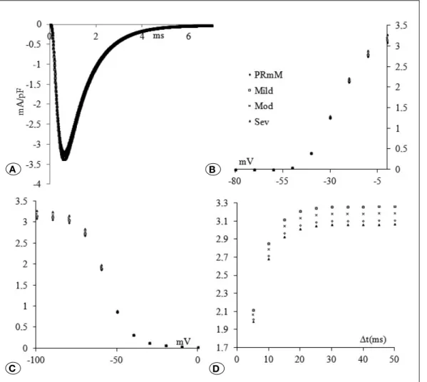
Effects of Hyponatremia
Effects of hyponatremic conditions on the Na+ channel kinetics are shown in Figure 1. Original current traces of +10 mV voltage clamped (from a holding potential of -100 mV) were given in accordance with the degree of the severity of the hyponatremia (Figure 1A-D). It is obvious that the decrease in [Na+] results in the rundown of the peak value of the current (Mild =-3.84%, Moderate =-7.30%, Severe =-11.48%) (Figure 1A). In panel B, activation characteristics of the currents for the different hyponatremic conditions are given (Figure 1A-D). Amongst tested potential differences, significance was found between the -20 mV – 0 mV when compared to physiological range values. The measured activation curve parameters following the fitting procedure (R2=0.99) are summarized in Table I. In parallel to the degree of significance of the hyponatremia, it was seen that activation curves start from more negative values and V50 values shift to more negative values together with a decrease in the steepness.
Inactivation results are summarized in Figure 1C. In a parallel manner to activation results, potential differences between –100 mV and -60 mV resembled a significant depression compared to the measured physiological values. The results of fitting parameters of the inactivation are given in Table I (R2=0.99). Except for the steepness of the fitted curves, none of the parameters were affected from the hyponatremia. Decrease in the [Na+]o leads to faster inactivation in the Na+ channels.
Results of the recovery from inactivation are summarized in Figure 1D. In a severity dependent manner, reactivation values were found to be significantly decreased. Results obtained from exponential fitting of reactivation values are given in Table I (R2=1.00). All the measured parameters were affected. The maximum point of the reactivation values were found to be depressed, together with a slowing down in the kinetics (time constant=tau).
Effects of Hypernatremia
Effects of mild, moderate and severe hypernatremic conditions on the Na+ channel currents are presented (Figure 2A). Peak values for these measured currents increase according to the degree of severity (Mild=1.20%, Moderate=3.52%,
Figure 1: Na ion channel kinetics under hyponatremic conditions. In the figure, the compared experimental sets are physiological range values (PR), mild (([Na+] o=131 mM), moderate
(mod) ([Na+]
o =121 mM)
and severe (sev) ([Na+] o =110 mM). A shows
the sample traces of Na currents of these experimental sets. B, C and D show activation, inactivation and reactivation profiles of the currents based on the records that were detailed in the methods section. Values are expressed as mA/pF in activation, inactivation and reactivation.
C D
reactivation values were found to be significantly increased. Results obtained from exponential fitting of reactivation values are given in Table II (R2=1.00). Among the measured parameters, no change was found at the starting point of the reactivation. However, the maximum values were found to be increased with faster kinetics only in severe case of the hypernatremia (time constant=tau).
█
DISCUSSION
The human body obtains Na through nutrition and loses it primarily in sweat and urine. In principle, the kidneys maintain a reliable level of Na in the body by adjusting the amount excreted in the urine. Na+ is mostly located in the circulating blood and in the fluid around cells. Besides, the regulation of the osmotic pressure keeping the fluids in a normal balance also plays a key role in normal nerve and muscle conduction facilities (18,23).
Small changes in plasma Na level may be seen in the metabolism. If the change is dramatic and persistent, its underestimation affects the prognosis, and causes coma and even death (1,9). It is reported that hyponatremia may affect the prognosis of some pathologies such as renal cell carcinoma, liver cirrhosis, congestive heart failure, head trauma and subarachnoid hemorrhage (8,11,19,28).
Severe=6.26%)(Figure 2A). Also, activation characteristics of the currents for the different hyponatremic conditions are demonstrated in Figure 2B. Amongst the tested potentials, a significant difference was found only at the 0 mV compared to the physiological range values. The results of the fitted parameters are given in Table II (R2=0.99). Although only the 0 mV value was significantly affected from the hypernatremia, our fitting results indicate a significant difference in all of the measured parameters. Matching the degree of significance of the hyponatremia, activation curves starts from more negative values and V50 values shift to more positive potentials together with the increase in the steepness.
Inactivation results, on the other hand, are summarized in Figure 2C. Similar to activation results, potential differences between –100 mV and -80 mV indicate a significant increase compared to the measured physiological values. The results of fitting parameters of the inactivation are given in Table II (R2=0.99). Except for the steepness of the fitted curves, none of the parameters were affected from the hyponatremia. Increase in [Na+]o results in slower inactivation for the mild and moderate states but faster for severe cases.
Additionally, results obtained from the reactivation protocol are summarized in Figure 2D. In a severity-dependent manner,
Figure 2: Na ion channel kinetics under hypernatremic conditions. In the figure, the compared experimental sets are physiological range values (PR), mild (([Na+]
o=147 mM),
moderate (mod) ([Na+] o =155
mM) and severe (sev) ([Na+] o
=165 mM).
A shows the sample traces of Na currents of these experimental sets.
B, C and D show activation, inactivation and reactivation profiles of the currents based on the records that were detailed in the methods section. Values are expressed as mA/pF in activation, inactivation and reactivation.
C D
Table I: Fitting Parameters for Activation, Inactivation and Reactivations of Ina under Hyponatremic Conditions. The Compared Experimental Sets in the Table are Physiological Range Values (PR), Mild (([Na+]
o=131 mM), Moderate (mod) ([Na+]o =121 mM) and Severe
(sev) ([Na+] o =110 mM) Activation Bottom V50 Slope R2 PR -0.043±0.000 -26.061±0.016 8.009±0.008 0.999 Mild -0.04047* -26.25 7.925* 0.999 Moderate -0.03773* -26.48* 7.812* 0.999 Severe -0.03456* -26.79* 7.674* 0.999 Inactivation Bottom V50 Slope R2 PR 1.525±0.003 -57.123±0.001 7.090±0.001 0.999 Mild 1.519* -57.122 7.092* 0.999 Moderate 1.524* -57.120* 7.090* 0.999 Severe 1.534* -57.120* 7.089* 0.999 Reactivation
Bottom Top Tau R2
PR 0.035±0.001 3.079±0.006 4.825±0.001 1
Mild 0.034* 2.995* 4.828* 1
Moderate 0.028* 2.902* 4.822* 1
Severe 0.033* 2.791* 4.821* 1
Table II: Fitting Parameters for Activation, Inactivation and Reactivations of Ina under Hypernatremic Conditions. The Compared Experimental Sets in the Table are Physiological Range Values (PR), mild (([Na+]
o=147 mM), Moderate (mod) ([Na+]o =155 mM) and Severe
(sev) ([Na+] o =165 mM) Activation Bottom V50 Slope R2 PR -0.043±0.000 -26.061±0.016 8.009±0.008 0.999 Mild -0.044* -25.91 8.076* 0.999 Moderate -0.046* -25.76* 8.147* 0.999 Severe -0.048* -25.60* 8.217* 0.999 Inactivation Bottom V50 Slope R2 PR 1.525±0.003 -57.123±0.001 7.090±0.001 0.999 Mild 1.522 -57.12 7.093* 0.999 Moderate 1.530 -57.12 7.087* 0.999 Severe 1.523 -57.12 7.091* 0.999 Reactivation
Bottom Top Tau R2
PR 0.032±0.001 3.069±0.006 4.825±0.001 1
Mild 0.032 3.130* 4.825 1
Moderate 0.034 3.192* 4.824 1
█
REFERENCES
1. Aleksandrowicz M, Dworakowska B, Dolowy K, Kozniewska E: Restoration of the response of the middle cerebral artery of the rat to acidosis in hyposmotic hyponatremia by the opener of large-conductance calcium sensitive potassium channels (BKCa). J Cereb Blood Flow Metab 37(9):3219-3230,2017 2. Arányi Z, Kovács T, Szirmai I, Vastagh I: Reversible nerve
conduction slowing in hyponatremia. J Neurol 251(12): 1532-1533, 2004
3. Ayaz E, Yanardag SB, Karabagli H, Ayaz M: Effects of the experiment conditions on the nerve action potential: A model study. Journal of Advanced Neuroscience Research 3(2): 61-69, 2016
4. Ayaz M, Ozdemir S, Ugur M, Vassort G, Turan B: Effects of selenium on altered mechanical and electrical cardiac activities of diabetic rat. Arch Biochem Biophys 426(1): 83-90, 2004
5. Ayaz M, Ozdemir S, Yaras N, Vassort G, Turan B: Selenium-induced alterations in ionic currents of rat cardiomyocytes. Biochem Biophys Res Commun 327(1): 163-173, 2005 6. Ayaz M: Biyofizik Sırları Çözülmüş. Konya: Nobel Tıp
Kitapevleri, 2013 (In Turkish)
7. Bataille S, Baralla C, Torro D, Buffat C, Berland Y, Alazia M, Loundou A, Michelet P, Vacher-Coponat H: Undercorrection of hypernatremia is frequent and associated with mortality. BMC Nephrol 15: 37-45, 2014
8. Biswas M, Davies JS: Hyponatraemia in clinical practice. Postgrad Med J 83: 373–378, 2007
9. Cairns SP, Buller SJ, Loiselle DS, Renaud JM: Changes of action potentials and force at lowered [Na ]o in mouse skeletal muscle: Implications for fatigue. Am J Physiol Cell Physiol 285(5): C1131-1141, 2003
10. Darmon M, Pichon M, Schwebel C, Ruckly S, Adrie C, Haouache H, Azoulay E, Bouadma L, Clec’h C, Garrouste-Orgeas M, Souweine B, Goldgran-Toledano D, Khallel H, Argaud L, Dumenil AS, Jamali S, Allaouchiche B, Zeni F, Timsit JF: Influence of early dysnatremia correction on survival of critically ill patients. Shock 41(5): 394-399, 2014
11. Dooling E, Winkelman C: Hyponatremia in the patient with subarachnoid hemorrhage. J Neurosci Nurs 36(3): 130-135, 2004
12. Fofi L, Dall’armi V, Durastanti L, Valenza A, Lorenzano S, Prencipe M, Toni D: An observational study on electrolyte disorders in the acute phase of ischemic stroke and their prognostic value. J Clin Neurosci 19(4): 513-516, 2012 13. Furukawa J, Miyake H, Kusuda Y, Fujisawa M: Hyponatremia
as a powerful prognostic predictor for Japanese patients with clear cell renal cell carcinoma treated with a tyrosine kinase inhibitor. Int J Clin Oncol 20(2): 351-357, 2015
14. Goldman DE: Potential, impedance and rectification in membranes. J Gen Physiol 2:37-49, 1943
15. Hodgkin AL, Horowicz P: The influence of K and Cl ions on the potential of single muscle fibres. J Physiol 148: 127-160, 1959
The kidney is usually involved in dysnatremic conditions. However, the most vulnerable organ is the brain due to its adaptation mechanisms to hypo/hypernatremic conditions. These may cause cerebral edema and increased intracranial pressure or loss of organic osmolytes of brain content and disruption of the tight junctions (20). The effect of dysnatremia on the peripheral nervous system has not been studied extensively (2,24). It is reported that hyponatremia results in a decrease in the nerve conduction, which can be explained by the lower electrochemical gradient due to reduced extracellular Na concentration. Although the correction of dysnatremia is very important, rapid correction causes central pontine and extrapontine myelinolysis and necrosis in several brain regions. The higher the speed of correction, the greater the damage (20).
Many of the clinical studies dealing with the hyponatremic conditions have reported decreased conduction velocities that are location-independent for the motor and sensorial nerves (2,25). Our previous and the new results have shown that hyponatremic situations, through modifying the axonal Na+ channels kinetics, result in the rundown of the INa. Once the current density is decreased, it also reflects itself in the measured compound action potentials -via affecting the single fiber action potential- in human conduction velocity measurements. Indeed, the degree of the hyponatremia-dependent effect seen in the in the sodium ion channel, is severity-dependent, being more effective in the recovery phase of the ion channel.
Hypernatremic conditions, on the other hand, have also affected the Na ion channel activity through modifying the kinetics of the channel. Unlike hyponatremia, the effect seen in the hypernatremia condition was through decreasing the response time (increased excitability) of the channel. However, the degree of the significance of the effect seen on the Na ion channel in the case of the hypernatremia was found to be less destructive compared to the hyponatremic condition. For each of the cases for the Na ion concentration effects, further investigations seem to be needed for better treatment strategies.
█
CONCLUSIONS
Na ion channel activity is much more susceptible to the changes in the extracellular Na ion concentrations in either way. Susceptibility of the Na channel kinetics -being much more pronounced for the hyponatremic state- makes hyponatremic conditions more dangerous for the patients. Although how the low extracellular Na ion concentration modifies the channel protein molecular biology needs further investigations, we highly recommend close monitoring of the serum Na level following surgery especially in intensive care units.
23. Schrier RW: Renal and Electrolyte Disorders. USA: Lippincott Williams & Wilkins, 2010
24. Serrano-Castro PJ, Alonso-Verdegay G, López-Martínez G, Arjona-Padillo A, Callejón JR, Olmedo VM, Guardado-Santervás P, Huete-Hurtado A, Olivares-Romero J, Fernández CN: Possible case of peripheral osmotic demyelination syndrome. J Neurol Neurosurg Psychiatry 79: 331–332, 2008 25. Stocker SD, Lang SM, Simmonds SS, Wenner MM, Farquhar
WB: Cerebrospinal fluid hypernatremia elevates sympathetic nerve activity and blood pressure via the rostral ventrolateral medulla. Hypertension 66(6): 1184-1190, 2015
26. Vandergheynst F, Gombeir Y, Bellante F, Perrotta G, Remiche G, Mélot C, Mavroudakis N, Decaux G: Impact of hyponatremia on nerve conduction and muscle strength. Eur J Clin Invest 46(4): 328-333, 2016
27. Waikar SS, Mount DB, Curhan GC: Mortality after hospitaliza-tion with mild, moderate, and severe hyponatremia. Am J Med 122(9): 857-865, 2009
28. Wunsch E, Naprawa G, Koziarska D, Milkiewicz M, Nowacki P, Milkiewicz P: Serum natremia affects health-related quality of life in patients with liver cirrhosis: A prospective, single centre study. Ann Hepatol 12(3): 448-455, 2013
29. Yanardag SB, Ayaz M: Naive attempt of quantum mechanics: Axonal membrane. Journal of Advanced Neuroscience Research 3(1): 19-23, 2016
16. Hodgkin AL, Huxley AF: A quantitative description of membrane current and its application to conduction and excitation in nerve. J Physiol 117(4): 500-544, 1952
17. Hodgkin AL, Katz B: The effect of sodium ions on the electrical activity of the giant axon of the squid. J Physiol 108:37-77, 1949
18. Humes HD: Kelley’s Essentials of Internal Medicine. USA: Lippincott Williams & Wilkins, 2001
19. Kawashima A, Tsujimura A, Takayama H, Arai Y, Nin M, Tanigawa G, Uemura M, Nakai Y, Nishimura K, Nonomura N; Osaka Renal Cell Carcinoma Clinical Study Collaboration: Impact of hyponatremia on survival of patients with metastatic renal cell carcinoma treated with molecular targeted therapy. Int J Urol 19(12): 1050-1057, 2012
20. Massieu L, Montiel T, Robles G, Quesada O: Brain amino acids during hyponatremia in vivo: Clinical observations and experimental studies. Neurochem Res 29(1): 73-81, 2004 21. Michal O, Magdalena MZ, Halina M, Magdalena BZ, Tadeusz
N, Elzbieta M, Marcin W: Hyponatremia effect in patients with alcohol dependence on their physical and mental health status. Alcohol 57: 49-53, 2016
22. Roquilly A, Mahe PJ, Latte DD, Loutrel O, Champin P, Di Falco C, Courbe A, Buffenoir K, Hamel O, Lejus C, Sebille V, Asehnoune K: Continuous controlled-infusion of hypertonic saline solution in traumatic brain-injured patients: A 9-year retrospective study. Crit Care 15(5): R260-268, 2011
![Figure 1: Na ion channel kinetics under hyponatremic conditions. In the figure, the compared experimental sets are physiological range values (PR), mild (([Na + ]](https://thumb-eu.123doks.com/thumbv2/9libnet/4899131.98038/3.914.86.796.545.1098/figure-channel-kinetics-hyponatremic-conditions-compared-experimental-physiological.webp)