Ankara Üniv Vet Fak Derg, 58, 229-232, 2011
Foraminal index on the dog femora
Mehmet Erkut KARA1, Figen SEVIL-KILIMCI1, Vedat ONAR2
1 Adnan Menderes University, Faculty of Veterinary Medicine, Department of Anatomy, Aydin; 2 Istanbul University, Faculty of
Veterinary Medicine, Department of Anatomy, Istanbul/Turkey.
Summary: Damages of the main nutrient artery during the fractures may lead to poor repair or bone infarcts. Therefore, knowledge about the localization and number of nutrient foramina of bones is important in some surgical procedures such as bone implants, fracture repair, bone grafts. Foraminal index is calculated as dividing the distance between proximal end of the bone and nutrient foramen to the total bone length. One hundred and twelve femora of the 56 mature dogs were used in this study. The general morphometric parameters of femur; the total length, the cranio-caudal and medio-lateral diameters of mid-femur were taken. The numbers of the primary and secondary nutrient foramina were determined and the foraminal index was calculated. One foramen nutricium was determined at the 88 bones while two foramen at the 24 bones. The mean foraminal index was 36.4%, between ranges of 31.8% - 55.1% in dog femora. Statistical difference of foraminal index was not determined between male and female dog femora. These data may be valuable for surgeons in cases of total hip arthroplasty, intramedullary pin, and external fixation and also on evaluating period of fracture repair after surgery.
Key words: Dog, femur, foraminal index
Köpek Femur’unda Foraminal İndeks
Özet: Kırık oluşumu veya tedavisi sırasında nutrient arterde oluşabilecek hasarlar, kemiğin zor iyileşmesi veya beslenme yetersizliğine bağlı nekroz oluşumuna yol açabilir. Bu nedenle uzun kemiklerde foramen nutricium’un yeri ve sayısının bilinmesi kemiğe çeşitli implantların uygulanması, kırık iyileşmesi, graft uygulanması gibi çeşitli ortopedik uygulamalar açısından önemlidir. Foraminal indeks; foramen nutricum’un kemiğin proximal ucuna olan mesafesinin kemik uzunluğuna oranı olarak ifade edilir. Bu çalışmada çeşitli ırklardan 56 erişkin köpeğe ait 112 adet femur kullanıldı. Çalışılan femur’ların genel morfometrik özellikleri için uzunluk ve çap ölçümleri alındıktan sonra, corpus femoris’in caudal yüzünde bulunan primer ve seconder for. nutricium’ların sayıları belirlendi ve foraminal indeks değerleri hesaplandı. İncelenen kemiklerin 88 tanesinde sadece bir adet foramen nutricium bulunurken, 24 kemikte iki adet delik tespit edildi. Foraminal index en düşük %31.8, en yüksek %55.1, ortalama olarak ise %36.4 olarak hesaplandı. Erkek ve dişi hayvanların femurları arasında indeksin istatistiksel farklılık göstermediği tespit edildi. Elde edilen bu değerler, köpeklerde total kalça protezi, intramedullar pin, plak veya eksternal fikzasyon gibi ortopedik uygulamalar sırasında ve bu tür uygulamalardan sonra kırık iyileşmesi sürecinin değerlendirilmesine ilişkin çalışmalarda faydalı olabilecektir.
Anahtar sözcükler: Femur, foraminal indeks, köpek
Introduction
Long bones have a nutrient artery, which pass through foramen nutricium and canalis nutricius of bone. When it reaches the marrow cavity, the artery is divided into proximal and distal branches and supply the bone marrow and the adjacent cortical bone (4, 5). In the metaphysial region, these branches anastomose with branches of metaphysial and epiphysial arteries that enter the bone toward its extremity. The mid-shaft region of the long bones relies mainly on the nutrient artery, the peripheral part on metaphysial arteries (4). The nutrient artery supplies about 50% of the total blood supply of the entire femur and about 70% of the total blood flow of the femoral shaft. In avoidable damages are resulted from the interruption of blood supply to long bones. For example,
necrosis of the marrow and some areas of the cortex are shown after induction of embolism in the nutrient arterial system (16). The surgeons can meet such situation in trauma or stabilization of fractured bone. Therefore, time course of healing in ceased at lacking of adequate blood supply. Interference with such a supply, which is caused with original trauma or result from technique used to stabilize the fracture, has a deleterious effect on bone healing (17). Therefore knowledge of the localization of foramen nutricium is important in orthopedic procedures such as in normal fracture repair (12) and applications of bone implants (15) or bone grafts (13).
The nutrient artery or foramen nutricium is at the center point of a long bone on fetus (5, 14). Because the distal part of the femur grows faster than proximal part
Mehmet Erkut Kara - Figen Sevil-Kilimci - Vedat Onar 230
the foramen nutricium moves through proximal part at of bones (14). Therefore knowledge of localization of the nutrient foramen can be also used to evaluate the bone growth. In order to describe the localization of the foramen nutricium foraminal index is calculated by dividing the distance between proximal end of the bone and foramen nutricium to the total bone length (2, 6, 9). Additionally, the bone growth shows gender differences (3), the foraminal index between male and female may be different.
A lot of studies related to blood supply of long bones or foramen nutricium in human (1, 2, 6, 9) and animals (8, 14, 16), but there is lack of information in range of the foraminal index with gender differences on dog femur. Therefore, the aim of this study is to provide information about localization of foramen nutricium in male and female dog femora.
Materials and methods
The location and number of foramen nutricium in 112 femora of the 56 mature dogs (34 male and 22 female) were determined in this study. The dogs were medium and large size. The bones were obtained from the collection of the Department of Anatomy, Faculty of Veterinary Medicine, Istanbul and Adnan Menderes Universities. This study was approved by the University Animal Care and Ethics Committee. The skeletally immature and unknown genders of bones were not used. The descriptive morphometric parameters of femur; the total length (between the proximal end of the great trochanter and the distal edges of the condyles), the cranio-caudal and medio-lateral diameters of mid-femur were taken with a digital caliper.
The number and location of foramen nutricium on the caudal surface of femoral shaft were noted for each bone examined. If there was more than one foramen, the largest (primer) foramen nutricium was recorded by calibration of the foramen with different sizes of needles. The foramina at the ends of bones and smaller than 24 gauge needle were excluded. In order to describe the location of main foramen nutricium, the foraminal index (FI) of femur was calculated by using the formula FI= (DNF/TL) x 100, where “DNF” is the distance from the proximal end of the bone to foramen nutricium and “TL” the total bone length (6, 9).
The femoral measurements and foraminal index on male and female dog femurs were compared using by the student’s t-test. Means, standard deviations, ranges and frequency were determined. Foramina number and localization between male and female dogs were compared using by Chi-square test. The statistical analyses were performed using a statistical package program (SPSS 13.0 for Windows). Statistical significance was described at p<0.05 level.
Results
There was only one foramen nutricium at the 78.6% and two foramina at the 21.4% of bones. The localization of the main foramen nutricium was observed on lateral lip of the facies aspera, distal end of the facies aspera, caudo-lateral side of the corpus femoris and medial lip of the facies aspera as 64.3%, 33%, 1.8% and 0.9%, respectively. When it was existed, the secondary foramen nutricium was observed at lateral lip of the facies aspera, caudo-medial side of the corpus femoris, distal end of the facies aspera, medial lip of the facies aspera and caudo-lateral side of the corpus femoris as 54.2%, 20.8%, 16.7%, 4.2% and 4.2%, respectively.
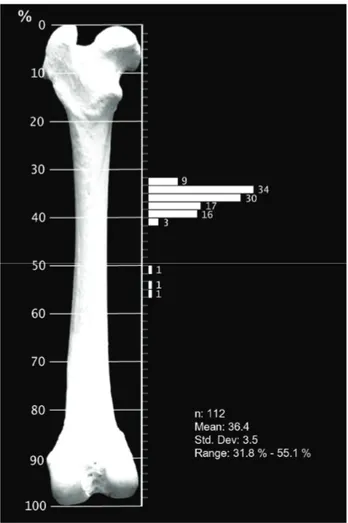
The mean foraminal index was 36.4% and ranges between 31.8% - 55.1% for main foramen nutricium. It is remarkable that the foraminal index were between 31.8% - 40.6% at 109 bones (97.3%) while 50.5%-55.1% at three bones (2.7 %) (Figure 1).
Figure 1. The histogram of the foraminal index on the dog femur.
Şekil 1. Köpek femur’unda foraminal index histogramı
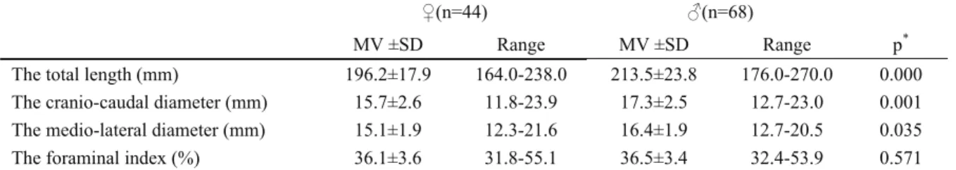
The foraminal index (Table 1), the localization and number of foramen nutricium between male and female dog femora were not different statistically, although there were statistical differences on descriptive morphometric parameters of femora (Table 1).
Ankara Üniv Vet Fak Derg, 58, 2011 231
Discussion and Conclusion
Because the main nutrient artery supplies a large portion of cortex and morrow (16), damages of this artery is especially important for orthopedic procedures. The femur fracture rates are more than double that of other long bones fractures and implant applications are common in dog (12). Therefore accurate knowledge of the location of foramen nutricium on femur may help to prevent intra-operative injures of this artery and orthopedic evaluations. The location of nutrient foramen might be also predisposed to medullary infarction on clinical practice of dog total hip arthroplasty. The depth of reaming and filing may contribute to femoral medullary infarction because of disruption of the nutrient artery (7, 15). When distance between the trochanter major and foramen nutricium greater than 79 mm dogs may predispose to femoral infarction on hip arthroplasty applications (15). In this study, the morphometric data of foramen nutricium was evaluated with foraminal index. It is especially important for canine medicine because there are about 300 breeds with various sizes (5). However there is lack of information about minimal and maximal values of foraminal index on dog femora. Evans (5) state that the largest nutrient foramen to enter the femur is found on the caudal surfaces at approximately the junction of the proximal and middle thirds of the dog femur. On the other hand, Riser (14) measured the length of proximal ends of femur from nutrient foramen on dog femur but they he not calculate foraminal index. According to these values, the foraminal index is calculated as about 41% at 1 year old of Greyhound. In this study the data obtained from femora of medium and large size of dogs. The statistical comparison foraminal index could not be made among dog breeds because of inadequate the number of dog in each breeds. The foraminal index was calculated in the range of 31.8% - 55.1%, but especially the foraminal index was widely seen between 31.8% - 40.6% for main foramen nutricium in dog femora. Therefore, surgeons should take into consideration of this range of foraminal index for some orthopedic applications such as total hip arthroplasty on dog.
On the other hand, morphological variation in the skeleton between males and females are well known (3, 10, 11). This may be resulted of different bone growth ratio between male and female (3). Therefore gender differences of foraminal index expected. In this study, however, there were no statistical differences for the foraminal index, the situation of the main foramen nutricium and foramina numbers between male and female dog femora. It can be expressed that the gender may not important factor for femoral supply on femora of adult dog for orthopedic viewpoints.
In conclusion, the main foramen nutricium was placed between 31.8% - 40.6% of adult dog femora without gender differences but variations can be seen on the location.
Acknowledgement
A part of this study was presented on the VI. National Veterinary Anatomy Congress, Turkey.
References
1. Bridgeman G, Brookes M (1996): Blood supply to the human femoral diaphysis in youth and senescence. J Anat, 188, 611-621.
2. Campos FF, Pellico LG, Alias MG, Valencia RF (1987): A study of the nutrient foramina in human long bones. Surg Radiol Anat, 9, 251-255.
3. Clark EM, Ness AR, Tobias JH, (2007) Gender differences in the ratio between humerus width and length are established prior to puberty. Osteoporos Int, 18, 463– 470.
4. Dyce KM, Sack WO, Wensing CJG (2002): Textbook of Veterinary Anatomy, Saunders, Philadelphia.
5. Evans HE (1993): Miller’s Anatomy of the dog, Saunders, Philadelphia.
6. Gümüsburun E, Yücel F, OzkanY, Akgün Z (1994): A study of the nutrient foramina of lower limb long bones. Surg Radiol Anat, 16, 409-412.
7. Haney DR, Peck JN (2009): Influence of canal preparation depth on the incidence of femoral medullary infarction with Zurich cementless canine total hip arthroplsty. Vet Surg, 38, 673-676.
8. Henderson RG (1978): The position of the nutrient forament in the growing tibia and femur of the rat. J Anat, 125, 593- 599.
Table 1. The femoral measurements and foraminal index on male and femaledog femora. Tablo 1. Erkek ve dişi kopek femur’larında foraminal index ve femur ölçümleri.
♀(n=44) ♂(n=68)
MV ±SD Range MV ±SD Range p*
The total length (mm) 196.2±17.9 164.0-238.0 213.5±23.8 176.0-270.0 0.000 The cranio-caudal diameter (mm) 15.7±2.6 11.8-23.9 17.3±2.5 12.7-23.0 0.001 The medio-lateral diameter (mm) 15.1±1.9 12.3-21.6 16.4±1.9 12.7-20.5 0.035 The foraminal index (%) 36.1±3.6 31.8-55.1 36.5±3.4 32.4-53.9 0.571
Mehmet Erkut Kara - Figen Sevil-Kilimci - Vedat Onar 232
9. Kizilkanat E, Boyan N, Ozsahin ET, Soames R, Oguz O (2007): Location, number and clinical significance of nutrient foramina in human long bones. Ann Anat, 189, 87-95.
10. Mahfouz MR, Merkl BC, Abdel Fatah EE, Booth R, Argenson JN (2007): Automatic methods for characterization of sexual dimorphism of adult femora: distal femur, Computer Methods in Biomechanics and Biomedical Engineering, 10, 447–456
11. Martiniaková M, Omelka R, Grosskopf B, Sirotkin AV and Chrenek P. (2008): Sex-related variation in compact bone microstructure of the femoral diaphysis in juvenile rabbits, Acta Veterinaria Scandinavica, 50, 1-6.
12. Piermattei DL, Flo GL, Decamp CE (2006) Handbook of Small Animal Orthopedıcs and Fracture Repair, Elsevier Inc.St Luis.
13. Ring D, Jupiter JB, Sanders RA, Quıntero J. Santoro VM, Ganz R, Martı RK (1997) Complex nonunıon of fractures of the femoral shaft treated by wave-plate osteosynthesıs. J Bone Joint Surg, 79, 289–94.
14. Riser WH (1975): Growth and development of the normal canine pelvis hip joints and femur from birth to maturity. Vet Pathol, 12, 264- 278.
15. Sebestyen P, Marcellin-Little D J, DeYoung B A (2000): Femoral medullary infarction secondary to canine total hip arthroplasty. Vet Surg, 29, 227-236.
16. Shim S S, Copp DH, Patterson F P (1968): Measurement of the rate and distribution of the nutrient and other arterial blood supply in long bones of the rabbit. J Bone Joint Surg, 50B, 178-183.
17. Sumner-Simith G (1991): Delayed Unions and Nonunions: Diagnosis, Patophysiology, and treatment. Vet Clin North Am Small Anim Pract, 21, 745–759.
Geliş tarihi: 01.07.2010 / Kabul tarihi: 24.03.2011 Address for correspondence:
Doç. Dr. M. Erkut Kara ADÜ Veteriner Fakültesi Anatomi, Anabilim Dalı Işıklı/Aydın, Türkiye e-posta: [email protected]