In vitro Study of Antibacterial Activity on Multi-Resistant
Bacteria and Chemical Composition of the Chloroform
Extract of Endemic Centaurea drabifolia subsp. cappadocica
Aysel Ugura*, Nurdan Saracb*, M. Emin Duruc and Yavuz Beyatlid
a
Departmentof Biology, Faculty of Arts and Sciences, Mugla University, Mugla TR 48121, Turkey
b
Medical Laboratory Program, Vocationary School of Health Care, Mugla University, Mugla TR 48700, Turkey
c
Departmentof Chemistry, Faculty of Arts and Sciences, Mugla University, Mugla TR 48121, Turkey
d
Department of Biology, Faculty of Science and Arts, Gazi University, Ankara TR 06500, Turkey [email protected]
Received: June 29th, 2009; Accepted: August 5th, 2009
The antimicrobial activity of the n-hexane, chloroform, ethyl acetate and ethanol extracts of the aerial parts of C. drabifolia S.M. subsp. cappadocica (DC.) Wagenitz (Asteraceae) was evaluated against microorganisms including multi-antibiotic resistant bacteria using the paper disc diffusion method. The chemical composition of the chloroform extract of this plant was determined by gas chromatography and gas chromatography-mass spectrometry. The chloroform extract exhibited significant antibacterial activity against all the bacteria tested, except Stenotrophomonas maltophilia MU63.The major compounds of the chloroform extract were spathulenol (14.1%), caryophyllene oxide (12.5%), octadecanol (10.2%), ethyl palmitate (7.7%), [Z,Z]-10,12-hexadecadienal (6.0%), 3-hydroxy p-anisaldehyde (5.9%) and pentacosane (5.8%).
Keywords: Centaurea drabifolia ssp. cappadocica, chemical composition, antimicrobial activity.
There is an increasing interest in medicinal plants as an alternative to synthetic drugs, particularly against microbial agents because of the growth of antibiotic resistance [1]. About 20,000 plant species used for medicinal purposes are reported by WHO [2].
Many members of the genus Centaurea have long been used in Anatolian folk medicine [3a-3c]. The aerial parts of the plants are known in Turkey as ‘peygamber cicegi, zerdali dikeni, coban kaldiran, and timur dikeni’ [3a,4]. C. drabifolia subsp.
cappadocica is an endemic species of Turkey. It is native to south Anatolia and the eastern part of central Anatolia, and grows on rocky slopes at altitudes of 1300-1600 m [4]. This species has no popular use described in the academic literature. The aim of this study was to identify the chemical composition and evaluate the antimicrobial activity of extracts of this species against different microorganisms including multi-resistant bacteria.
Ethanol, n-hexane, chloroform and ethyl acetate extracts of C. drabifolia ssp. cappadocica were investigated for their antimicrobial activities. Nine standard test microorganisms (Micrococcus luteus NRRL B-4375, Bacillus subtilis ATCC 6633,
Streptococcus mutans CNCTC 8/77, Staphylococcus
aureus ATCC 25923, Enterobacter aerogenes RSKK 720, Pseudomonas aeruginosa ATCC 27853,
Escherichia coli ATCC 25922, Candida albicans
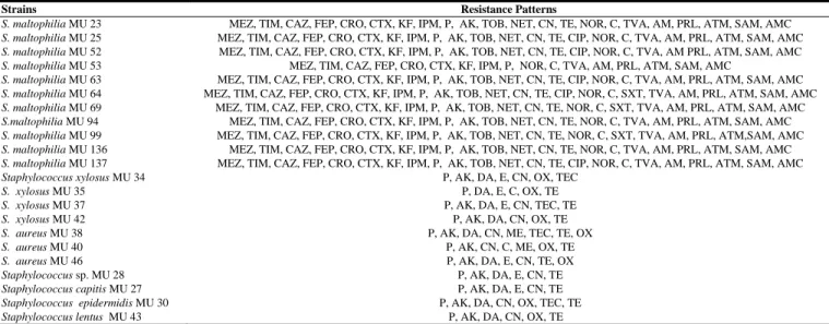
ATCC 10239, Candida tropicalis RSKK 665), and multi-resistant strains of S. maltophilia and various species of Staphylococcus were used. The antibiotic resistance patterns of the multi-antibiotic resistant bacteria are shown in Table 1.
The ethyl acetate and ethanol extracts inhibited all of the Gram-negative bacteria, including the multi-antibiotic resistant strains of S. maltophilia. The chloroform extract inhibited all S. maltophilia strains, except for S. maltophilia MU 63.
NPC
Natural Product Communications
Vol. 4
2009
No. 9
1267 - 1270
1268 Natural Product Communications Vol. 4 (9) 2009 Ugur et al.
Table 1: Antibiotic resistance patterns of S. maltophilia and Staphylococcus species.
Strains Resistance Patterns
S. maltophilia MU 23 MEZ, TIM, CAZ, FEP, CRO, CTX, KF, IPM, P, AK, TOB, NET, CN, TE, NOR, C, TVA, AM, PRL, ATM, SAM, AMC
S. maltophilia MU 25 MEZ, TIM, CAZ, FEP, CRO, CTX, KF, IPM, P, AK, TOB, NET, CN, TE, CIP, NOR, C, TVA, AM, PRL, ATM, SAM, AMC
S. maltophilia MU 52 MEZ, TIM, CAZ, FEP, CRO, CTX, KF, IPM, P, AK, TOB, NET, CN, TE, CIP, NOR, C, TVA, AM PRL, ATM, SAM, AMC
S. maltophilia MU 53 MEZ, TIM, CAZ, FEP, CRO, CTX, KF, IPM, P, NOR, C, TVA, AM, PRL, ATM, SAM, AMC
S. maltophilia MU 63 MEZ, TIM, CAZ, FEP, CRO, CTX, KF, IPM, P, AK, TOB, NET, CN, TE, CIP, NOR, C, TVA, AM, PRL, ATM, SAM, AMC
S. maltophilia MU 64 MEZ, TIM, CAZ, FEP, CRO, CTX, KF, IPM, P, AK, TOB, NET, CN, TE, CIP, NOR, C, SXT, TVA, AM, PRL, ATM, SAM, AMC
S. maltophilia MU 69 MEZ, TIM, CAZ, FEP, CRO, CTX, KF, IPM, P, AK, TOB, NET, CN, TE, NOR, C, SXT, TVA, AM, PRL, ATM, SAM, AMC
S.maltophilia MU 94 MEZ, TIM, CAZ, FEP, CRO, CTX, KF, IPM, P, AK, TOB, NET, CN, TE, NOR, C, TVA, AM, PRL, ATM, SAM, AMC
S. maltophilia MU 99 MEZ, TIM, CAZ, FEP, CRO, CTX, KF, IPM, P, AK, TOB, NET, CN, TE, NOR, C, SXT, TVA, AM, PRL, ATM,SAM, AMC
S. maltophilia MU 136 MEZ, TIM, CAZ, FEP, CRO, CTX, KF, IPM, P, AK, TOB, NET, CN, TE, NOR, C, TVA, AM, PRL, ATM, SAM, AMC
S. maltophilia MU 137 MEZ, TIM, CAZ, FEP, CRO, CTX, KF, IPM, P, AK, TOB, NET, CN, TE, CIP, NOR, C, TVA, AM, PRL, ATM, SAM, AMC
Staphylococcus xylosus MU 34 P, AK, DA, E, CN, OX, TEC
S. xylosus MU 35 P, DA, E, C, OX, TE
S. xylosus MU 37 P, AK, DA, E, CN, TEC, TE
S. xylosus MU 42 P, AK, DA, CN, OX, TE
S. aureus MU 38 P, AK, DA, CN, ME, TEC, TE, OX
S. aureus MU 40 P, AK, CN, C, ME, OX, TE
S. aureus MU 46 P, AK, DA, E, CN, TE, OX
Staphylococcus sp. MU 28 P, AK, DA, E, CN, TE
Staphylococcus capitis MU 27 P, AK, DA, E, CN, TE
Staphylococcus epidermidis MU 30 P, AK, DA, CN, OX, TEC, TE
Staphylococcus lentus MU 43 P, AK, DA, CN, OX, TE
MEZ: Mezlocillin (75 µg), TIM: Ticarcillin+clavulanic acid (75+ 10 µg), CAZ: Ceftazidime (30 µg), FEP: Cephepim (30 µg), CRO: Ceftriaxone (30 µg), CTX: Cefotaxime (30 µg), KF: Cephalothin (30 µg), IPM: Imipenem (10 µg), P: Penicillin (10 U), AK: Amikacin (30 µg), TOB: Tobramycin (10 µg), NET: Netilmicin (30 µg), CN: Gentamicin (10 µg), TE: Tetracycline (30 µg), NOR: Norfloxacin (10 µg), C: Chloramphenicol (30 µg), TVA: Trovafloksasin (10 µg), AM: Ampicillin (10 µg), PRL: Piperacillin (100 µg), ATM: Aztreonam (30 µg), SAM: Sulbactam + Ampicillin (10 µg + 10 µg), AMC: Amoxicillin + Clavulanic acid (20 µg + 10 µg), CIP: Ciprofloxacin (5 µg), SXT: Trimetoprim+sulfamethoxazole (1.25 µg + 23.75 µg), DA: Clindamycin (2 µg); E: Erythromycin (15 µg); ME: Methicillin (5µg); OX: Oxacillin (1µg); TEC: Teicoplanin (30 µg).
Multidrug resistance is common and increasing among Gram-negative nonfermenters, and a number of strains have now been identified that exhibit resistance to essentially all commonly used antibiotics, including antipseudomonal penicillins and cephalosporins, aminoglycosides, tetracyclines, fluoroquinolones, trimethoprim-sulfamethoxazole, and carbapenems [5]. S. maltophilia is one of the important members of this group.
S. maltophilia has received much attention in the last
decade because of its role as a pathogenic microorganism in an increasing number of clinical syndromes [6], such as bacteremia, infections of the respiratory and urinary tracts, skin and soft tissue infections, biliary tract infection, meningitis, serious wound infections, conjunctivitis and endocarditis[7]. The treatment of infections caused by this microorganism is difficult because S. maltophilia is frequently resistant to most of the widely used antibiotics [8]. This in vitro study provided evidence that the extracts of C. drabifolia subsp. cappadocica are potentially rich source of antibacterial agents against multi-antibiotic resistant S. maltophilia.
The chloroform extract was active against all the tested Gram-positive bacteria, including the multi-antibiotic resistant strains of Staphylococcus. The antibacterial activities of the chloroform extract on the Gram-positive bacteria were higher than on Gram-negative bacteria.
Staphylococci are among the most commonly encountered pathogens in clinical practice. S. aureus is a major cause of nosocomial infections, food poisoning, osteomyelitis, pyoarthritis, endocarditis, toxic shock syndrome, and a broad spectrum of other disorders [9]. In recent years, several species of coagulase negative staphylococci (CNS) have been recognized as opportunistic pathogens and have been implicated in human infections and disease, especially in immune-compromised and seriously ill patients. In recent years, there has been an alarming increase in nosocomial staphylococcal infections by strains with multiple drug resistance [10].
The ethyl acetate and ethanol extracts had antibacterial activity on all of the tested Gram-negative bacteria, including the multi-antibiotic resistant strains of S. maltophilia. The ethanol extract also inhibited the Gram-positive bacteria, except S.
capitis MU 27. The chloroform extract inhibited the
all of the Gram-positive and Gram-negative bacteria, except S. maltophila MU 63.
The results indicate that the chloroform extract of
C. drabifolia subsp, cappadocica has a capacity to
inhibit the growth of multi-antibiotic resistant strains. For this reason, the chemical composition of its chloroform extract was determined. Twenty compounds were detected using GC and GC-MS (Table 4). The percentage composition of the chloroform extract was determined with a Class-GC
Chemical composition of Centaurea drabifolia subsp. cappadocica Natural Product Communications Vol. 4 (9) 2009 1269
Table 2: Antimicrobial activities of the various extracts of C. drabifolia subsp. cappadocica on Gram-negative bacteria.
Inhibition zone (mm) Strains n-Hexane extract Chloroform extract Ethanol extract Ethyl acetate extract E. aerogenes RSKK 720 - 11 15 11 P. aeruginosa ATCC 27853 - 16 15 13 E. coli ATCC 25922 11 13 14 12 S. maltophila MU 23 15 11 10 14 S. maltophila MU 25 13 11 15 19 S. maltophila MU 52 14 10 15 16 S. maltophila MU 53 13 11 19 18 S. maltophila MU 63 11 - 14 12 S. maltophila MU 64 - 12 16 13 S. maltophila MU 69 - 9 10 15 S. maltophila MU 94 - 12 11 11 S. maltophila MU 99 12 12 15 19 S. maltophila MU 136 - 11 13 14 S. maltophila MU 137 14 9 12 16 (-) : No activity
Table 3: Antimicrobial activities of the various extracts of C. drabifolia subsp. cappadocica on Gram-positive bacteria and yeasts.
Inhibition zone (mm) of the extracts Strains n-Hexane extract Chloroform extract Ethanol extract Ethyl acetate extract M. luteus NRRL B- 4375 9 13 12 12 B. subtilis ATCC 6633 10 14 14 10 S. aureus ATCC 25923 11 17 14 - S. mutans CNCTC 8/77 - 16 10 10 S. capitis MU 27 - 22 - 16 Staphylococcus sp. MU 28 - 15 15 17 S. epidermidis MU 30 - 19 17 - S. xylosus MU 34 - 21 14 11 S. xylosus MU 35 - 16 13 19 S. xylosus MU 37 - 16 14 10 S. aureus MU 38 - 14 16 - S. aureus MU 40 14 16 12 13 S. xylosus MU 42 9 16 16 18 S. lentus MU 43 14 20 12 10 S. aureus MU 46 11 17 15 14 C. albicans ATCC 10239 - - - - C. tropicalis RSKK 665 - - - - (-) : No activity
computer program. The major compounds were spathulenol (14.1%), caryophyllene oxide (12.5%), octadecanol (10.2%), ethyl palmitate (7.7%), [Z,Z]-10,12-hexadecadienal (6.0%), 3-hydroxy-p-anisaldehyde (5.9%) and pentacosane (5.8%).
The components of the extract were separated into five classes: oxygenated monoterpene hydrocarbons (11.3%), sesquiterpene hydrocarbons (5.5%), oxygenated sesquiterpenes (30.0%), aromatic alcohols (3.8%) and others (49.4%). Of the oxygenated sesquiterpenes, spathulenol (14.1%) and caryophyllene oxide (12.5%) were the main components.
Sesquiterpenes have been reported to have potent antimicrobial activity and play a critical role in plant defense mechanisms [11]. Spathulenol and caryophyllene oxide are also known to exhibit antibacterial activity [12a,12b,13]. In this study, these are the most abundant constituents of the chloroform extract.
Table 4: Chemical composition of chloroform extract of C. drabifolia subsp. cappadocica
No Compoundsa RIa Percentage (%) Methods 1 2-Amino, p-cymene 842 4.9 b 2 5-Amino, 2-methoxyphenol 916 3.8 b 3 cis-7-Decen-1-al 973 4.9 b 4 [E-E]-2,4-Decadienal 1024 1.7 a, b 5 3-Hydroxy, p-anisaldehyde 1082 5.9 a, b 6 β-cyclocitral 1135 2.9 a, b 7 Vanillin 1197 2.5 a, b 8 [E-E]-2,4-Dodecadienal 1256 1.5 b 9 β-Caryophyllene 1298 2.6 a, b 10 cis-α-Bisabolene 1307 2.8 a, b 11 Caryophyllene oxide 1324 12.5 a, b 12 Spathulenol 1355 14.1 a, b 13 [Z,Z]-10,12-Hexadecadienal 1478 6.0 b 14 2-Methyl hexadecanol 1527 1.6 b 15 Ethyl palmitate 1619 7.7 a, b 16 [Z]-9-Octadecenal 1674 3.0 b 17 Hexahydro farnesyl acetone 1746 3.4 a, b
18 Heneicosane 1867 1.9 a, b
19 Pentacosane 2103 5.8 b
20 Octadecan-1-ol 2149 10.2 a, b
TOTAL 100.0
a: co-injection with authentic compounds, b: MS,
a : In DB-5 fused silica capillary column
To our knowledge, this is the first study of the antimicrobial activity and chemical composition of extracts of C. drabifolia subsp. cappadocica. Our results indicate that the chloroform extract of this species has a capacity to inhibit the growth of pathogenic bacteria, especially multi-antibiotic resistant strains of S. aureus. These extract may be useful as alternative antimicrobial agents for multi-antibiotic resistant Staphylococci.
Experimental
Plant material and extraction: C. drabifolia ssp.
cappadocica (Asteraceae) was collected at the
flowering stage from Mugla, Turkey. A voucher specimen (Herbarium No: O.V. 4448) has been deposited in the Herbarium of University of Mugla, Turkey. The air dried and powdered aerial parts of C. drabifolia ssp. cappadocica were extracted successively with n-hexane, chloroform, ethyl acetate and ethanol in a Soxhlet apparatus. Solvents of all the extracts were removed under low vacuum using rotary evaporation. Crude extracts were maintained at +4ºC until investigated for antimicrobial activity. Microorganisms: Three negative, 4 Gram-positive and multi-antibiotic resistant bacteria, and 2 yeasts were used. The strains of MU coded were obtained from Mugla University Culture Collection. Disc diffusion assay: The antibacterial activity was based on a disc diffusion method [14-16] using bacterial cell suspensions whose concentration was equilibrated to 0.5 McFarland standard after the bacteria cultured. Each bacterial suspension (100 μL)
1270 Natural Product Communications Vol. 4 (9) 2009 Ugur et al.
was spread on a Mueller–Hinton agar plate. Sterile paper discs (6 mm diameter) were impregnated with 20 μL of each extract dissolved in the solvent used for extraction at 25 mg/mL. The discs were allowed to dry and then placed on the inoculated agar. The plates were incubated at appropriate temperature and time for microorganisms. Discs of n-hexane, chloroform, ethanol, and ethyl acetate were used as controls. After the incubation time, the zones of inhibition were measured. The experiment was performed in triplicate.
Gas chromatography (GC) and GC-MS analysis: GC and GC/MS analyses were performed under the experimental conditions as reported earlier [13].
Identification of the components was based on GC retention indices and computer matching of MS with those of standards (NIST & Wiley, and a personal library of 320 spectra), as well as by comparison with the fragmentation patterns of MS reported in the literature [17] and, whenever possible, by co-injection with authentic compounds.
Acknowledgements - This work was supported by
Mugla University Research Funds. The authors wish to thank Associate Prof. Dr. Omer Varol, Department of Biology, Faculty of Science and Art, University of Mugla, for the identification of the plant material collected.
References
[1] Tavares AC, Gonçalves MJ, Cavaleiro C, Cruz MT, Lopes MC, Canhoto J, Salgueiro LR. (2008) Essential oil of Daucus carota
subsp. halophilus: Composition, antifungal activity and cytotoxicity, Journal of Ethnopharmacology, 119, 129–134.
[2] Gulluce M, Aslan A, Sokmen M, Sahin F, Adiguzel A, Agar G, Sokmen A. (2006) Screening the antioxidant and antimicrobial
properties of the lichens Parmelia saxatilis, Platismatia glauca, Ramalina pollinaria, Ramalina polymorpha and Umbilicaria
nylanderian. Phytomedicine, 13, 515–521.
[3] (a) Baytop T. (1999) Turkiye’de Bitkiler ile Tedavi (Gecmiste ve Bugun). Istanbul, Nobel Tip Kitabevleri; (b) Yesilada E, Sezik E,
Honda G, Takaishi Y, Takeda Y, Tanaka T. (1999) Traditional medicine in Turkey IX: Folk medicine in northwest Anatolia.
Journal of Ethnopharmacology, 64, 195-210; (c) Sezik E, Yesilada E, Honda G, Takaishi Y, Takeda Y, Tanaka T. (2001)
Traditional medicine in Turkey X. Folk medicine in central Anatolia. Journal of Ethnopharmacology, 75, 95-115.
[4] Wagenitz G. (1975) Centaurea L. In: Davis PH. (Ed.), Flora of Turkey and the East Aegean Islands Vol. 5, Edinburgh University
Press, Edinburgh, pp. 465-585.
[5] McGowan JE Jr. (2006) Resistance in nonfermenting Gram-negative bacteria: multidrug resistance to the maximum. American
Journal of Infection Control, 34 (5 suppl 1), S29-37.
[6] Robin T, Janda JM. (1996) Pseudo-, Xantho-, Stenotrophomonas maltophilia: An emerging pathogen in search of a genus. Clinical
Microbiology Newsletter, 18, 9-13.
[7] Denton M, Kerr KG. (1998) Microbiological and clinical aspects of infection associated with Stenotrophomonas maltophilia.
Clinical Microbiology Reviews, 11, 57-80.
[8] Krueger TS, Clark EA, Nix DE. (2001) In vitro susceptibility of Stenotrophomonas maltophilia to various antimicrobial
combinations. Diagnostic Microbiology & Infectious Disease, 41, 71-78.
[9] Rubin RJ, Harrington CA, Poon A, Dietrich K, Grene JA, Moiduddin A. (1999) The economic impact of Staphylococcus infection
in New York City hospitals. Emerging Infectious Diseases, 5, 9-17.
[10] Kloos WE, Bannerman TL. (1995) Staphylococcus and Micrococcus. In: Murray PR, Baron EJ, Pfaller MA, Tenover FC, Yolken
RH. (Eds)., Manual of Clinical Microbiology. 6th ed, ASM Press, Washington, D.C. USA, pp. 282–298.
[11] Oliva MM, Demo MS, Lopez AG, Lopez ML, Zygadlo J. (2005) Antimicrobial activity and composition of Hyptis mutabilis
essential oil. Journal of Herbs Spices & Medicinal Plants, 11, 59-65.
[12] (a) Ulubelen A, Topcu G, Eris C, Sonmez U, Kartal M, Kurucu S, Bozok-Johansson C. (1994) Terpenoids from Salvia sclarea.
Phytochemistry, 36, 971-974; (b) Tzakou O, Skaltsa H. (2003) Composition and antibacterial activity of the essential oil of Satureja parnassica subsp. parnassica. Planta Medica, 69, 282-284.
[13] Ugur A, Sarac N, Duru ME. (2009) Antimicrobial activity and chemical composition of Senecio sandrasicus on antibiotic resistant
Staphylococci. Natural Product Communications, 4, 579-584.
[14] Bauer AW, Kirby WM, Sherris JC, Turck M. (1966) Antibiotic susceptibility testing by a standardized single disk method.
American Journal of Clinical Pathology, 45, 493-496.
[15] Collins CH, Lyne PM, Grange JM. (1995) Microbiological Methods. 7th. ed. London, Butterworths.
[16] Murray PR, Baron EJ, Pfaller NA. (1995) Manual of Clinical Microbiology, ASM Press, Washington, D.C.
[17] Adams RP. (2001) Identification of Essential Oils Components by Gas Chromatography/ Quadrupole Mass Spectroscopy. Allured