MOLECULAR ANALYSIS OF
SENESCENCE-ASSOCIATED PROTEIN PHOSPHATASES
DUSP10 AND MTMR11
A THESIS SUBMITTED TO
THE DEPARTMENT OF MOLECULAR BIOLOGY AND
GENETICS AND THE INSTITUTE OF ENGINEERING AND
SCIENCE OF BILKENT UNIVERSITY
IN PARTIAL FULFILLMENT OF THE REQUIREMENTS
FOR THE DEGREE OF
MASTER OF SCIENCE
BY
SUNA PELİN GÜLAY
JULY 2009
I certify that I have read this thesis and that in my opinion it is fully adequate, in scope and in quality, as a thesis for the degree of Master of Science.
___________________________ Assoc. Prof. Dr. Rengül Atalay
I certify that I have read this thesis and that in my opinion it is fully adequate, in scope and in quality, as a thesis for the degree of Master of Science.
___________________________ Prof. Dr. Mehmet Öztürk
I certify that I have read this thesis and that in my opinion it is fully adequate, in scope and in quality, as a thesis for the degree of Master of Science.
___________________________ Assist. Prof. Dr. Ali Osmay Güre
I certify that I have read this thesis and that in my opinion it is fully adequate, in scope and in quality, as a thesis for the degree of Master of Science.
___________________________ Assist. Prof. Dr. Ayşe Elif Erson
Approved for the Institute of Engineering and Science
___________________________ Director of Institute of Engineering and Science
ABSTRACT
MOLECULAR ANALYSIS OF SENESCENCE-ASSOCIATED PROTEIN PHOSPHATASES DUSP10 AND MTMR11
Suna Pelin Gülay
MSc. in Molecular Biology and Genetics Supervisor: Assoc. Prof. Rengül Atalay
July 2009, 129 Pages
Liver cancer is the fifth most common cancer in the world. Until recently, tumor cells were thought to proliferate indefinitely. In a previous study, our group showed spontaneous induction of replicative senescence in p53- and p16INK4a-deficient HCC (hepatocellular carcinoma) cell clones. The gene expression profiling was later done for these different clones, in an attempt to find novel therapeutic targets in HCC. Since protein kinases are known to be very important in disease formation and carcinogenesis, their partners in signaling, protein phosphatases should also be important in these processes. Hence analysis and targeting of protein phosphatases genes with differential expression between immortal and senescent clones might prove beneficial for HCC therapeutics. Among the phosphatase genes with differential expression patterns, we focused on two most upregulated genes in senescent clones with respect to immortal clones, DUSP10 and MTMR11. After gathering detailed information on these genes and their products by bioinformatics analysis, we confirmed the upregulation of the two genes in our senescent clones compared to our immortal clones by semi-quantitative RT-PCR. We then checked DUSP10 and MTMR11 expression in HCC and breast cancer cell lines to see if a differential expression of these genes are observed in different subtypes of these cell lines. Other experiments on MTMR11 focused on discovery of novel transcripts of this gene in HCC and breast cancer cell lines and checking the amounts of different transcripts in different subtypes of these cell lines, to form a bridge between MTMR11 transcript variants and carcinogenesis, however we did not observe differential expression. Two microarray studies comparing non-tumor and HCC tissues have listed MTMR11 as upregulated in HCC. Hence, upregulation of this gene in senescent clones may not be significant in hepatocarcinogenesis or replicative senescence, and further experiments should be performed. Considering DUSP10, we checked the subcellular localization of this protein in HCC cell lines by immunostaining, to see if the two subtypes (well-differentiated and poorly-differentiated) of HCC cell lines differed in DUSP10 localization. We observed some cell lines having only nuclear or only cytoplasmic DUSP10, whereas most had both nuclear and cytoplasmic DUSP10. This lead the way for us to explore the factors that may be important in changing this protein’s localization, as this may be a type of regulation on this protein, and may change during carcinogenesis or upon induction of senescence. For this purpose, we checked to see if DUSP10 changed its localization in aging MRC-5 cell passages compared to young, proliferating ones and in premature senescence-induced cells compared to normal ones. Interestingly, it was found that upon replicative senescence induction, but not premature senescence, DUSP10 localized more to the cell nucleus which indicated a connection between DUSP10
localization and replicative senescence. We also checked to see if DUSP10 changed its localization upon disruption of the MAPK pathways it participates in, by kinase inhibitor experiments. Interestingly, it was found that DUSP10 localized significantly more to the cell nucleus upon inhibition of JNK pathway but not p38 pathway, in well-differentiated subtype of HCC cell lines. DUSP10 localization did not change significantly in poorly-differentiated subtype of HCC cell lines. Although JNKs, which seem to regulate DUSP10 through its localization according to this study, act as oncogenes in HCC, the significance of the change in DUSP10 localization should be characterized further before stating that DUSP10 can be a putative tumor suppressor. However, our other results indicate a relationship between DUSP10 localization and replicative senescence, which is promising because DUSP10 has emerged from our group’s microarray data as a replicative senescence-associated gene, and this connection should be analyzed further.
ÖZET
HÜCRE YAŞLANMASIYLA İLİNTİLİ DUSP10 VE MTMR11 FOSFATAZ GENLERİNİN MOLEKÜLER ANALİZİ
Suna Pelin Gülay
Moleküler Biyoloji ve Genetik Yüksek Lisansı Tez Yöneticisi: Doç. Dr. Rengül Atalay
Temmuz 2009, 129 Sayfa
Karaciğer kanseri (hepatosellüler karsinom) dünya çapında en sık görülen beşinci kanser türüdür. Yakın bir zamana kadar, tümör hücrelerinin sonsuz bölünme kapasitesine sahip olduğu düşünülmekteydi. Grubumuzun önceki bir çalışmasında p53 ve p16INK4a proteinlerine sahip olmayan karaciğer kanseri hücre klonlarında kendiliğinden gelişen bir hücre yaşlanması olayı gösterildi. Elde edilen senesant ve ölümsüz klonlarda değişen gen ifadelerinin incelenmesi karaciğer kanseri için tedavi amaçlı yeni hedef gen ve proteinlerinin bulunmasini sağlayabilirdi. Biz de bu amaçla, yakın zamanda hücre içi sinyal iletiminde ve bunun zarar görmesi sonucunda oluşan hastalıklarda önemleri anlaşılmaya başlanmış olan fosfataz genlerine odaklandık ve çalışmamız için senesant klonlarda ifadesi ölümsüz klonlara göre en yüksek anlamlı artışı gösteren DUSP10 ve MTMR11 fosfataz genlerini seçtik. DUSP10, mitojenler tarafindan aktive edilen protein kinazlardan (MAPK) JNK ve p38’i deaktive eder. MTMR11 ise myotubularin fosfataz ailesindendir. Bu genlerin ve gen ürünlerinin detaylı biyoinformatik analizinden sonra, revers transkriptaz polimeraz zincir reaksiyonu (RT-PZR) ile senesant klonlarda ölümsüz klonlara göre ifadelerinin arttığını doğruladık. Değişik kökenli ve değişik farklılaşma oranlarına sahip karaciğer ve meme kanseri hücre hatlarında, RT-PZR ile DUSP10 ve MTMR11 genlerinin ifadesine baktik. Amacımız bu genleri kanser oluşumu ve hücre yaşlanmasıyla ilişiklendirmekti. MTMR11 konusundaki diğer deneyler, elimizdeki hücre hatlarında bu genin yeni ve değişik transkript formlarinin bulunmasina ve bu farklı transkriptlerin miktarının karaciğer ve meme kanseri hücre hattı alt-türlerinde değişiminin saptanmasına odaklandı. Bu konuda farklı alt-türleri farklı transkriptler ile ilişiklendiremedik. Başka iki mikrodizilim çalışmasında da MTMR11’in karaciğer kanserinde ifadesi artmış bir gen olarak gösterildiğini de göz önünde bulundurursak, grubumuzun mikrodizilim çalışmasında MTMR11’in ifadesinin senesant klonlarda artmış olması, bu gen ve ürününün kanserleşme veya senesansta fonksiyonel bir rolü olduğu anlamına gelmiyor olabilir. Bunu ileriki çalışmalar gösterecektir. DUSP10 için ise, yine değişik karaciğer kanseri hücre hattı alt-türlerinde (epitel veya mezenkim kökenli) bir fark yakalayabilmek için, bu proteinin hücre içindeki lokalizasyonunu immüno-boyama ile tespit ettik. Bu proteinin bazı hücre hatlarında sadece çekirdekte veya sadece sitoplazmada, çoğundaysa her iki yerde de olduğunu gözlemledik. Bir proteinin hücre içi lokalizasyonunun değişimi bir çeşit kontrol mekanizması olabileceği, bu da bu proteini kanserleşme veya hücre yaşlanmasıyla ilişkilendirebileceği için, hücre içi DUSP10 lokalizasyonunu değiştirebilecek faktörleri incelemeye yöneldik. Öncelikle telomere bağlı olan ve olmayan hücre yaşlanması tiplerinde bu proteinin lokalizasyonunun değişimine baktık. Bunun için genç MRC-5 fibroblast hücre hattı pasajları ile yaşlanmakta olan pasajları
karşılaştırdık. İlginç bir şekilde, DUSP10’un yaşlanmakta olan hücrelerde çekirdeğe lokalize olmaya başladığını gözlemledik. Değişik kimyasallarla erken (prematüre) hücre yaşlanmasına sebep olunduğunda ise DUSP10’un lokalizasyonunun değişmediğini gördük. Mikrodizileme çalışmasının yapıldığı klonlarda gözlemlenen hücre yaşlanmasının telomere bağlı olduğu düşünüldüğünde aldığımız sonuç DUSP10’un bu tip hücre yaşlanmasında kontrolünün değiştiğini göstermesi açısından umut vericiydi. Bunlara ilaveten, DUSP10 lokalizasyonunun p38 ve JNK sinyal yolaklarının kimyasal ilaçlarla engellenmesi üzerine değişip değişmediğine de baktık. İlginç bir şekilde, DUSP10’un JNK’nin inhibisyonu sonucunda anlamlı bir şekilde çekirdeğe lokalize olduğunu, bu sonucun p38’in inhibisyonu sonucunda ise oluşmadığını gördük. Ayrıca bu gözlemimizi epitel kökenli hepatosellüler karsinom hücre hatlarında yapabildik. Mezenkim kökenli hücre hatlarında ise DUSP10 lokalizasyonunda bir değişiklik göremedik. Bu çalışmada DUSP10 lokalizasyonunu kontrol ettiği görülen JNK’ların hepatosellüler karsinomda onkoprotein olarak işlev gördükleri bilinmektedir. Yine de DUSP10’un tümör inhibisyonuyla veya telomere bağlı hücre yaşlanması ile olan ilişkisinin kesinleştirilmesi için bu proteinin hücre içi lokalizasyonundaki değişiminin öneminin başka deneyler aracılığıyla daha iyi anlaşılması gerekmektedir.
ACKNOWLEDGEMENTS
First of all, I would like to express my gratitude for my thesis advisor Assoc. Prof. Dr. Rengül Atalay for her guidance throughout this study. Being her student was a true privilege since she was always accessible for questions and discussions, and very supportive during hard times.
I would like to thank my co-advisor and the founder of KANİLTEK project, Prof. Dr. Mehmet Öztürk, for his guidance throughout this study and for providing me with inspiration.
I would like to thank the entire MBG faculty. I had the chance to interact with almost all of them through the courses I took or the ones I was involved in as a teaching assistant, and I learned a lot from each. I would also like to thank Dr. Hani Al-Otaibi for providing me with experimental support in the earlier parts of my research.
I would like to express my deepest thanks to Şerif Şentürk who has provided me with experimental support right from the beginning of my study until the very end, always being there when I needed to ask something or borrow a reagent.
I would like to thank all the past and present members of the MBG lab for creating a friendly, comfortable and helpful atmosphere to do research in. Many thanks go to the members of Molecular Oncology Group, especially to Ayça Arslan-Ergül, Mine Mumcuoğlu, Sevgi Bağışlar, Tülin Erşahin, Eylül Harputlugil, Gökhan Yıldız, Bilge Kılıçoğlu, Ebru Bilget-Güven and the past member Nilgün Taşdemir, as all of these people has done at least one thing to make my life easier in one or more aspects. Same is true for Duygu Akbaş-Avcı, Ender Avcı and Ceyhan Ceran, as I have enjoyed their friendships very much.
I am indebted to Füsun Elvan, Sevim Baran, Abdullah Ünnü and Pelin Telkoparan for their help in various ways, and especially for providing an ideal environment for research by making everything run smoothly.
Last but of course not the least, my deepest gratitude goes to my family for their patience, unconditional love and support throughout my life, and I dedicate this thesis to them.
This work was supported by the KANİLTEK project from State Planning Office (coordinator M. Ozturk). Additionally, during my MSc research, I was personally supported by TÜBİTAK with scholarship 2228.
The confocal microscopy images were taken at Central Laboratory Molecular Biology and Biotechnology R&D Center at Middle East Technical University, Ankara, with the help of Dr. Can Özen.
TABLE OF CONTENTS
SIGNATURE PAGE………..II ABSTRACT………...III ÖZET………...V DEDICATION……… VII ACKNOWLEDGEMENTS………...VIII TABLE OF CONTENTS………...X LIST OF TABLES………XV LIST OF FIGURES………XVI CHAPTER 1. INTRODUCTION………...11.1 Protein phosphorylation and its regulation by kinases and phosphatases…………1
1.1.1 Kinase gene family………2
1.1.2 Phosphatase gene family………...3
1.1.3 Deregulation of protein kinases and phosphatases in cancer and other diseases………...5
1.2 Hepatocellular carcinoma………...10
1.2.1 Aetiologies of hepatocellular carcinoma………...………..11
1.3 Protein kinases and phosphatases implicated in hepatocarcinogenesis…………..13
1.3.1 Growth factor receptor tyrosine kinases………...14
1.3.2 Kinases and phosphatases of PI3K/PTEN/AKT/mTOR pathway…………...16
1.3.3 Mitogen-activated protein kinase (MAPK) pathways……….16
1.4 Cellular senescence and liver cirrhosis………...18
1.4.1 Replicative senescence and telomere shortening………20
1.4.2 Premature (stress-induced) senescence………...21
1.5.1 Induction of spontaneous replicative senescence in stable clones derived from
parental HCC cell lines………....22
1.5.2 Mechanism of spontaneous replicative senescence in stable clones derived from parental HCC cell lines………22
1.6 Gene expression changes between senescent and immortal Huh7 clones……….23
1.6.1 Gene expression profiling of senescent and immortal Huh7 clones…………...23
1.6.2 Analysis of genes differentially expressed between senescent and immortal clones………23
1.6.3 Identification of DUSP10 and MTMR11 as senescence-associated genes…….24
1.6.4 Cellular activities of DUSP10……….26
1.6.5 Cellular activities of MTMR11………...27
CHAPTER 2. OBJECTIVES AND RATIONALE……….28
CHAPTER 3. MATERIALS AND METHODS….……….30
3.1 MATERIALS……….30
3.1.1 Reagents………..30
3.1.2 Enzymes and nucleic acids………..30
3.1.3 Oligonucleotides………..30
3.1.4 Electrophoresis, photography and spectrophotometer………30
3.1.5 Tissue culture reagents and cell lines………..31
3.1.6 Antibodies and chemiluminescence………31
3.2 SOLUTIONS AND MEDIA………..32
3.2.1 General solutions……….32
3.2.2 Microbiological media, reagents and antibiotics……….32
3.2.3 Tissue culture solutions………...33
3.2.4 SDS (Sodium Deodecyl Sulfate)-PAGE (Polyacrylamide Gel Electrophoresis) solutions………...33
3.2.5 Immunoblotting solutions………...34
3.2.6 Immunofluorescence and immunoperoxidase solutions……….34
3.2.7 SABG assay solutions……….34
3.3.1 General methods……….35
3.3.1.1 Preparation of competent cells……….35
3.3.1.1.1 Supercompetent E. coli preparation………..35
3.3.1.1.2 Electrocompetent E. coli preparation………....35
3.3.1.2 Transformation of E. coli……….36
3.3.1.2.1 Transformation by heat-shock method……….36
3.3.1.2.2 Electroporation of bacteria………....36
3.3.1.3 Long term storage of bacterial strains………..37
3.3.1.4 Purification of DNA……….37
3.3.1.4.1 Purification of plasmid DNA using MN (Macherey-Nagel) miniprep kit…37 3.3.1.4.2 Large-scale plasmid DNA purification……….37
3.3.1.5 Quantification and qualification of nucleic acids……….38
3.3.1.6 Restriction enzyme digestion of DNA……….38
3.3.1.7 Gel electrophoresis of nucleic acids……….38
3.3.2 Computer analyses………..38
3.3.3 Vector construction……….39
3.3.4 Tissue culture techniques………39
3.3.4.1 Cell lines……….………..39
3.3.4.2 Thawing cell lines………40
3.3.4.3 Growth conditions of cells………...40
3.3.4.4 Cryopreservation of cells………..40
3.3.5 Extraction of total RNA from tissue culture cells and tissue samples…………41
3.3.6 First strand cDNA synthesis………41
3.3.7 Primer design for expression analysis by semi-quantitative PCR………...41
3.3.8 Fidelity and DNA contamination control in first strand cDNAs……….42
3.3.9 Expression analysis of a gene by semi-quantitative PCR and GAPDH normalization………43
3.3.10 Crude total protein extraction………43
3.3.11 Western blotting………43
3.3.12 SABG assay………..45
3.3.13 Immunostaining……….45
3.3.13.1 Immunofluorescence of cell culture monolayers………...45
CHAPTER 4. RESULTS………..….………47
4.1 DUSP10 (MKP-5)……….47
4.1.1 Bioinformatics analysis of DUSP10………...47
4.1.1.1 Sequence and gene information………...47
4.1.1.2 Protein information………..48
4.1.1.2.1 General information………..48
4.1.1.2.2 Protein domains and motifs………..49
4.1.1.2.3 Protein structure………51
4.1.1.3 Gene homologs and protein sequence conservation……….52
4.1.1.3.1 Orthologs of DUSP10………...52
4.1.1.3.2 Paralogs of DUSP10………..55
4.1.1.4 DUSP10 in gene expression profiling arrays………...58
4.1.1.5 Expression data………59
4.1.2 Analysis of DUSP10 expression change between senescent and immortal Huh7 clones………60
4.1.3 DUSP10 expression data……….61
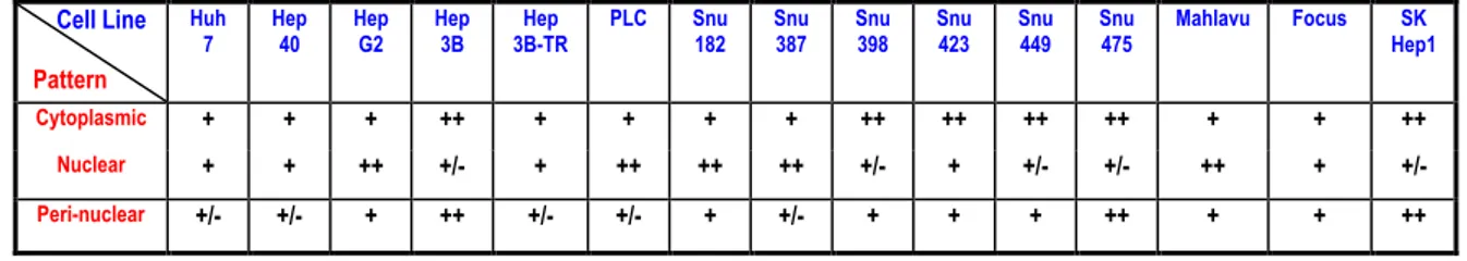
4.1.4 Subcellular localization of DUSP10 in hepatocellular carcinoma cell lines…...63
4.1.4.1 Localization of DUSP10 changes between different cell lines………63
4.1.4.2 DUSP10 does not co-localize with calnexin, an ER-membrane protein, in cell lines showing peri-nuclear / cytoplasmic DUSP10 staining………73
4.1.4.3 DUSP10 co-localizes with beta-actin, a cytoskeletal protein, in cell lines showing peri-nuclear / cytoplasmic DUSP10 staining……….76
4.1.5 Assesment of factors that may have an effect on DUSP10 localization……….77
4.1.5.1 Effect of senescence induction on DUSP10 localization……….77
4.1.5.1.1 Nuclear translocation is seen in late passage MRC-5 fibroblasts compared to early passage cells………77
4.1.5.1.2 Camptothecin and TGF-β, senescence-inducing agents, do not have an effect on DUSP10 localization………...78
4.1.5.2 Effect of JNK and p38 MAPK inhibitors on the subcellular localization of DUSP10………80
4.1.6 Ectopic overexpression studies for DUSP10………..92
4.2 MTMR11………...93
4.2.1.1 Sequence and gene information………...93
4.2.1.2 Protein information………..94
4.2.1.2.1 General information………..94
4.2.1.2.2 Protein domains and motifs………..94
4.2.1.2.3 Protein structure………96
4.2.1.3 Gene homologs and protein sequence conservation……….97
4.2.1.3.1 Orthologs of MTMR11……….97
4.2.1.3.2 Paralogs of MTMR11……….100
4.2.1.4 MTMR11 in gene expression profiling arrays………...102
4.2.1.5 Expression data………..104
4.2.2 Analysis of MTMR11 expression change between senescent and immortal Huh7 clones………..105
4.2.3 MTMR11 expression data……….106
4.2.4 MTMR11 transcript variants in different cell lines………...106
4.2.4.1 MTMR11 transcript variants in senescent and immortal clones………107
4.2.4.2 MTMR11 transcript variants in HCC cell lines……….108
4.2.4.3 MTMR11 transcript variants in breast cancer cell lines……….108
4.2.5 Sequencing of the extra bands in RT-PCR experiments………...109
CHAPTER 5. DISCUSSION..………..….………..113
CHAPTER 6. FUTURE PERSPECTIVES………119
LIST OF TABLES
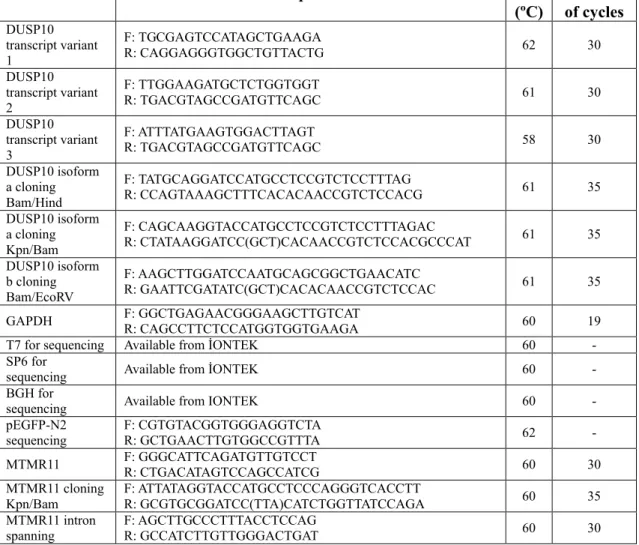
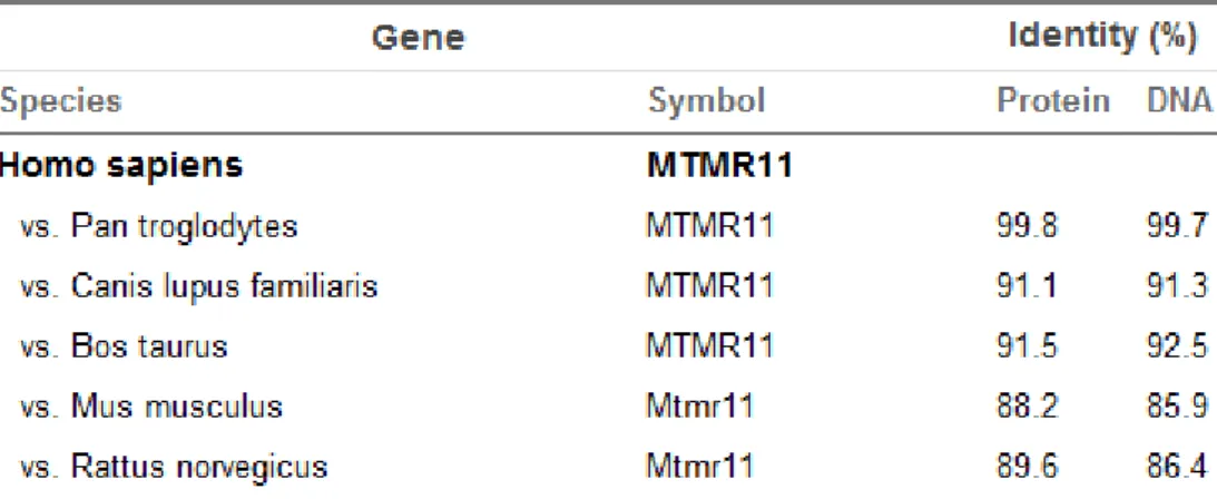
Table 1.1: Kinases and phosphatases that are implicated in various diseases………...8 Table 1.2: Differentially expressed phosphatases in significant lists………..26 Table 3.1: Antibody dilution table………...31 Table 3.2: Primer list for PCR and sequencing reactions………42 Table 4.1.1: Orthologs of human DUSP10 and multiple alignment pair-wise similarity scores between DUSP10 protein and DNA sequences of different species…………52 Table 4.1.2: DUSP10 staining patterns of 15 HCC cell lines according to immunoperoxidase staining………..63 Table 4.1.3: DUSP10 staining patterns of 15 HCC cell lines according to immunofluorescence………68 Table 4.2.1: Orthologs of human MTMR11 and multiple alignment pair-wise similarity scores between MTMR11 protein and DNA sequences of different species………..97
LIST OF FIGURES
Fig.1.1: Frequently phosphorylated residues in humans………2
Fig.1.2: Protein phosphatase tree………...4
Fig.1.3: Cell signaling circuitry……….5
Fig.1.4: Risk factors and mechanisms of hepatocarcinogenesis………..11
Fig.1.5: Major growth factor receptor signaling pathways important in HCC………15
Fig.1.6: Mitogen-activated protein kinase pathways………17
Fig.1.7: Stimuli that cause cells to undergo senescence………...19
Fig.1.8: Molecular mechanisms of senescence………20
Fig.4.1.1: Genomic context of DUSP10 gene obtained from NCBI Gene…………..47
Fig.4.1.2: Locus, transcripts and protein isoforms of DUSP10 obtained from NCBI Gene……….48
Fig.4.1.3a: Motif Scan results of DUSP10 isoform a………..50
Fig.4.1.3b: Motif Scan results of DUSP10 isoform b………..50
Fig.4.1.3: NCBI Conserved Domain result for DUSP10 isoform a……….51
Fig.4.1.5a: NPS consensus secondary structure prediction for DUSP10 isoform a….51 Fig.4.1.5b: NPS consensus secondary structure prediction for DUSP10 isoform b....51
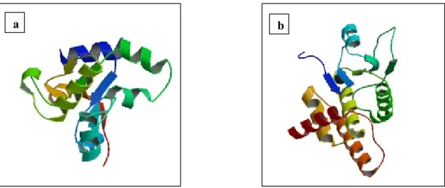
Fig.4.1.6: a. 3D structure of DUSP10 rhodanese domain, b. 3D structure of DUSP10 catalytic domain………...52
Fig.4.1.7: ClustalW2 multiple sequence alignment of DUSP10 orthologs except the Danio rerio ortholog……….53
Fig.4.1.8: Phylogenetic tree of DUSP gene family with branch lengths………..56
Fig.4.1.9: Cluster of MKPs according to substrate preference………57
Fig.4.1.10: Conserved motifs in MKPs………57
Fig.4.1.11: DUSP10 expression in Wurmbach microarray data………..58
Fig.4.1.12: Expression data of DUSP10 based on the GNF Expression Atlas 2 Data from U133A and GNF1H Chips………..59
Fig.4.1.13a: DUSP10 is upregulated in senescent and revertant clones compared to the immortal clone………..60 Fig.4.1.13b: DUSP10 is upregulated in senescent and revertant clones compared to the immortal clone………61 Fig.4.1.14a: DUSP10 transcript variant 1 mRNA levels and isoform a protein levels in HCC cell lines………..62 Fig.4.1.14b: DUSP10 transcript variant 1 mRNA levels in selected breast cancer cell lines………..62 Fig.4.1.15a: DUSP10 transcript variant 2 is not expressed in HCC cell lines……….62 Fig.4.1.15b: DUSP10 transcript variant 3 is not expressed in HCC cell lines……….63 Fig.4.1.16: DUSP10 immunostaining in HCC cell lines………..64 Fig.4.1.17: DUSP10 immunofluorescence in HCC cell lines………..69 Fig.4.1.18: DUSP10 and calnexin co-staining in Snu423………74 Fig.4.1.19a: Confocal image of DUSP10/Calnexin costaining, the plane where DUSP10 staining is the highest (40x), b: the plane where Calnexin staining is the highest (40x)……….75 Fig.4.1.20a. Confocal image of DUSP10/Calnexin costaining, the plane where DUSP10 staining is the highest (80x), b. (160x)………..75 Fig.4.1.21: DUSP10/Actin and DUSP10/Calnexin staining in Snu182 cell line (40x)……….76 Fig.4.1.22: Effect of replicative senescence on DUSP10 subcellular localization (40x)……….78 Fig.4.1.23: Effect of camptothecin on DUSP10 subcellular localization (40x)……...79 Fig.4.1.24: Effect of TGF-β on DUSP10 subcellular localization (40x)……….80 Fig.4.1.25a: Simplified JNK pathway and its possible feedback regulatory mechanism………81 Fig.4.1.25b: Simplified p38 pathway and its possible feedback regulatory mechanism………81 Fig.4.1.26a: Effect of JNK and p38 inhibitors on DUSP10 localization in Hep3B cell line (40x)………..82 Fig.4.1.26b: Effect of JNK and p38 inhibitors on DUSP10 localization in Snu182 cell line (40x)………..84 Fig.4.1.27: Effect of JNK inhibitor V on DUSP10 localization in Huh7, HepG2 and Mahlavu cell lines (40x)………...86
Fig.4.1.28a: Change in nuclear DUSP10 localization percentage in Huh7 cell line due
to JNK inhibitor V………....87
Fig.4.1.28b: Change in nuclear DUSP10 localization percentage in HepG2 cell line due to JNK inhibitor V……….87
Fig.4.1.28c: Change in nuclear DUSP10 localization percentage in Hep3B cell line due to JNK inhibitor V……….88
Fig.4.1.28d: Change in nuclear DUSP10 localization percentage in Snu182 cell line due to JNK inhibitor V……….88
Fig.4.1.28e: Change in nuclear DUSP10 localization percentage in Mahlavu cell line due to JNK inhibitor V……….89
Fig.4.1.29: Effect of the used concentration of JNK inhibitor V on c-Jun phosphorylation in Huh7 cells………..90
Fig.4.1.30: Phosphorylated and total JNK or p38 amounts in HepG2, Hep3B, Snu182 and Mahlavu cell lines………..90
Fig.4.1.31: Confocal microscopy results on inhibitor treatment of selected HCC cell lines (40x)……….91
Fig.4.1.32: DUSP10 protein amounts upon JNK inhibitor V treatment in the cell lines employed………..92
Fig.4.1.33: No significant overexpression of DUSP10 is seen upon transient transfection………...92
Fig.4.2.1: Genomic context of MTMR11 gene………93
Fig.4.2.2a: Motif Scan results of MTMR11 isoform a……….95
Fig.4.2.2b: Motif Scan results of MTMR11 isoform b………95
Fig.4.2.3a: NPS consensus secondary structure prediction for MTMR11 isoform a………96
Fig.4.2.3b: NPS consensus secondary structure prediction for MTMR11 isoform b………....96
Fig.4.2.4: ClustalW2 multiple sequence alignment of MTMR11 (transcript variant 2) orthologs………...98
Fig.4.2.5: Phylogenetic tree of myotubularin phosphatase gene family with branch lengths………101
Fig.4.2.6: Conserved motifs in myotubularin phosphatases………..102
Fig.4.2.7a: MTMR11 expression in Wurmbach microarray data………..103
Fig.4.2.8: Expression data of DUSP10 based on the GNF Expression Atlas 2 Data
from U133A and GNF1H Chips………104
Fig.4.2.9a: MTMR11 is upregulated in senescent and revertant clones compared to the immortal clone………105
Fig.4.2.9b: MTMR11 is upregulated in senescent and revertant clones compared to the immortal clone………..105
Fig.4.2.10: MTMR11 mRNA levels in HCC cell lines………..106
Fig.4.2.11: RT-PCR primer pairs for MTMR11 used in our studies and the lengths of the PCR products produced………....106
Fig.4.2.12: MTMR11 transcript variants in the established clones ………...107
Fig.4.2.13: MTMR11 transcript variants in HCC cell lines………...108
Fig.4.2.14: MTMR11 transcript variants in breast cancer cell lines………..108
Fig.4.2.15a: 340bp band sequencing results………..109
Fig.4.2.15b: 600bp band sequencing results………..110
Fig.4.2.15c: 800bp band sequencing results………..111
Fig.4.2.15d: 900bp band sequencing results………..112
CHAPTER 1. INTRODUCTION
1.1 Protein phosphorylation and its regulation by kinases and phosphatases
Reversible post-translational modification is a very important process in regulating protein activity and thus crucial in cell signaling pathways. Many types of post-translational modifications exist, including but not limited to phosphorylation, ubiquitination, acetylation, methylation, glycosylation and myristoylation. Historically, phosphorylation is the best studied of these modifications mostly due to the relative ease of detecting protein phosphorylation in vitro and in vivo (see Hunter T, 2007). Phosphoproteomic analysis has shown the extent of protein phosphorylation in mammalian cells and the temporal dynamics of this post-translational modification through mass spectrometry. A recent study has detected 6600 phosphorylation sites in 2244 proteins of HeLa cell line and has further found that 14% of these sites are modulated at least 2-fold by epidermal growth factor (EGF) within 20 minutes (Olsen JV et al, 2006). These phosphoproteins include enzymes (specifically kinases), ubiquitin ligases, guanidine nucleotide exchange factors (GEFs), GTPase activating proteins (GAPs), transcription factors and other transcriptional regulators.
Phosphorylation is a well-characterized biochemical process in which a phosphate group is added through a phosphoester bond (O-phosphate) to the hydroxyl side chain of serine, threonine or tyrosine residues (Fig.1.1). The relative ratio of phosphorylation of these aminoacids in mammalian cells is about 1000:100:1 respectively. Of lower frequency, N-phosphorylation of histidine residues is also observed (see Kowluru A, 2008). The low frequency of phosphorylation of a specific residue does not show its insignificance in signaling as tyrosine phosphorylation is a very important process regulating activities of various growth factor receptors (see Arena S et al, 2005).
Phosphates are negatively charged groups and their addition to a protein can cause a conformational change in that protein, regulating its binding affinities to other proteins and its substrates thus controlling its function in a signaling cascade. In addition to this allosteric regulation, the phosphate groups may exert steric effects on
proteins such as shielding their positive charges as seen in the case of histones (see Grant PA, 2001). In addition, when coupled to ubiquitination, protein phosphorylation may also be a step in the degradation of a protein. Hence, protein phosphorylation can be considered as a molecular switch, turning the protein activity on or off. Additionally, it is a rapid and reversible process, all of which must have contributed to its selection during evolution as a key mechanism for the control of cell homeostasis (see Hunter T, 2007; Arena S et al, 2005).
Fig.1.1: Frequently phosphorylated residues in humans: This image was retrieved from
http://www.ionsource.com/Card/phos/phos.h1.gif on July 15th, 2009.
The key players in protein phosphorylation are kinases and phosphatases. Kinases are in charge of transferring phosphate groups from ATP molecules to their substrates, while phosphatases remove the phosphate groups from their substrates, reversing the action of kinases (see Arena S et al, 2005).
1.1.1 Kinase gene family
Kinases represent 1.5-2.5% of all eukaryotic genes, thus confirming the prominent role of these enzymes in controlling key cellular functions. There are 478 classical kinases which can be characterized by the presence of a conserved catalytic domain. A kinome tree can be seen in www.sciencemag.org/cgi/data/298/5600/1912/DC2/1.
Kinases can be grouped into seven according to the sequence similarity between protein kinase domains: AGC (containing PKA, PKG, PKC families), CAMK (calcium/calmodulin dependent protein kinase), CK-1 (casein kinase 1), CMGC (containing CDK, MAPK, GSK3, CLK families), STE (homologs of yeast Sterile 7, Sterile 11, Sterile 20 kinases), TK (tyrosine kinase) and TKL (tyrosine kinase-like). There are also 40 atypical kinases including lipid kinases. Serine-threonine kinases comprise 67% of all kinases whereas tyrosine kinases comprise 17%. Tyrosine kinase-like kinases and atypical kinases comprise 8% of all kinases each. Despite their
relatively low number, tyrosine kinases are involved in key signaling pathways, including the transduction of extracellular stimuli to the cell nucleus. Tyrosine kinases can further be divided into two subgroups: Receptor tyrosine kinases (RTKs) which include membrane-spanning receptors, such as EGFR, FGFR and c-MET, characterized by an extracellular ligand-binding domain and an intracellular kinase domain, and non-receptor tyrosine kinases (NRTKs) which include cytoplasmic proteins, such as SRC, ABL and Janus kinases, generally involved in the intracellular signaling cascades. There are also a number of dual-specificity kinases (phosphorylating both serine-threonine and tyrosine residues) including mitogen-activated protein kinase kinases (MAPKKs, also known as MKKs) and cdc2-like kinases (CLKs), listed under different kinase groups (see Arena S et al, 2005).
1.1.2 Phosphatase gene family
Phosphatases generally counteract kinases in phosphorylation process by dephosphorylating proteins. They are newly acquiring a central importance in the control of proliferation, differentiation, cell adhesion and motility. There are 153 protein phosphatases in the human genome, and they can be classified into three: Protein serine-threonine phosphatases (PSTPs), protein tyrosine phosphatases (PTPs) and dual specificity phosphatases (DSPs) which include both serine-threonine and tyrosine dephosphorylating phosphatases such as MAPK phosphatases (MKPs), and protein and lipid dephosphorylating phosphatases such as myotubularin related phosphatases (MTMRs). 54% of all phosphatases are PTPs, 27% are DSPs and 19% are PSTPs. In contrast with the kinome, in the phosphatome serine-threonine phosphatases are lower in number which is because of the relatively low number of PSTP catalytic subunits. However, PSTP function is mostly determined by regulatory subunits, exceeding 50 subunits for some PSTPs such as protein phosphatase 1 (PPP1). Hence, the reactions PSTPs take part in are still numerous, including glycogen synthesis, muscle contractility, protein synthesis, stress response and regulation of circadian rhythm. PSTPs can be divided into three subgroups: PPPs (PPP1-7), PPMs (magnesium-dependent phosphatases, PPM1A-M and PPM2C) and FCP1 (dephosphorylation of RNA polymerase II carboxy-terminal-domain). Like TKs, PTPs can be divided into two subgroups: Receptor tyrosine phosphatases (RPTPs, further grouped according to protein structure and domains: R1-8, containing
phosphatases PTPRA-Z and PTPN5/STEP) and non-receptor tyrosine phosphatases (NRPTPs, further grouped according to protein structure and domains: NT1-9, containing phosphatases PTPN1-23 except PTPN5). RPTPs are generally implicated in processes that involve cell and matrix contact since they resemble cell-adhesion molecules in their extracellular segment. NRPTPs are generally found to be associated with a variety of TKs and hence are important in regulation of signal transduction. Finally, DSPs can be divided into many families of various functions according to their conserved domains, the major ones being: DUSPs (DUSP1-DUSP26 and DUPD1) (which bear yet another subfamily within: MAPK phosphatases/MKPs), Slingshots (SSH1-3), PRLs (PRL1-3), PTENs, MTMRs (MTM1 and MTMR1-13, this family includes pseudophosphatases with inactive catalytic sites that are important in regulation through binding) and CDC25 (CDC25A-C). The phosphatase tree is shown in Fig.1.2. PTENs and MTMRs have both lipid and protein phosphatase activity, however their preferred substrates are lipids (see Arena S et al, 2005; Tonks NK, 2006; Alonso A et al, 2004).
Fig.1.2: Protein phosphatase tree.
DSPs PSTPs PTPs RPTPs NRPTPs FCP1 PPM1A-M PPP1-7 PPPs PPMs R1-8 PPM2C PTPRA-Z PTPN5/STEP NT1-9 PTPN1-23, except PTPN5 DUSPs DUPD1 DUSP1-26 Slingshots SSH1-3 PRLs PRL1-3 PTENs MTMRs MTM1 MTMR1-13 CDC25 CDC25A-C MKPs
1.1.3 Deregulation of protein kinases and phosphatases in cancer and other diseases
Through phosphorylation, the cell regulates complex functions such as proliferation, differentiation, adhesion, metabolism and apoptosis (Fig.1.3). It is therefore not surprising that aberrant phosphorylation correlates strongly with the development of cancer and other complex diseases. Aberrant phosphorylation occurs through inactivation or hyperactivation of kinases and phosphatases.
Fig.1.3: Cell signaling circuitry: The proteins known to be functionally altered in cancer are
highlighted in red. Kinases are circled in purple and phosphatases in pink (there is only one phosphatase in this scheme, PTEN, which is a dual specificity phosphatase acting mainly on PIP3). The
abundance of protein kinases in signaling pathways resemble their importance for the cell (modified from Hanahan D, Weinberg RA, 2000).
Kinases are more studied than phosphatases in diseases and mostly are products of protooncogenes in case of cancer (Table 1.1). Kinase products of tumor suppressor genes also exist. For example, deletion of LKB1 (STK11) has been found to cause Peutz-Jeghers syndrome characterized by polyp formation in gastrointestinal tract. Downregulation of LATS1 (large tumor suppressor kinase 1) has been associated with an aggressive phenotype in breast cancers and molecular alterations of this gene have
been found in soft tissue sarcoma. Role of LATS1 in formation of soft tissue sarcomas has also been supported with knockout mice studies. MKK4 has been found to be mutated or deleted in a variety of human cancer cell lines, including pancreatic, testis, breast and colon cancer cell lines (Teng DH et al, 1997). Other well known examples of tumor suppressor kinases include CHEK2 and ATM which are important in DNA damage response, loss of function mutations of which cause Li-Fraumeni syndrome (characterized as a cancer predisposition syndrome) and ataxia telangiectasia (characterized by an increased sensitivity to ionizing radiation, this disease is neurodegenerative, it affects many parts of the body causing severe disability and susceptibility to cancer), respectively. Death-associated protein kinase 1 (DAPK1) is also a tumor suppressor candidate as its expression is lost in some human B-cell lymphoma, bladder, breast and renal carcinoma cell lines (Kissil JL et al, 1997). Interestingly, all these tumor suppressor and candidate kinases are either serine-threonine or dual-specificity kinases. Many growth factor receptors (RTKs) are known to be encoded by protooncogenes or candidates (including EGFR, PDGFR, FGFR). Additionally, there are well known examples of protooncogenes among NRTK genes, including SRC and ABL. Other than cancer, kinases also take part in formation of a variety of diseases ranging from metabolic diseases such as diabetes to severe chronic immunodeficiencies and neurodevelopmental disorders (Table 1.1).
Gene Name Cancer Other Disease Effect in Cancer
LIMK1 Williams syndrome
INSR Leprechaunism, diabetes
PDGFRA
Atypical CML, gastrointestinal stromal tumors (GIST), pulmonary artery intimal sarcoma, glioblastoma, osteosarcoma, dermatofibrosarcoma protuberans
Hypereosinophilic syndrome (HES), systemic mastocytosis
(SM) Oncogenic
PDGFRB CMML, GIST, atypical CML, AML Oncogenic
ABL CML, ALL, AML, GIST Oncogenic
ALK
Non-Hodgkins lymphoma, anaplastic large cell lymphoma (ALCL), inflammatory myofibroblastic tumor
Oncogenic
ABL2 (ARG) AML Oncogenic
RPS6KA3 Coffin-Lowry syndrome
ATM Ataxia-telangiectasia Tumor suppressive
CHEK2 Li-Fraumeni syndrome Tumor suppressive
AMPK Wolff-Parkinson-White syndrome
EIF2AK3 Wolcott-Rallison syndrome
MET
Papillary renal cancer, HCC, HPRCC, HNSCC, gastric cancer, malignant melanoma, SCLC, musculoskeletal tumors,
Oncogenic
FGFR1 Atypical CML Craniosynostosis, Eosinophilia-myalgia syndrome (EMS) Oncogenic
FGFR3 Multiple myeloma, T lymphoma Oncogenic
FLT3 AML Oncogenic
CSF1R (c-FMS) AML Myelodysplastic syndrome Oncogenic
NTRK1 Papillary thyroid carcinoma Oncogenic
NTRK3 AML, congenital fibrosarcoma, mesoblastic nephroma, secretory breast carcinoma
Oncogenic
JAK3 X-linked SCID
ZAP70 Autosomal recessive SCID
RET
Familial medullary thyroid cancer, radiation-associated papillary thyroid carcinoma, multiple endocrine neoplasia types 2A and 2B, neuroblastoma
Hirschsprung disease Oncogenic
ROS1 Glioblastoma, astrocytoma Oncogenic
BTK X-Linked agammaglobulinaemia
DMPK Myotonic muscular dystrophy
VEGFR-1 and -2 NSCLC and breast, prostate, renal, colorectal cancers (through
overexpression of their ligands) Oncogenic
KIT AML, GIST, seminoma, SCLC, sarcomas Systemic mastocytosis (SM), myelodysplastic syndrome Oncogenic
PIK3CA NSCLC, colorectal, brain and breast cancer Oncogenic
CDKs CLL, Non-Hodgkins’ lymphoma, breast and lung cancer Oncogenic
SYK Myelodysplastic syndrome
RAF1 Kidney cancer, melanoma Oncogenic
P38 Rheumatoid arthritis
BRAF Melanoma, colorectal cancer Oncogenic
EGFR (ERBB1)
Glioblastoma, NSCLC, colorectal cancer, squamous cell carcinoma of head/neck (SCCHN), pancreatic cancer, ovarian cancer
Oncogenic
ERBB2 (HER-2) Breast, lung, ovarian cancer Oncogenic
ERBB3 Soft tissue clear-cell sarcoma Oncogenic
AKT2 Ovarian and pancreatic cancer Oncogenic
JAK2 Colorectal, lung, brain and breast cancer, AML, ALL, atypical CML
Polycythemia vera,
myeloproliferative diseases Oncogenic
PP2A* SV40 transformation Tumor suppressive
PRL-3* Colorectal cancer, HCC Oncogenic
PTPN11* AML, CML, ALL
Polycythemia vera, Noonan syndrome, LEOPARD
syndrome Oncogenic
PTPRG* Colorectal cancer Tumor suppressive
PTPN14* Colorectal cancer Tumor suppressive
PTPN13* Colorectal cancer Tumor suppressive
PTPRT* Colorectal, gastric, brain cancer Tumor suppressive
PTEN* Glioma, prostate, breast, endometrial and colorectal cancer
Cowden syndrome, Bannayan-Zonana syndrome, Lhermitte-Duclos disease
Tumor suppressive
PPM1D* Breast and ovarian cancer Oncogenic
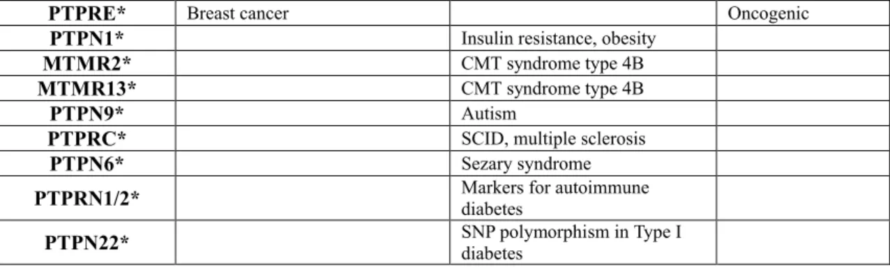
PTPRE* Breast cancer Oncogenic
PTPN1* Insulin resistance, obesity
MTMR2* CMT syndrome type 4B
MTMR13* CMT syndrome type 4B
PTPN9* Autism
PTPRC* SCID, multiple sclerosis
PTPN6* Sezary syndrome
PTPRN1/2* Markers for autoimmune diabetes
PTPN22* SNP polymorphism in Type I diabetes
Table 1.1: Kinases and phosphatases that are implicated in various diseases: Various kinases and
phosphatases, and the diseases they have a role in are shown. Also their effect in cancer (oncogenicity vs. tumor suppression) has been noted. (*) shows phosphatases (see Cohen P, 2001; Krause DS, Van Etten RA, 2005; Arena S et al, 2005; Janssens V et al, 2005; Gallego M, Virshup DM, 2005; Ventura J-J, Nebreda AR, 2006).
Since phosphatases reverse the action of kinases, they can be considered as equally significant in disease formation, although their importance has been started to be discovered only recently. In the case of cancers, phosphatases are generally expected to be products of tumor suppressor genes. However just like there are tumor suppressor kinases, there are phosphatases encoded by protooncogenes and candidates (Table 1.1). For example, wild-type p53-induced phosphatase 1 (Wip1 or PPM1D) has been shown to be amplified and overexpressed in multiple cancer types, including breast and ovarian carcinomas. This phosphatase is known to suppress important tumor suppressors such as p53, ATM, p16INK4A and ARF, hence its overexpression may mediate carcinogenesis through inactivation of these proteins (see Lu X et al, 2008). Phosphatases of regenerating liver (PRLs), especially PRL-1 and PRL-3 have been associated with tumor progression, metastasis and angiogenesis. PRL-3 has been found to be amplified in colorectal cancer (see Arena S et al, 2005; Bessette DC et al, 2008). PTPN11 (Shp2) has been found to be mutated in polycythemia vera (a myeloproliferative disorder characterized by overproduction of red blood cells), Noonan syndrome (a genetic disease affecting the whole body characterized by congenital heart malformation and neurological problems) and LEOPARD syndrome (“lentigines, electrocardiogram abnormalities, ocular hypertelorism, pulmonic stenosis, abnormalities of genitalia, retardation of growth, and deafness,” a multisystem disease). Additionally, somatic mutations of PTPN11 have been found in approximately 35% of patients with sporadic JMML (juvenile myelomonocytic leukemia) (see Chan G et al, 2008). Furthermore PTPN11 overexpression has been implicated in primary adult AML, CML and ALL (Xu R et al, 2005). PTPRE has been
implicated in aiding Neu (HER-2 or ERBB2) -induced mammary tumorigenesis (see Berman-Golan D et al, 2008). In addition to oncogenic phosphatases, there are also tumor suppressor and candidate phosphatases. One of the best known tumor suppressor phosphatase is PTEN, a dual specificity phosphatase that can act both on proteins and lipids, and its main target is PIP3. Deletion of PTEN has been observed in
a variety of cancers including glioma, prostate, breast, endometrial and colorectal cancer. Another well known tumor suppressor phosphatase is PP2A, as it is known that small t antigen of SV40 tumor virus targets this phosphatase system to promote tumor formation. This is thought to occur by activation of PIP3 signaling pathway and
stabilization of c-myc oncoprotein as both are targets of PP2A under normal physiological conditions (see Gallego M, Virshup DM, 2005). Just like kinases, phosphatases take part in a number of other diseases ranging from insulin resistance and obesity to myopathies and sensory neuropathies such as Charcot-Marie-Tooth (CMT) disease (Table 1.1).
Although there are many kinases and phosphatases whose role in cancer is well known as already mentioned, not everything is black and white for the functions of rest of these enzyme families in cancer development and progression. For example, there are controversial data on the role of MKPs in carcinogenesis. This is only normal because although ERK mainly shows oncogenic effect, the effects of JNK and p38 MAPKs changes according to the cancer type and stage. They act to suppress some cancers mainly through induction of apoptosis, whereas in some other cancers in which inflammation contributes to carcinogenesis, they act in favor of cancer development. Another complication especially important for JNK is that the duration and magnitude of signal mediated through it causes differential results such as proliferation versus apoptosis. Hence negative regulators of these two MAPK subtypes act either as oncoproteins or tumor suppressors depending on the cancer type, stage, the duration and magnitude of signaling (see Keyse SM, 2008; Krishna M, Narang H, 2008).
In an additional study, researchers have employed RNA interference to downregulate each known kinase and phosphatase in HeLa cell line, and have found that 73 kinases and 72 phosphatases are necessary for survival and growth and 12 other phosphatases function in sensitizing cells to apoptosis (MacKeigan JP et al, 2005), further
suggesting that kinases and phosphatases are vital for cells and that knowledge on these enzymes may lead to new treatment options for a large variety of diseases.
1.2 Hepatocellular carcinoma
Liver cancer is the fifth most common cancer in men and the eighth in women worldwide. It is also one of the leading causes of cancer-related deaths worldwide, ranking third in men and sixth in women (American Cancer Society, 2007). This high lethality rate is attributable in part to a resistance to existing anticancer agents, a lack of biomarkers that can detect surgically resectable incipient disease, and underlying liver disease that limits the use of chemotherapeutic drugs. Primary liver cancers include hepatocellular carcinoma, intrahepatic bile duct carcinoma (cholangiocarcinoma), hepatoblastoma, bile duct cystadenocarcinoma, haemangiosarcoma and epitheloid haemangioendothelioma, of which hepatocellular carcinoma is the most common (see Farazi PA, DePinho RA, 2006).
Hepatocarcinogenesis proceeds through a multi-step histological course. The discussed risk factors act to promote rounds of hepatocyte necrosis and regeneration that pave the way for development of a chronic liver disease. Liver injury and exposure to various cytokines, in a chronic liver disease condition, provoke stellate cell activation, which is associated with cellular proliferation and the robust synthesis of extracellular matrix components such as collagen, therefore contributing to liver fibrosis. Meanwhile the destruction-proliferation cycles also promote hepatocyte proliferative arrest due to telomere shortening (telomerase reactivation has also been associated with hepatocarcinogenesis, although the exact timing is not known). These changes lead to cirrhosis which is characterized by regenerative nodules surrounded by fibrous scar tissue (collagen). These nodules transform into hyperplastic nodules of regenerating hepatocytes which represent a potential first step towards HCC. Hyperproliferation of hepatocytes in these nodules causes accumulation of mutations and chromosome aberrations that leads to genomic instability, and results in pre-malignant dysplastic nodules characterized by accumulation of lipids or glycogen inside the cell and nuclear crowding. Also abnormal liver architecture is seen at this stage. The dysplastic nodules can evolve into HCC due to high genomic instability and loss of p53. HCC has the capacity to invade the surrounding fibrous stroma and
vessels, and occasionally has metastatic potential. HCC can be further classified into well differentiated, moderately differentiated and poorly differentiated tumors, the last being the most malignant form of primary HCC. (see Goodman ZD, 2007; Farazi PA, DePinho RA, 2006).
1.2.1 Aetiologies of hepatocellular carcinoma
Fig.1.4: Risk factors and mechanisms of hepatocarcinogenesis (see Farazi PA, DePinho RA, 2006).
Hepatitis B virus (HBV) is the main causal factor of HCC globally, due to the fact that it is one of the most common diseases in the world, with an estimated 350 million chronically infected carriers worldwide. HBV is a single-stranded DNA virus of the hepadnavirus family that is integrated into the host genome. The integration of virus DNA may activate cellular proto-oncogenes or cause microdeletions that can target cancer-relevant genes, although no consistent integration sites have been observed (see Lok AS, 2000). HBx protein has transcriptional activation activity that has been shown to alter the expression of growth-control genes, such as src tyrosine kinases, Ras, Raf, ERK and JNK (Tarn C et al, 2001). HBx protein has also been shown to bind and inactivate p53 by sequestration in vitro and to block p53 mediated apoptosis in vivo. ER stress and oxidative stress might also result due to viral-ER physical interactions (see Farazi PA, DePinho RA, 2006).
Hepatitis C virus (HCV) is known to be the greatest risk factor for development of HCC, increasing the rate of HCC formation in patients with HCV approximately 17-fold (Degos F et al, 2000). It is an RNA virus of the flaviviridae family. It has no reverse transcriptase activity and hence does not integrate itself into the host genome. Still, HCV shows a higher propensity to yield chronic infection which might relate to immune evasion by this virus, and that HCV shows a higher propensity to promote liver cirrhosis (see Farazi PA, DePinho RA, 2006). HCV core proteins have transcription regulatory functions on many different host genes, including the proto-oncogene c-myc (Ray RB et al, 1995). HCV core proteins are also known to inhibit multiple activators of apoptosis, including Fas and tumor necrosis factor-α (TNF-α) (Marusawa H et al, 1999; Jin X et al, 2006). These effects of HCV core proteins may be exerted through the constitutive activation of ERK signaling cascade (Hayashi J et al, 2000). In addition to the core proteins, HCV nonstructural protein NS5A have been shown to interact with and inactivate p53 by sequestration to the perinuclear membrane (Majumder M et al, 2001). ER-stress and oxidative-stress-mediated mechanisms might also be possible for HCV-induced HCC (see Farazi PA, DePinho RA, 2006). Additionally HCV might be impairing immune system by interfering with T-cell activation (see Pachiadakis I et al, 2005).
Chronic alcohol intake has been implicated in causing production of pro-inflammatory cytokines through monocyte activation, in increasing intestinal permeability to bacteria/lipopolysaccharides, leading to Küpffer cell activation. Küpffer cells are specialized macrophages located in the liver and they release many chemokines and cytokines upon activation (including TNF-α, interleukin-1β, interleukin-6 and prostaglandin E) with adverse effects on hepatocyte survival. Chronic ethanol exposure causes hepatocytes to show increased sensitivity to the cytotoxic effects of TNF-α which may be responsible for chronic hepatocyte destruction-regeneration cycles leading to stellate cell activation, fibrosis, cirrhosis and ultimately HCC (see Farazi PA, DePinho RA, 2006; McKillop IH et al, 2006). Alcohol also damages the liver through oxidative stress mechanisms that result upon hepatic ethanol metabolism (see McKillop IH et al, 2006).
Aflatoxins are mycotoxins from Aspergillus flavus and Aspergillus parasiticus. Hepatic cytochrome p450 metabolizes aflatoxin (AFB1) and converts it to its highly
reactive exo-8,9-epoxide form. This epoxide reacts with guanine nucleotides in the hepatocyte DNA to form a number of adducts, which lead to heritable mutations that result in hepatocarcinogenesis (see McKillop IH et al, 2006). The main mechanism of AFB1 in hepatocarcinogenesis has been found to be GC→TA transversion at the third
position of codon 249 of p53 gene (resulting in an Arg→Ser alteration in the protein) (Puisieux A et al, 1991).
Other aetiological factors in HCC can be listed as follows:
Non-alcoholic fatty liver disorders (NAFLD) and non-alcoholic steatohepatitis (see Farrell GC, Larter CZ, 2006).
Type 2 diabetes and its associated hyperinsulinemia and hyper-IGF-1 production (El-Serag HB et al, 2004).
Certain metabolic disorders such as hereditary haemochromatosis, porphyria cutanea tarda, Wilson’s disease and primary biliary cirrhosis (see McKillop IH et al, 2006; Farazi PA, DePinho RA, 2006).
Possibly use of oral contraceptives through their estrogen and progesterone components (see Maheshwari S et al, 2007; El-Serag HB, Rudolph KL, 2007).
1.3 Protein kinases and phosphatases implicated in hepatocarcinogenesis
Disruption of cell signaling is frequently seen in hepatocarcinogenesis. Mutations and other regulatory problems in p53 tumor suppressor, Wnt/β-Catenin pathway, growth factor receptors (including ErbB receptor family and c-MET) and their associated pathways, pRb tumor suppressor, Ras proteins and associated pathways, Janus kinases / signal transducers and activators of transcription (JAK/STAT) pathway are observed.
Deregulation of protein phosphorylation in important signaling pathways is very important in initiation and progression of HCC. For example, in a recent study, PRL-3 has been found to be significantly upregulated in HCC tumor tissues compared to the paired noncancerous liver tissues. The mRNA level of PRL-3 in tissues was also correlated with serum α-fetoprotein level, vascular invasion and metastasis in this study. Furthermore, a significant correlation between PRL-3 mRNA expression and
MMP-2, MMP-9 and E-cadherin has been found (Zhao WB et al, 2008). Another example is the tyrosine kinase p60c-src (SRC), which is overactivated in hepatoma cells and this is thought to account for the desensitization of liver tumor cells to TRAIL and CD95. SRC works with EGFR in desensitizing cells to apoptosis (De Toni EN et al, 2007). Another recent example may be DUSP1 (MKP-1). The expression of this MAPK phosphatase, a negative regulator of ERK, is dysregulated in liver tumors with poor prognosis. This is suggested to be through increased ubiquitination of DUSP1 by S-phase kinase-associated protein 2 (SKP2)/CDC28 protein kinase 1b (CKS1) ubiquitin ligase complex which leads to increased proteasomal degradation of DUSP1 (Calvisi DF et al, 2008). A final example comes from a very recent study on Rho-associated, coiled-coil containing protein kinase 2 (ROCK2) which has been found to be overexpressed in about 54% of the 41 HCC samples and this overexpression has correlated with a more aggressive phenotype. Further experiments have also associated ROCK2 with increased invasiveness in HCC, which is in accordance with the fact that ROCKs normally regulate actin cytoskeleton and cell motility (Wong CC et al, 2009).
In addition to these specific and novel examples, there are a number of important signaling pathways known to be deregulated in HCC, in which kinases and phosphatases play major roles (Fig.1.5).
1.3.1 Growth factor receptor tyrosine kinases
ErbB family of receptor tyrosine kinases consists of ERBB1 (EGFR), ERBB2 (HER2/neu), ERBB3 and ERBB4. EGFR has been found to be overexpressed in 4-70% of HCC cases, HER2 in 0-30%, ERBB3 in 84% and ERBB4 in 61% (see Breuhahn K et al, 2006; Höpfner M et al, 2008; Farazi PA, DePinho RA, 2006).
Fig.1.5: Major growth factor receptor signaling pathways important in HCC: Predominantly
dysregulated components of signaling pathways are highlighted in dark gray. Seldom regulated components (eg. Smad 2/3), molecules not expressed by tumor cells (eg. HGF), and distinct protein family members dysregulated in HCC (eg. FZD-7) are highlighted in light gray (see Breuhahn K et al, 2006).
Considering IGF signaling, expression of IGF-2R has been found to be reduced in 63% of human HCC. Loss of heterozygosity at the igf-2r locus has also been observed in HCC and its premalignant lesions coupled to inactivating mutations of the second allele in 25% of the cases. Furthermore, several missense mutations are reported in HCC which target the extracellular domain of IGF-2R (see Breuhahn K et al, 2006; Höpfner M et al, 2008).
Mesenchymal-epithelial transition factor (MET) is a proto-oncogene encoding for a membrane receptor tyrosine kinase, of which hepatocyte growth factor (HGF) is the only known ligand. Overexpression of the MET receptor has been observed in 20-48% of tumor samples. This induction in HCC cells may be attributed to genomic alterations (7q gains have been observed in 16.8% of HCCs), tumor hypoxia or HGF-dependent transcriptional activation of MET (Breuhahn K et al, 2006; Höpfner M et al, 2008; Farazi PA, DePinho RA, 2006).
There are different data on the expression of TGFβ receptors, which are serine-threonine kinases, in HCC. There are studies that have not detected a change in
expression levels of the receptors. In another study, upregulation of TGF-βRI levels has been detected in 60% of the cases. In yet another one, downregulation of TGF-βRI levels has been detected in 80% of the cases. In some studies, including this latter study, downregulation of TGF-βRII levels has been detected in 37-70% of the cases. Overall, most studies document a reduction of the receptors in up to 70% of HCCs. (Breuhahn K et al, 2006).
1.3.2 Kinases and phosphatases of PI3K/PTEN/AKT/mTOR pathway
A major activation of this pathway can be either due to higher activity of receptor tyrosine kinases or to the reduced expression of PTEN, a negative regulator of the pathway. 10q loss is a genomic alteration that occurs in 17-27% of HCC cases, and this region also contains the PTEN gene. Furthermore, somatic missense mutations, frame shifts, splice site mutations and loss of promoter activity of PTEN have also been observed in HCC tissues and cell lines (Kawamura N et al, 1999; Ma DZ et al, 2005; Fujiwara Y et al, 2000). Downstream of this pathway, mTOR, a serine-threonine kinase implicated in the regulation of translation, is overexpressed in %50 of HCC cases (n=314), as found in a recent broad study. Additionally, chromosomal gains in RICTOR were observed in 25% of patients (Villanueva A et al, 2008).
1.3.3 Mitogen-activated protein kinase (MAPK) pathways
The mitogen-activated protein kinase (MAPK) family consist of five subgroups, including the extracellular signal-regulated kinase homologs 1 and 2 (ERK1/2), big MAPK-1 (BMK-1/ERK5), c-Jun N-terminal kinase homologs 1, 2 and 3 (JNK1/2/3), stress-activated protein kinase 2 (SAPK-2) homologs α, β, and δ (p38α/β/δ), and ERK6 (p38γ). These kinases are activated through dual phosphorylation of T and Y residues located in their activation loop by their corresponding MAPK kinases (MKKs). MAPKs are implicated in diverse cellular processes such as cell growth and proliferation, differentiation, adhesion, apoptosis and stress response (see Pimienta G, Pascual J, 2007). Major MAPK pathways and their components are shown in Fig.1.6.
Fig.1.6: Mitogen-activated protein kinase pathways (see Zhang Y, Dong C, 2005).
Extracellular signal-regulated kinases (ERKs) or classical MAP kinases are widely expressed and are involved in functions including the regulation of cell proliferation and differentiation. Many different stimuli, including growth factors, cytokines, virus infection, ligands for heterotrimeric G protein-coupled receptors, transforming agents, and carcinogens, activate the ERK pathway. Main isoforms are ERK1 and ERK2 (Krishna M and Narang H, 2008).
c-Jun N-terminal kinases (JNKs), originally identified as kinases that phosphosphorylate c-Jun on Ser63 and Ser73 within its transcriptional activation domain, are responsive to stress stimuli, such as cytokines, ultraviolet irradiation, heat shock, and osmotic shock. They are involved in cell differentiation, proliferation and apoptosis. Main isoforms are JNK1 and JNK2 which are ubiquitiously expressed, and JNK3 which is only found in brain, heart and testes (Krishna M and Narang H, 2008).
Similar to JNK pathway, p38 MAP kinases are activated by a variety of cellular stresses including osmotic shock, heat shock, inflammatory cytokines, lipopolysaccharides (LPS), ultraviolet light and growth factors. They are involved in cell differentiation, apoptosis and Ras-induced senescence. Main isoforms are p38α, p38β, p38γ (also known as ERK6) and p38δ (Krishna M and Narang H, 2008).
Other MAPKs are: ERK5, found recently, is activated both by growth factors and stress stimuli, and participates in regulation of cell proliferation, and, atypical MAPKs ERK3/4 and ERK7/8 which are cytoplasmic proteins and possess a C-terminal extension. Atypical MAPKs are also recently found and not much is known on them (Krishna M and Narang H, 2008).
Proteins of HBV, HCV and hepatitis E virus target and modulate multiple steps along MAPK pathway. In one study, it has been shown that HCV E2 protein, one of the two envelope glycoproteins of hepatitis C virus, activates the MAPK pathway in human hepatoma Huh-7 cells and promotes cell proliferation (Zhao LJ et al, 2005). Furthermore, increased levels of ERK have been observed in HCC and are known to correlate with tumor progression (Tsuboi Y et al, 2004). In another study, expression of Spred protein (Sprouty-related protein with Ena/vasodilator-stimulated phosphoprotein homology-1 domain), an inhibitor of the Ras/Raf-1/ERK pathway has been found to be deregulated in human HCC. Also in this study, ectopic overexpression of Spred has been observed to cause inhibition of ERK activation both in vivo and in vitro, resulting in reduced cancer cell proliferation and low secretion of MMP2 and MMP-9 (Yoshida T et al, 2006).
1.4 Cellular senescence and liver cirrhosis
Liver cirrhosis is the greatest clinical risk factor in hepatocarcinogenesis regardless of the aetiology underneath (HBV, HCV, aflatoxin or alcohol). In fact when compared to other cancers, HCC is characterized by an underlying cirrhosis condition, and 45-90% of all HCCs worldwide occur in the setting of liver cirrhosis. Cirrhosis has life-threatening complications other than HCC, ascites (fluid retention in the abdominal cavity) being the most common one, leading to an increased risk of infection and a poor long-term outcome (see Goodman ZD, 2007).
As already mentioned, liver cirrhosis is characterized by abnormal nodules of regenerating hepatocytes surrounded by fibrous scar tissue. Telomere shortening and senescence have been shown as the general markers of human liver cirrhosis, also correlating with progression of fibrosis in cirrhosis (Wiemann SU et al, 2002).
Cellular senescence was initially defined as the loss of proliferative capacity of cells in culture typically after cultures have been passaged 50 or more times (Sherwood SW et al, 1988; Hayflick L, 1965). Cellular senescence, the state of stable cell cycle arrest, can be provoked by a variety of stimuli, such as telomere shortening, DNA damage, activation of certain oncogenes and oxidative stress (Fig.1.7). Senescence is associated with a number of gross cellular changes including cell-cycle arrest (see Collado M et al, 2005; Herbig U and Sedivy JM, 2006), increase in cell size and size heterogeneity, and increase in the frequency of cells with chromosomal aberrations, including polyploidy (Sherwood SW et al, 1988). Senescent cells display SABG (senescence-associated β-galactosidase) activity at pH 6.0 and this activity can be used as a marker to identify senescent cells. Other characteristic features of senescent cells include presence of p16INK4a, senescence-associated DNA-damage foci and senescence-associated heterochromatin foci (see Ozturk M et al, 2008). Cellular senescence acts as a barrier to cancer, preventing damaged cells from undergoing aberrant proliferation (see Chen JH et al, 2007, Campisi J, 2005), although it probably also has deleterious effects on organisms such as tissue aging (see Ozturk M et al, 2008). Two well-established tumor suppressor proteins, pRb and p53, have been shown to play key roles in cellular senescence (Fig.1.8) (see Ben-Porath I and Weinberg RA, 2004).
Fig.1.8: Molecular mechanisms of senescence (see Funayama R, Ishikawa F, 2007).
1.4.1 Replicative senescence and telomere shortening
Telomeres progressively shorten with age in somatic cells in culture and in vivo because DNA replication results in the loss of sequences at the 5' ends of double-stranded DNA. Whereas somatic cells do not express the enzyme telomerase which adds repeated telomere sequences to chromosome ends, telomerase activity is detected in immortalized and cancer cells in vitro and in primary tumor tissues. This represents an important difference between normal cells and cancer cells, suggesting that telomere shortening causes cellular senescence (see Oshimura M and Barrett JC, 1997). This form of senescence is called as replicative or telomere-dependent senescence. There is accumulating evidence that when only a few telomeres are shortened, they form end-associations, resulting in a DNA damage response which leads to replicative senescence (see Shay JW and Wright WE, 2004). This DNA damage signal leads to the activation of cell cycle checkpoint pathways involving p53, p16INK4a, and/or retinoblastoma (pRb) proteins (see Campisi J, 2005; Dimri GP, 2005). Inactivation of p53 and p16INK4a genes (see Shay JW and Bacchetti S, 1997) and reactivation of hTERT gene expression (see Sherr CJ and McCormick F, 2002) cause cells to gain replicative immortality (see Ozturk M et al, 2008).