GENETIC AND EPIGENETIC TARGETS IN LIVER
CANCER
A THESIS SUBMITTED TO
THE DEPARTMENT OF MOLECULAR BIOLOGY AND GENETICS AND THE INSTITUTE OF ENGINEERING AND SCIENCE OF
BİLKENT UNIVERSITY
IN PARTIAL FULFILLMENT OF THE REQUIREMENTS FOR THE DEGREE OF DOCTOR OF PHILOSOPHY
BY
HALUK YÜZÜGÜLLÜ AUGUST, 2010
I certify that I have read this thesis and that in my opinion it is fully adequate, in scope, and in quality, as a thesis for the degree of Doctor of Philosophy.
_____________________ Prof. Dr. Mehmet Öztürk
I certify that I have read this thesis and that in my opinion it is fully adequate, in scope, and in quality, as a thesis for the degree of Doctor of Philosophy.
______________ Prof. Dr. Kuyaş Buğra
I certify that I have read this thesis and that in my opinion it is fully adequate, in scope, and in quality, as a thesis for the degree of Doctor of Philosophy.
________________ Prof. Dr. İhsan Çalış
I certify that I have read this thesis and that in my opinion it is fully adequate, in scope, and in quality, as a thesis for the degree of Doctor of Philosophy.
____________________ Assoc. Prof. Dr. İhsan Gürsel
I certify that I have read this thesis and that in my opinion it is fully adequate, in scope, and in quality, as a thesis for the degree of Doctor of Philosophy.
________________
Assist. Prof. Dr. Uygar Tazebay
Approved for the Institute of Engineering and Science
____________________ Prof. Dr. Levent Onural Director of the Institute of Engineering and Science
ABSTRACT
Genetic and Epigenetic Targets in Liver Cancer
Haluk Yüzügüllü
Ph.D. in Molecular Biology and Genetics Supervisor: Prof. Dr. Mehmet Öztürk
August, 2010, 143 Pages
Hepatocellular carcinoma (HCC) kills nearly 600.000 people each year and the only effective therapy for this cancer is liver transplantation or tumor ablation only when the tumor is small enough. These tumors are surprisingly resistant to conventional therapies such as chemotherapy and radiotherapy. Moreover, as HCC is almost always associated with cirrhosis, the treatment with cytotoxic agents is dangerous as they will also affect hepatic functions of the diseased liver. Therefore, there is urgent need to find novel therapeutic approaches against HCC in order to diminish death toll. Our overall goal is to discover “druggable target genes” in HCC. In other words, we wish to identify novel genes and novel mechanisms involved in these cancers in order to use them as potential therapeutic targets.
During my thesis work, I developed different approaches to find new mechanisms and novel targets:
1- Deciphering the role of canonical Wnt signaling in HCC: We classified human HCC cell lines into "well-differentiated" and "poorly differentiated" subtypes, based on the expression of hepatocyte lineage, epithelial and mesenchymal markers. Poorly differentiated cell lines lost epithelial and hepatocyte lineage markers, and overexpressed mesenchymal markers. Also, they were highly motile and invasive. We compared the expression of 45 Wnt pathway genes between two subtypes. Likewise, six Frizzled receptors, and canonical Wnt3 ligand were expressed in both subtypes. In contrast, canonical ligand Wnt8b and noncanonical ligands Wnt4, Wnt5a, Wnt5b and Wnt7b were expressed selectively in well- and poorly differentiated cell lines, respectively. Canonical Wnt signaling activity, as tested by a TCF reporter assay was detected in 80% of
well-suggesting a repressive mechanism. We tested Wnt5a as a candidate antagonist. It strongly inhibited canonical Wnt signaling that is activated by mutant β-catenin in HCC cell lines.
2. Systematic screening of protein kinases and phosphatases as potential therapeutic targets: There is evidence of aberrant activation of several signaling cascades in HCC, and a multikinase inhibitor, sorafenib, has shown survival benefits in patients with advanced HCC. We used siRNAs to screen a large number of kinases and phosphatases to identify related genes involved in HCC cell survival. A total of 7 kinases and 5 phosphatases were identified as strong candidate targets.
3-Screening of a set of selected epigenetic regulators as potential therapeutic targets: Recent studies have indicated that senescence arrest or senescence escape could be regulated by epigenetic changes on chromatin. We wanted to identify key histone methylation and acetylation changes associated with senescence or senescence escape, and select key histone modifying enzymes, as potential targets for “pro-senescence” interventions (therapeutic interventions that allow senescence induction in cancer cells). We identified ATAD2 as an epigenetic target, and found ATAD2 gene overexpressed in HCC compared to normal liver. We also found a stepwise increase of ATAD2 protein expression in late stages with respect to pre-neoplastic and early stage during hepatocellular carcinogenesis. ATAD2 knockdown using siRNAs in cancer cells leads to increase in global histone acetylation and inhibit cell proliferation and induce caspase-3 dependent apoptosis in HCC and induce senescence in MRC5 cells. Its potentiator role (coactivator role with estrogen, androgen and myc targets) indicates ATAD2 as a potential therepeutic target for HCC.
ÖZET
Karaciğer kanserinde Genetik ve Epigenetik Hedefler
Haluk Yüzügüllü
Moleküler Biyoloji ve Genetik Doktorası Tez Yöneticisi: Prof. Dr. Mehmet Öztürk
Ağustos, 2010, 143 sayfa
Karaciğer kanseri en sık rastlanan birincil kanserlerden olup, yılda 600.000 civarında ölüme sebep olmaktadır. Şu andaki en etkili tedavi yöntemi karaciğer transplantasyonudur ve tümör ablasyon yöntemi sadece tümör yeterince küçük ise uygulanabilmektedir. Kanserli hücreler kemoterapi ve radyoterapi gibi yöntemlere oldukça dayanıklıdırlar. Dahası, karaciğer kanseri sürekli siroz üzerine geliştiği için karaciğerin diğer hepatik fonksiyonlarını yerine getirmesini de engellediği için, sitotoksik ajanlar kullanmak oldukça tehlikelidir. Bu nedenle karaciğer kanserini yenmek için daha yeni ve geçerli terapatik yöntemlere ihtiyac duyulmaktadır.
Bizim amacımiz karaciğer kanserinde yeni hedeflenebilir genleri keşfetmektir. Başka bir değişle, biz bu çalışma ile karaciğer kanserinde yeni hedefler ve yeni mekanizmalar keşfederek bunları potansiyel terapötik hedefler olarak kullanmayı amaçladık.
Tez çalışmalarım sırasında, yeni hedefler ve yeni mekanizmalar keşfetmek için değişik yöntemler kullanmaya çalıştım:
1- Karaciğer kanserinde kanonik Wnt sinyallenme yolağının rolü: karaciğer kanserı hücrelerini hepatosit spesifik, epitel ve mezenkimal markörler kullanarak “iyi diferansiye” ve “kötü diferansiye” hücre hatları olarak iki gruba ayırdık. Kötü diferansiye hücre hatlarında epitel ve hepatosit spesifik markörlerin negatif hale geldiğini, mezenkimal markörleri anlatmaya başladığını gösterdik. Wnt sinyal yolağında görev alan 45 genin, gen anlatım seviyelerini iki grupta inceledik. 6 Frizzled almaç geninin ve wnt3 ligandının her iki grupta da anlatıldığını gösterdik. Kanonik Wnt ligandı wnt8b`nin sadece iyi diferansiye grupta
sinyallenmesinin sadece iyi diferansiye hücrelerde seçici olarak aktif olduğunu değil, aynı zamanda kötü diferansiye tümörlerde de baskılandığını gösterdik. Kanonik olmayan hücre hattı, Snu449 da mutant beta-kateninin ektopik anlatımının TCF aktivitesini arttıramaması bu gruptaki hücrelerde baskılayıcı bir mekanizma olduğuna işaret etti. Kanonik olmayan wnt5a genini aday olarak test ettik. Wnt5a geninin mutant beta-kateninin aktive ettiği TCF aktivitesini baskıladığını gösterdik. Bazı kanser tiplerinde, APC, Axin ve beta-katenin gibi genlerde oluşan mutasyonlarla Wnt-beta-katenin sinyallenmesinin, Wnt ligandlarının olmadığı ortamda anormal olarak aktifleşip, beta-kateninin sitoplazma ve çekirdekte birikmesine sebep olup hedef genlerin aktivasyonuna yol açtığı biliniyor. Bu nedenle beta-kateninin hücre zarı, sitoplazma ve çekirdekteki fraksiyonlarını tanıyabilen monoklonal antikorlar üretip, bu antikorları kanserli dokularda test ederek klinik değerlerini karakterize ettik. 2- Kinaz ve fosfataz genlerini potansiyel terapatik hedef olarak sistematik olarak
taranması: Karaciğer kanserinde değişik sinyallenme yolaklarının aktif hale geldiği ve bu hastalarda multikinaz inhibitorü olan sorafenib ilacının, ileri derece hastaların ömürlerini uzattığı biliniyor. Bu amaçla yeni hedefler bulmak ve hücre yaşlanması indüklenmesi yönünde terapatik hedef gösterebilmek için çok fazla sayıda kinaz ve fosfatazları hedef alan RNA müdehalesi yöntemi ile tarama gerçekleştirdik. Toplamda 7 kinaz ve 5 fosfataz genini güçlü adaylar olarak gösterildi.
3- Seçilen epigenetik hedeflerin potansiyel terapötik hedef olarak taranması: Yakın dönemde yapılan çalışmalar, hücre yaşlanması ve bundan kaçışın kromatin üzerindeki epigenetik değişiklikler ile düzenlendiğini gösterdi. Bu amaçla hücre yaşlanması ve bundan kaçış ile uyumlu histon asetillenmesi ve metillenmesi değişiklikleri tanımladık, bundan sorumlu histon enzimlerini bulup, bunları “hücre yaşlanması öncüleyici” hedefler (kanserli hücreleri hücre yaşlanmasına götüren) olarak göstermeyi hedefledik. ATAD2 genini epigenetik hedef olarak tanımlayıp, karaciğer kanserinde normal hepatositlere göre daha fazla gen ifadesi olduğunu gösterdik. ATAD2 proteinin öncü-neoplastik ve ilk aşama karaciğer
Kanserli hücrelerde siRNA tekniği kullanarak, ATAD2 proteinini azaltıp ve buna bağlı global histon asetillenmesinin arttığını, hücre çoğalmasının azaldığını, hücreleri apoptoza götürdüğünü ve MRC5 akciğer fibroblastlarında ise hücre yaşlanmasını indüklediğini gösterdik. ATAD2 geninin potansiyel artırıcı (östrojen, androjen ve myc hedefleri üzerinde) etkisinden dolayı karaciğer kanserinde aday terapötik hedef olduğunu işaret etmektedir.
ACKNOWLEDGMENTS
I would like to begin thanking to my thesis advisor Prof. Mehmet Öztürk for all that I have learnt from him. Without his support, guidance, and patience, none of this would be possible. I am also thankful that he let me learn new techniques, introduce me to new networks and gave me more freedom in the lab. He was a lot more than a supervisor for me.
I would like to thank Dr. Rengül Çetin-Atalay, for her knowledge, experience, support and patience.
I would like to thank TUBITAK for financial supports for 2211 and 2214 programs. I would like to thank also to all members of Equipe 2 and 5, especially Dr. Jean Luc Coll for their reagents and technical helps. To Ralph Meuwissen for his career advices, discussions, and share his experience.
I also want to thank Dr. Stephan Dimitrov for his career and scientific advices.
I am also grateful to all the other researchers and fellows in Bilkent University for providing me such a wonderful scientific and friendly environment. It will definitely be an unforgettable memory for me.
I thank all the current and former members of Ozturk Lab, for their help and experience they shared. Especially I am grateful to Nuri Ozturk for his invaluable discussion and scientific help. I want to thank also Hani Al Otaibi, Khemais Benhaj, Pelin Telkoparan and Bilge Kilic for their both technical help and advice.
I would like thank my family and especially to my parents for their never-ending support and care. I am away from them for almost 14 years and I hope it is worth it.
And throughout my journey, she is the one that believed me and always supported me even in very difficult times. A fellow and beloved, Özge, she has been always with me and I believe I cannot even thank her enough for her help and support both intellectually and technically and the wonderful meaning she gave to my life…
TABLE OF CONTENTS SIGNATURE PAGE………...….I ABSTRACT...III ÖZET...V ACKNOWLEDGEMENTS ...VII TABLE OF CONTENT...VIII LIST OF TABLES ...XV LIST OF FIGURES ...XVI ABBREVIATIONS...XX
CHAPTER 1. INTRODUCTION……….………1
1.1 Hepatocellular malignancy...1
1.2 Aetiological factors of hepatocellular carcinoma...1
1.2.1 Viral induced hepatocarcinogenesis……….………....2
1.2.2 Alcohol and cirrhosis...2
1.2.3 Aflatoxin B1 induced hepatocarcinogenesis...3
1.3 Genetic and Epigenetic events in HCC……...3
1.4 Role of β-catenin In HCC...5
1.4.1 Structure of beta-catenin protein and interaction partners...7
1.5 Protein Kinase and Phosphatases………...10
1.5.1 Role of Kinases and Phosphatases in cancer…………...13
1.5.2 Role of kinases and Phosphatases in liver cancer………...13
1.6 Role of H19/Igf2 locus in HCC…………...13
1.7 Senescence of hepatocytes and chronic liver disease…...13
1.8 Histone modifications………...15
1.8.1 Histone modifications and cancer………. ...18
1.8.2 H3K4 Methylation…...19
1.8.2.1 MLL and SMYD3 – H3K4 methyltransferases...19
1.8.3 H3K9 Methylation………...19
1.8.5 H3K36 methylation...21 1.8.5.1 Setd2 methyltransferases...21 1.8.5.2 FBXL11 Histone demethylases...21 1.8.6 H4K20 methylation...21 1.8.6.1 Suv420H1 methyltransferases...22 1.9 DNA Methylation………22
1.9.1 DNA Methylation and Cancer……….22
1.10 Role of ATAD2 gene in cancer………..23
CHAPTER 2. OBJECTIVES AND RATIONALE………...24
CHAPTER 3. MATERIALS AND METHODS…………...……….26
3.1 MATERIALS...26 3.1.1 Reagents...26 3.1.2 Bacterial Strains………..27 3.1.3 Enzymes………..27 3.1.4 Nucleic Acids………..27 3.1.5 Oligonucleotides……….28
3.1.6 Electrophoresis and photography, luciferase assay, ELISA readings and spectrophotometer ………..28
3.1.7 Tissue culture reagents………29
3.1.8 Antibodies and chemiluminescence………...29
3.2 SOLUTIONS AND MEDIA...29
3.2.1 General Solutions………29
3.2.2 Microbiological media, reagents and antibiotics…...………30
3.2.3 Tissue culture solutions………..31
3.2.4 BES Transfection solutions………31
3.2.5 Antibiotics……….…………..31
3.2.6 Immunoblotting solutions……….………..32
3.2.7 RNA Study solutions………..32
3.3 METHODS………..……32
3.3.1 General Methods………....32
3.3.1.1 Long term storage of bacterial strains………..32
3.3.1.2 Purification of plasmid DNA using Qiagen miniprep kit………33
3.3.1.3 Large Scale Plasmid DNA purification……….………..33
3.3.1.4 Quantification and qualification of nucleic acids ………...33
3.3.1.5 Restriction enzyme digestion of DNA………....33
3.3.1.6 Gel electrophoresis of nucleic acids ………...34
3.3.1.6.1 Horizontal agarose gels of DNA samples……….34
3.3.2 Computer analyses………...……….… 34
3.3.3 Characterization of Ig subtype of the anti-β-catenin antibodies…………34
3.3.4 Tissue culture techniques……...………34
3.3.4.1 Cell lines and stable clones………..34
3.3.4.2 Cryopreservation of cell lines………..35
3.3.4.3 Thawing of cell lines…….………...35
3.4 Growth conditions of cells……….36
3.5 Transfection and Virus Production ………..… 36
3.6 Transduction of cells with viruses ………...………..…...37
3.7 Transfection and Colony-forming ability assay .……….….37
3.8 Western Blotting…………..………..……38
3.9 Senescence-associated beta-galactosidase (SABG) assay …………..….38
3.10 Indirect immunofluorescence ………...38
3.11 Cell cycle analysis and bromodeoxyuridine (BrdU) incorporation assay ……….………..38
3.12 Detection of Apoptosis using Annexin V staining………39
3.13 Detection of Histone acetylation levels using FACs………39
3.14 Treatment of cell lines with TGF-beta and Doxorubucin ………....39
3.15 RNAi screening protocol ………..39
3.18 Xenograft models ………..41
3.19 Optical imaging of mice bearing tumors ………..41
3.20 Transient transfection of eukaryotic cells using “Lipofectamine Reagent”……….41
3.21 Extraction of total RNA from tissue culture cells and tissue samples ………41
3.22 First strand cDNA synthesis………..41
3.23 Primer design for expression analysis by semi-quantitative PCR……….42
3.24 Fidelity and DNA contamination control in first strand cDNAs………...42
3.25 Expression analysis of a gene by semi-quantitative PCR………..42
3.26 Protocol for quantitative PCR………42
3.27 Crude total protein extraction………44
3.28 Nuclear extract isolation protocol………..45
3.29 Quantification of Proteins using Bradford Assay………..45
3.30 Luciferase assay……….48
3.31 Immunohistochemical studies for β-catenin Mabs………49
3.32 Immunohistochemistry analysis of ATAD2 using tissue microarray……50
3.33 Statistical analysis………..50
CHAPTER 4. RESULTS………..51
4.1 Canonical Wnt signaling is antagonized by noncanonical Wnt5a in hepatocellular carcinoma cells ………..51
4.1.1 Classification of hepatocellular carcinoma cell lines into "well-differentiated" and "poorly "well-differentiated" subtypes ………51
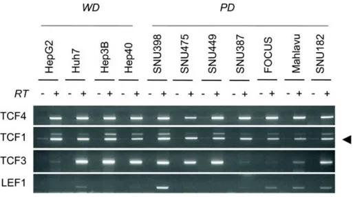
4.1.2 Expression TCF/LEF family of transcription factors………56
4.1.3 Expression of Frizzled receptors and LRP co-receptors………56
4.1.5 Autocrine canonical Wnt signaling in well differentiated hepatocellular
carcinoma cell lines………59
4.1.6 Canonical Wnt signaling is repressed in poorly differentiated hepatocellular carcinoma cells………...61
4.1.7 WNT5A inhibits canonical Wnt signaling in HCC cells………...64
4.2 Selective monoclonal antibodies dirested against C-terminal domain of β-Catenin………...65
4.2.1 Antibody Isotyping………66
4.2.2 Selective immunoreactivity of 4C9 and 9E10 antibodies against cellular β-Catenin………...66
4.2.3 9E10 Mab immunoreactivity with primary tissue and tumor samples…..69
4.3 Targeting human kinome and phosphatome in hepatocellular carcinoma cells using RNAi………73
4.3.1 Validation of Kinase and Phosphatase target genes………..74
4.3.2 In vitro Screening………..76
4.3.3 Development of in vivo screening method………79
4.4 Epigenetic targets in liver cancer………...80
4.4.1 Immunostaining of Histone modifying Enzymes in normal hepatocytes and immortal Huh7 cells and DNA damage induced senescence model………..82
4.4.2 Histones are hypomethylated in HCC compared to cirrhosis and normal hepatocytes in vivo………84
4.4.3 Validation and targeting of histone modifying enzymes responsible for aberrant histone codes………86
4.4.4 Validation of Knock-down of enzymes in HCC cells………...89
4.4.5 Knockdown of enzymes failed to induce apoptosis, senescence and inhibition of growth of cells………..91
4.4.6 Knockdown of enzymes failed to affect main p53 and Rb pathways…...92
4.4.7 Role of Histone 4 K20 methylation and Suv420H1 enzyme in liver cancer……….92
4.4.9 Suv420H1 enzyme is the responsible enzyme for trimethylation of H4K20………94 4.4.10 Suv420H1 failed to change the DNA Damage response of cells to Doxorubucin treatment………..98
4.4.11 Suv420H1 failed to regulate the imprinting of H19 gene in HCC cells…99 4.5 Role of ATAD2 gene in Liver Cancer……….101 4.5.1 Identification of ATAD2………..101 4.5.2 ATAD2 is highly expressed in immortal HCC cells………...102 4.5.3 ATAD2 expression is associated with high grade and increased risk of lymph node invasion of tumor cells……….104 4.5.4 Inhibition of ATAD2 expression induces apoptosis in liver cancer cells and affects the growth of normal cells……….107 4.5.5 Mechanism of Apoptosis in liver cancer cells……….110 4.5.6 Survivin did not rescue loss of ATAD2 induced apoptosis in liver cancer cells………..112
4.5.7 RNAi targeting of ATAD2 inhibited cell proliferation and colony formation of non-liver cells……….113
4.5.8 Loss of ATAD2 induced senescence in MRC5 cells………...116 4.5.9 RNAi knockdown of ATAD2 using tet-inducible lentivirus system…...116 4.5.10 Downregulation of ATAD2 expression by tet-inducible RNAi suppressed tumorigenecity of HCC cells in vivo………...119
4.5.11 Loss of ATAD2 increased histone acetylation and increased p300 histone acetyltransferase levels………120
4.5.12 ATAD2 knockdown induced HAT expression in Liver Cancer cells….122
CHAPTER 5. DISCUSSION………..…………..……….123
5.1 Canonical Wnt signaling is antagonized by noncanonical Wnt5a in hepatocellular carcinoma cells ………123 5.2 Selective monoclonal antibodies directed against C-terminal domain of β-Catenin………..126
5.3 Screening of human kinome and Phosphatome
………...128
5.4 Histone methylation and Role of histone modifying enzymes in liver cancer ………...129
5.5 Role of ATAD2 in hepatocellular carcinoma………..130
CHAPTER 6. FUTURE PERSPECTIVES………..131
LIST OF TABLES
Table 1.1 Mutations in tumor suppressor genes and oncogenes in
HCC……….4
Table 1.2 Kinase distribution by major groups in human and model systems ………..………..10
Table 1.3 Phosphatase distribution by major groups in human and model systems..12 Table 1.4 Histone modifying enzymes and residues modified……….….17
Table 3.1 PCR primer list………. 43
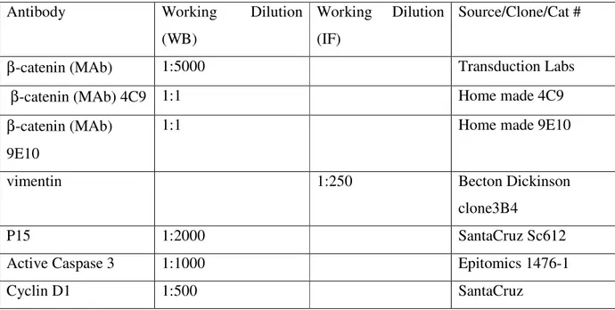
Table 3.2 Antibodies used in this study and their dilution in various protocols ………45
Table 4.1.1: Well-differentiated and poorly differentiated HCC cell lines according to
hepatocyte lineage, epithelial and mesenchymal markers, and in vitro motility and invasiveness assays ………...55
Table 4.2.1: Distribution of membrane form of β-catenin expression in the tumor
samples………...70
Table 4.2.2: Distribution of different forms of ß-catenin expression in 54 colorectal
cancer samples using 9E10 antibody……….73
Table 4.3.1: Validation of kinases obtained in vitro and in vivo………...74
Table 4.3.2: Validation of Phosphatase genes obtained in vitro and in vivo………….75 Table 4.3.3: in vitro Screening results of kinases………..76 Table 4.3.4: in vitro Screening results of phosphatases……….77 Table 4.5.1: Immunohistochemistry analysis of ATAD2 protein in HCC samples….106
LIST OF FIGURES
Figure 1.1: Multistage process of hepatocarcinogenesis………....4 Figure 1.2: Overview of the canonical Wnt signaling pathway ………6 Figure 1.3: Schematic view of β-catenin protein domains, principal interacting proteins and monoclonal antibody epitopes……….. 8
Figure 1.4: DNA damage and p53 activation play a central role in different senescence
pathways ………...15
Figure 1.5: Recruitment of Proteins to Histones……….. 17
Figure 4.1.1: Expression analysis of a-fetoprotein (AFP), E-cadherin and five mesenchymal cell markers in HCC cell lines ……….………..52
Figure 4.1.2: Immunocytochemical analysis of mesenchymal marker vimentin protein in
AFP+ and AFP- HCC cell lines……… ………53
Figure 4.1.3: Expression of hepatocyte lineage markers HNF-4α and HNF-1α in HCC cell lines correlate with low motility……….54
Figure 4.1.4: Comparative analysis of TCF/LEF transcription factors in hepatocellular
carcinoma cell lines. ………..56
Figure 4.1.5: Comparative analysis of Frizzled receptors and LRP co-receptors in hepatocellular carcinoma cell lines………... 57
Figure 4.1.6: Comparative analysis of canonical Wnt ligands in hepatocellular carcinoma
cell lines. ……….………..58
Figure 4.1.7: Comparative analysis of noncanonical (top) and unclassified (bottom) Wnt
ligands in hepatocellular carcinoma cell lines………. .58
Figure 4.1.8: Frequent constitutive activation of canonical Wnt signaling in well-differentiated, but not in poorly differentiated hepatocellular carcinoma cell lines……. 60
Figure 4.1.9: Expression analysis of genes inhibiting canonical Wnt signaling
downstream to β-catenin in HCC cells………. 61
Figure 4.1.10: Ectopic expression of mutant β-catenin induces high canonical Wnt activity in well-differentiated, but not in poorly differentiated hepatocellular carcinoma cells forms differentially ………...62
Figure 4.1.12: Wnt5a inhibits canonical Wnt signaling activity in Huh7 and HepG2 cells.
………65
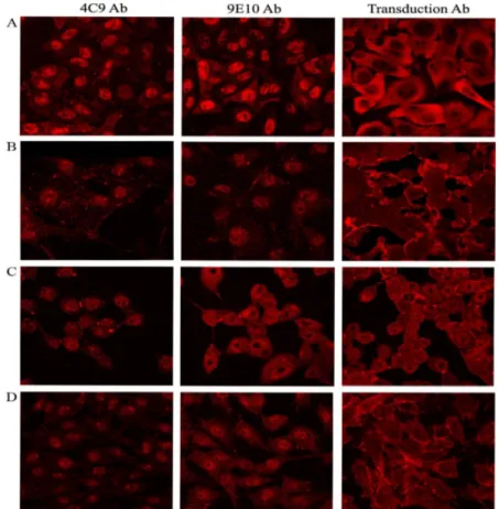
Figure 4.2.1: Confocal microscopy of 4C9, 9E10 and commercial β-catenin antibodies in β-catenin mutant cell lines……….68
Figure 4.2.2 Confocal microscopy of 4C9, 9E10 and commercial β-catenin antibodies in poorly differentiated SNU449.cl8 cells……….68
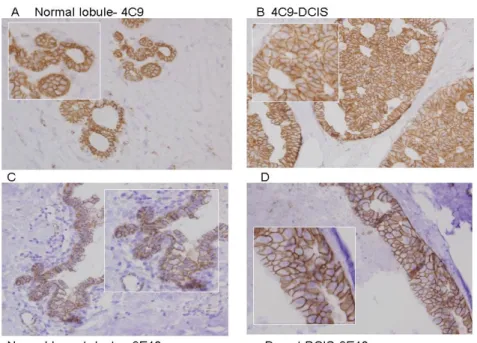
Figure 4.2.3: Immunohistochemistry analysis of 4C9 and 9E10 in paraffin embedded normal and breast cancer samples……… 69
Figure 4.2.4: Immunohistochemistry analysis of 9E10 in paraffin embedded carcinoma samples……….. 72
Figure 4.3.1: Normalized survival percentage of Kinase genes……….. 77
Figure 4.3.2: Normalized survival percentage of Phosphatase genes………...78
Figure 4.3.3: Map showing the effective kinases in the kinome tree………..78
Figure 4.3.4: in vivo imaging of Alexa-RAFT RGD targeted Huh7 xenograft tumors..79
Figure 4.4.1: SABG staining of HepG2 and Huh-7 cells and the rate of SABG-positive cells………....80
Figure 4.4.2: Immunostaining of Normal hepatocytes, Huh7 and LD-Doxorubucin treated Huh7 cells with methylation specific histone lysine antibodies………....81
Figure 4.4.3: Immunostaining of Normal hepatocytes, Huh7 and LD-Doxorubucin treated Huh7 cells with EZH2, SETD8 and Suv420H1 enzyme antibodies………..83
Figure 4.4.4: Immunostaining of Normal hepatocytes, Huh7 and LD-Doxorubucin treated Huh7 cells with SETD2, WHSC1 and FBXL11 enzyme antibodies……….84
Figure 4.4.5: Histone methylation in in-vivo………..85
Figure 4.4.6: Bar-chart representations of the results of immunohistochemistry counts of indicated histone residues. ………86
Figure 4.4.7: RT-PCR analysis of histone modifying enzymes in HCC cell lines…….87
Figure 4.4.8: RT-PCR analysis of histone modifying enzymes in tumor and non-tumor pairs………88
Figure 4.4.9: FBXL11 Knockdown in hepG2 and Huh7 cell lines……….90 Figure 4.4.10: Knockdown of enzymes failed to induce apoptosis and induce tumor
Figure 4.4.11: H4K20 methylation in HCC cells and tumor and non-tumor pairs….…..93
Figure 4.4.12: H4K20Me3 levels in Huh7 cells and MRC5 cell lines……….…....94
Figure 4.4.13: H4K20 methylation status in Huh7 cells after LD-Dox treatment……....95
Figure 4.4.14: Effect of Suv420H1 overexpression in HCC cells………96
Figure 4.4.15: Suv420H1 did not induce apoptosis in HCC cells……….97
Figure 4.4.16: Effect of Suv420H1 ectopic expression in Hek293T cells and DNA damage response of cells did not change………...98
Figure 4.4.17: New polyclonal Suv420H1 antibody (suv0032)………...…99
Figure 4.4.18: The polymorphism analysis in H19 gene………100
Figure 4.5.1: Identification of ATAD2………..102
Figure 4.5.2: ATAD2 expression is up in HCC cells………….………...103
Figure 4.5.3: ATAD2 expression status in malignant and normal cells………105
Figure 4.5.4: immunohistochemistry images of HCC samples……….106
Figure 4.5.5: ATAD2 knockdown inhibited cell growth of HCC cells………..107
Figure 4.5.6: Colony formation experiment of Hep3b and HepG2 cell lines after transfection of ATAD2 siRNAs………..108
Figure 4.5.7: Apoptosis was detected in Hep3b and HepG2 cells but not Huh7 and Hep40 cells………...109
Figure 4.5.8: ATAD2 Knockdown is shown in Hep3b and HepG2 cells……….110
Figure 4.5.9: Expression levels of Survival kinases after ATAD2 KD in Hep3b and HepG2 cells………..111
Figure 4.5.10: Epithelial and Liver specific markers and tumor suppressor genes after ATAD2 knockdown……….112
Figure 4.5.11: Survivin failed to rescue loss of ATAD2 induced apoptosis in Hep3b cells………..113
Figure 4.5.12: ATAD2 KD inhibited growth and colony formation of Hela cells…….114
Figure 4.5.13:Key cell cycle regulatory protein expression after ATAD2 KD in Hela cells……….115
Figure 4.5.15:shRNA lentiviral vectors downregulated ATAD2 levels when induced by
doxycycline ……….118
Figure 4.5.16: Xenograft models and imagining of shATAD2 and ShCTRL hep3b and
huh7 cells……….119
Figure 4.5.17: ATAD2 KD leads to increase in global histone acetylation increase….121
Figure 4.5.18: Key histone acetyltransferase and deacetylase expression after ATAD2
Knockdown………..122
ABBREVIATIONS
AD activation domain
AFB1 aflotoxinB1
AFP alpha-feto protein
APC Adenomatous polyposis coli BRCA breast cancer gene, early onset
BrdU Bromodeoxyuridine
CDK Cyclin Dependent Kinase
CK Casein Kinase
CK19 Cytokeratin 19
CO2 carbon dioxide
CRC colorectal cancer
C-terminus carboxy terminus
Dkk Dickkopf
DNA Deoxyribonucleic acid
DOX Doxorubucin
Dox Doxycycline
Dsh Dishevelled (in Drosophila) Dvl Dishevelled (in vertebrates)
EtBr ethidium bromide
FAP familial adenomatous polyposis FBS fetal bovine serum
Fz Frizzled
g gram
GAPDH glyceraldehyde-3-phosphate dehydrogenase GSK3β glycogen synthase kinase 3 beta
HB Hepatoblastoma
HBV Hepatitis B Virus
HbX hepatitis B virus X protein HBXAg Hepatitis B virus X antigen HCC Hepatocellular Carcinoma
HCV Hepatitis C Virus
HDAC histone deacetylase HRP horse radish peroxidase
hTERT human Telomerase Reverse Transcriptase ICR Imprinting Control Regions
Ig immunoglobulin
IGF2 insulin-like growth factor 2
IGF2R1 IGF2 Receptor
IGFBP IGF-binding proteins
JNK Jun Kinase
Kan kanamycin
Kb Kilo base
kDa kilo dalton
LB Luria-Bertani media
LD Low dose
LEF lymphoid enhancer-binding factor 1 LOH loss of heterozygosity
LTR long terminal repeat
M6P/IGF2R mannose-6-phosphate/insulin-like growth factor 2 receptor
MAb monoclonal antibody
MAP/ERK mitogen activated protein/extracellular signal-regulated kinase MAPK Mitogen Activated Protein Kinase
MDM2 Mouse Double Minute 2
MET met proto-oncogene (hepatocyte growth factor receptor)
mg milligram
µg microgram
MgSO4 Magnesium Sulfate
MHBSt carboxyterminal truncated middle hepatitis B surface protein
ml milliliter
µl microliter
MMLV Murine Maloney Leukemia Virus
MQ MilliQ water
NaCl Sodium Chloride
NaOH Sodium Hydroxide
NEAA Non-essential Amino Acid nm nanometer (1/109 of a meter) NS3 Nonstructural Protein 3 NS5A Nonstructural Protein 5A N-terminus amino terminus
O/N over night
OD Optical Density
PAb polyclonal antibody
PAGE Polyacrylamide gel electrophoresis PBS Phosphate Buffered Saline
PBS-T Phosphate Buffered Saline with Tween-20 PCR Polymearase chain reaction
PP2A protein phosphatase 2A
PPARγ peroxisome proliferators antigen receptor γ
pRb Retinoblastoma protein
PS presenescent
RD regulatory domains
RFLP Restriction length fragment polymorphisms
RNA Ribonucleic acid
RNAi RNA interference
RTK receptor tyrosine kinases
S senescent
S/N supernatant
SABG senescence associated beta galactosidase SAP shrimp alkaline phosphatase
SCD small cell-dysplasia SDS Sodium Dodecyl Sulfate
SDS-PAGE SDS- Polyacrylamide Gel Electrophoresis sFRPs secreted Frizzled-related proteins
shRNA short hairpin RNA
siRNA small interfering RNA
Smad homolog of mothers against decapentaplegic (MAD) SWI/SNF switching-defective and sucrose nonfermenting TBS Tris Buffered Saline
TBS-T Tris Buffered Saline with Tween-20 TCF T-cell specific, HMG-box
TEMED N, N, N, N-tetramethyl-1, 2 diaminoethane TGFα transforming growth factor alpha
TGF-β Transforming growth factor Tris Tris (hydroxymethyl)-methylamine
WIF-1 Wnt-inhibitory factor-1
X-Gal 5-bromo-4-chloro-3-indolyl-b-D-galactoside
CHAPTER 1. INTRODUCTION
1.1 Hepatocellular malignancy
Hepatocellular carcinoma (HCC) is believed to be originated from either hepatocytes or their progenitors. It is the one of the most frequent neoplasms worldwide and causes around 600,000 deaths per year, and the incidence is steadily increasing in Europe [1]. More than 80% of HCC occur in sub-Saharan Africa and Eastern Asia. The disease incidence is decreasing in these areas due to massive Hepatitis B virus (HBV) vaccination and the control of dietary aflatoxin intake, whereas it is still increasing in low-rate area including Europe. The increase has been associated with hepatitis C virus (HCV) infection. Hepatocellular carcinoma is classified into 4 different groups; well, moderately, poorly differentiated and undifferentiated tumors, respectively. HCC arises as a well differentiated cancer and continues with a stepwise dedifferentiation process.
Indeed, well-differentiated histology is generally seen in early stage and advanced HCC is associated with dedifferentiation. Well-differentiated and moderately differentiated cells are alike hepatocytes, and they are smaller in size and they have irregular trabecular or pseudoglandular architechtural patterns. Poorly differentiated and undifferentiated HCC cells have scanty cytoplasms and pleomorphism [2]. The progenitor cells in HCC like other epithelial tumors evolve during tumor progression and become autonomous later. This is observed with changes in their morphology and behavior; cuboidal shape and polarity is lost, and become more autonomous. Finally, they invade the underlying tissue and form distant metastases. The progressive loss phenotypic and biochemical of hepatocytes is defined as "dedifferentiation" [3]. Poorly differentiated and undifferentiated HCCs are associated with portal veneous invasion and the tumor invasiveness is a crucial factor in determining the long-term clinical outcome for the patient [4-5].
1.2 Aetiological factors of Hepatocellular Carcinoma
HCC occurs after years of damage to liver with inflammatory conditions resulting with chronic hepatitis and/or cirrhosis. This leads to death of hepatocytes and attack of inflammatory
cirrhosis inducing factors together are responsible for about 70% - 90% of all HCCs [7-8]. Apart from these factors, there are other aetiological factors including long term use of oral contraceptives, hereditary haemochromatosis diseases, Non-alcoholic fatty liver disorders (NAFLD) and diabetes [6, 9-11].
HCC is a slow process, and usually requires between 10-30 years from the initiation step to the fully malignant phenotype. This process is co-occurring with damage and death of hepatocytes with structurally aberrant chromosomes and genes often as a result of telomerase reactivation with marked genomic instability. The alteration of genes and chromosomes are irreversible and leads to heterogeneous malignant phenotype. [6]. Chronic inflammation, massive cell death and necrosis, fibrosis and cirrhosis, cycles of regeneration, and finally dysplasia and HCC occurs [12]. Recent studies indicate that cirrhosis is associated with senescence arrest of hepatocytes, from which HCC cells must emerge through the bypass of senescence control [13].
1.2.1 Viral induced Hepatocarcinogenesis
The two main hepatitis viruses are hepatitis B virus (HBV) and hepatitis C virus (HCV). HBV infects each year around 2 billions individuals, causes 320,000 deaths each year, and 30-50% of each attributable to HCC. Hepatitis C virus (HCV) infects each year 170,000 individuals, 20% of which develops cirrhosis [14-15]. HBV is a double stranded DNA virus and a member of
hepadnaviridae family. HBV can integrate into the host genome and may lead to chromosomal
instabilities and DNA microdeletions, may target telomerase reverse transcriptase enzyme (hTERT) and also encodes a protein so called protein x (HBx) [16-20]. The role of this protein is not fully characterized yet. HCV is a positive stranded RNA virus, a member flaviviridae family, thus cannot integrate into the genome. HCV is different from the HBV that it may yield a higher chronic infection rates and may cause high rates of cirrhosis (5-20%) [21-22]. HCV infected hepatocytes can escape from apoptosis by immune cells via using their HCV core protein and non structural protein NS5A interacting with TNF-α and IFN-α [23-25].
1.2.2 Alcohol and cirrhosis
Chronic alcohol intake or ethanol exposure affect hepatocyte cell signalling pathways that regulate chronic hepatocyte destruction–regeneration, stellate cell activation, cirrhosis and
eventually HCC [26]. Alcohol damages liver cells and induces oxidative stress which contributes to development of fibrosis and cirrhosis and further on HCC itself.
1.2.3 Aflatoxin B1-induced hepatocarcinogenesis
AflatoxinB1 (AFB1) was shown to have a well-defined genotoxic effect in hepatocarcinogenesis. Consumption of food contaminated with aflatoxins, toxic metabolites of some species of Aspergillus fungi, is a specific mutagen. Aflatoxin is metabolized specifically in liver to aflatoxin B1-8,9-epoxide, a toxic product that induces a G to T mutation of the p53 gene at codon 249 [27-28]. Aflatoxin B1 exposure coexisting with HBV infection, increases the risk of HCC to a 5–10-fold compared with exposure to only one of these factors [29].
1.3 Genetic and epigenetic events in HCC
HCC is a multistep process going through rounds of hepatocyte turnover due to damage and regeneration because of the aetiological factors as stated earlier [5]. This multistep process includes chronic liver injury that produces inflammation, cell death, cirrhosis and regeneration, DNA damage, dysplasia, and finally well differentiated to poorly differentiated HCC (figure 1.1). Telomere shortening is accompanied starting with chronic liver disease and cirrhosis, followed by genomic instability and telomerase reactivation and occasionally structural aberrations in genes and chromosomes. During this multistep process, a number of molecular alterations must occur, but only a limited number of is known currently. These aberrations can be listed as tumor suppressor genes and oncogenes including TP53, β-catenin, ErbB receptor family members, MET and its ligand hepatocyte growth factor (HGF), p16(INK4a), E-cadherin. Alterations in microRNA expression may also be implicated in hepatocellular carcinogenesis [30-32]. The table showing the mutation types and rates of tumor suppressors and oncogenes are listed in the table 1.1.
Figure 1.1: Multistage processes of hepatocarcinogenesis [8, 13] (modified from ref. 8 and 13).
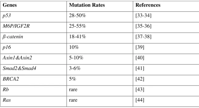
Table 1.1: Mutations in tumor suppressor genes and oncogenes in HCC
Genes Mutation Rates References
p53 28-50% [33-34] M6P/IGF2R 25-55% [35-36] β-catenin 18-41% [37-38] p16 10% [39] Axin1&Axin2 5-10% [40] Smad2&Smad4 3-6% [41] BRCA2 5% [42] Rb rare [43] Ras rare [44]
Recent advances showed that liver cancer cells harbor aberrant epigenetic abnormalities, suggesting that together with genetic events, epigenetics promote cancer progression. For instance, the CDKN2A gene encoding p16Ink4a and ARF proteins are not mutated whereas they are epigenetically silenced. More than 50% of HCCs display de novo methylation of the promoter of CDKN2A gene, resulting in loss of gene expression [13, 45]. Epigenetic mechanisms also contribute to strong expression of genes like TGF-α and IGF-2, promoting of growth of hepatocytes during hepatocarcinogenesis [46] .
Characterization of events involved in HCC dedifferentiation and invasiveness are ill known. Hepatocyte nuclear factors and E-cadherin of epithelial markers were downregulated in HCC [3, 47] and this is associated with tumor invasion and metastasis. In contrast, HCC invasiveness and/or metastasis are positive for mesenchymal markers such as snail [48], twist [49] and vimentin [50]. These changes represent the epithelial-mesenchymal transition (EMT) in HCC, as shown by in vitro studies [51-56]. Terminally differentiated hepatocytes express hepatocyte nuclear factor-4α (Hnf-4α) [57-58], and its expression is downregulated during HCC progression in mice [59]. This process is characterized by accumulation of genetic events like chromosomal gains and losses, as well as p53 mutations [6] and identified as dedifferentiation. A rare exception to this picture is the CTNNB1 gene that encodes β-catenin, a key component of the Wnt/β-catenin (canonical Wnt) signaling pathway.
1.4 Role of ββ-catenin In HCC ββ
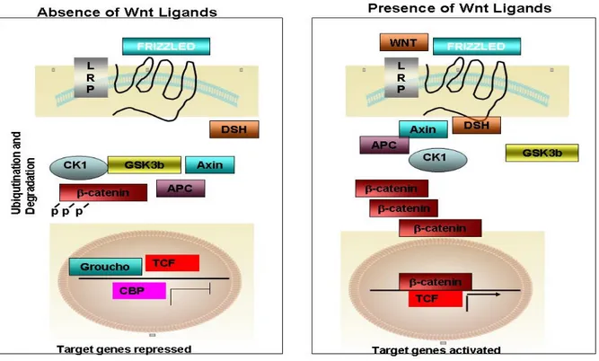
In the presence of wnt/canonical signaling ligands, these ligands binds to Frizzled receptor, disheveled is recruited to membrane and inhibits the GSK3b kinase and β-catenin is no longer phosphorylated and accumulated inside the cytoplasm and is translocated to nucleus, further activating several targets including MYC, cyclin-D1 and MMP7 (figure 1.2) [60].
β-catenin mutations were found to be associated with a subset of low grade (well-differentiated) HCCs with good prognosis and chromosome stability [40, 61-65]. Among 366 HCCs studied by Hsu et al., β-catenin mutations were associated with grade I histology. Another
Figure 1.2: Overview of the canonical Wnt signaling pathway, (Modified from ref. 60) A) In the absence of Wnt ligands, the cytoplasmic β-catenin is degraded in the destruction complex, composed of APC, axin/conductin, and GSK-3β. First, β-catenin is phosphorylated on Ser 45 residue by CKIα (priming), primed β-catenin is further phosphorylated at Thr 41, Ser 37, and Ser 33 residues by GSK-3β. Phosphorylation of these amino acids triggers ubiquitination of β-catenin by β-TrCP and degradation in proteasomes. Therefore, the cytoplasmic level of β-catenin is kept low in the absence of Wnt. The LEF/TCFs cannot activate the target genes without β-catenin. The WIF-1, sFRP, and/or Dkk can inhibit the Wnt signaling by binding to Wnt ligands or LRP5/6. B) In the presence of Wnt, a) Wnt signaling triggers CKIε-dependent phosphorylation of Dvl (Dsh) and enhances the binding of Dvl (Dsh) to Frat-1. Frat-1 binds to 3β in the Axin complex, resulting in a reduction of the phosphorylation of β-catenin by GSK-3β; or b) Wnt triggers the phosphorylation of LRP6 by CK1γ and GSK3β, which provides a docking site for Axin and recruits it to the plasma membrane. Wnt also enhances the binding of Dvl (Dsh) to Frizzled, and Dvl (Dsh) bound to Frizzled is necessary for the formation of a complex between Axin and LRP6, resulting in reduced phosphorylation of β-catenin and Axin by GSK3β. Therefore, β-catenin escapes from phosphorylation and subsequent ubiquitination, and accumulates in the cytoplasm. The accumulated cytoplasmic β-catenin goes into the nucleus, where it binds to LEF/TCFs and activates the transcription of target genes
study with similarly high number of tumors (n = 372) also showed that mutant nuclear β-catenin is associated with non-invasive tumor and but not with portal vein tumor thrombi [63]. In
signaling activity in primary tumors is possible indirectly by using target genes [62]. For instance, using glutamine synthetase (encoded by canonical Wnt signaling target GLUL gene) as a marker for canonical Wnt signal activity, Audard et al. showed that 36% HCCs displayed canonical Wnt activation. These tumors are compatible with well-differentiated morphology. The association of β-catenin mutation and nuclear translocation with well-differentiated tumor grade was also reported using several transgenic mouse models of HCC [62, 66]. Liver tumors from c-myc and c-c-myc/TGF-β1 transgenic mice showed activated beta-catenin. However, it was very rare with faster growing and histologically more aggressive HCCs in the same model. Thus nuclear translocation of β-catenin and activation of canonical Wnt signaling are early events mostly affecting well-differentiated HCCs.
Mutations of β-catenin gene were initially identified in colorectal cancers, leading to aberrant β-catenin protein accumulation and constitutive activation of canonical Wnt signaling. APC gene mutations in colorectal cancer and AXIN1 in HCC also activate canonical Wnt signaling by β-catenin protein accumulation. Therefore, both β-catenin, APC or AXIN1 mutations are considered to display active or constitutive canonical Wnt signalling [67]. Only a subset of HCC is associated with activated of canonical Wnt signaling as opposed to colorectal cancers. Interestingly, oncogenic β-catenin expressing transgenic mice develop only hepatomegaly [68-69], in contrast to intestinal polyposis and microadenoma in intestinal cells [70].
Taken together, Wnt pathway and β-catenin mutations may play a dual role in HCC, both in the initiation of HCC by close association of β-catenin mutation with well differentiated HCC and later stages as well [5].
1.4.1 Structure of beta-catenin protein and interaction partners
The protein β-catenin has a dual role; one in the establishment and maintenance of cell– cell adhesion at adherens junctions, and the other in the regulation of gene expression through Wnt signaling [71-72]. Both of these roles are conserved throughout evolution [73]. The protein is composed of short N-terminal, C-terminal domains interrupted with central core domain consisting of 12 armadillo repeats (Figure 1.3). Both structural and signal regulatory roles are
binding, together with E-cadherin binding to the central core domain is required for adhesion role of β-catenin [78]. The BTrCP binding is necessary for its subsequent degradation of β-catenin after phosphorylation of specific sites at the same N-terminal domain by glycogen synthase kinase 3 beta (GSK-3β). This phosphorylation event is facilitated by a multiprotein complex including Axin which bind to β-catenin through armadillo repeat domain [74, 79], and (APC). The presence of Wnt ligands or active Wnt signalling efficiently decreases the destruction of β-catenin via inhibition of GSK-3β. LEF/TCF factors (TCF) bind to the armadillo repeat domain for its function in transcriptional activation of Wnt target genes. ICAT and Chibby proteins for inhibitory roles compete with TCF for binding to the same core domain [80-81].
Figure 1.3 Schematic view of ββ-catenin protein domains, principal interacting proteins and monoclonal ββ
antibody epitopes [Yuzugullu et al, unpublished data].
The C-terminal domain of β-catenin has not been of great interest until recently. This acidic tail is the primary transactivation domain and TBP, CBP and p300 [82], as well as Teashirt [83], Chibby [80] are all associated through this domain. The C-terminal domain, together with the last three armadillo repeats of the core domain has been recently involved in nuclear import-export of the β-catenin protein [84]. Indeed the last 70 amino acids of β-catenin appear to play a crucial role in cytonuclear shuttling of β-catenin [85]. A relatively new and interesting aspect of
homology (PDZ)-interaction domain proteins. The PDZ domain is a specific protein-interaction module with an interaction ‘pocket’ that can be filled by a PDZ motif ‘ligand’. The PDZ motifs are consensus sequences that usually located at the extreme intracellular carboxyl terminus. The last 4 amino acids of β-catenin (DTDL) which is highly conserved among all metezoans excluding C. elagans [73] is a consensus PDZ motif D(S/T)XL (X denotes any residue) [86]. Proteins bind to these PDZ motifs and thus can target directly or indirectly, cluster and cycle the receptors, ion channels as well as transporters. PDZ proteins can link the signaling pathways and different components. Such roles implicate their role in many diseases. They control the pre- and post synaptic proteins in neurotransmission and plasticity. At the synapse, PDZ proteins have a role in neurotransmitter vesicle docking and receptor cycling. Cell movements, migration, polarity, and neighbouring cell interactions, as well as cell phenotypes including morphology are controlled with PDZ machinery. Beta-catenin interacts with several PDZ proteins including synaptic scaffolding molecule (S-SCAM; [87-90], its non-neuronal isoform called membrane-associated guanylate kinase with inverted domain organization-1 (MAGI-1; [91], Na+/H+ exchanger regulatory factor-1/EBP50 (NHERF-1; [92-93] and NHERF-2; [94], Tax-1 interacting protein (TIP-1; [95-96], LIN-7 [97].
Cadherins and catenins play important roles in synapse assembly. The PDZ domain of β-catenin is involved in the localization of a reserved pool of synaptic vesicles [98]. If beta-β-catenin is lost in hippocampal pyramidal neurons, this leads to misregulation of response in response to prolonged repetitive stimulation and a corresponding decrease of vesicles. Beta-catenin modulation of vesicle localization is mediated by its PDZ binding domain [99]. Epithelial and neuronal beta-catenin interacts with LIN-7 PDZ proteins. This event is Ca2+ dependent and is mediated direct linking of LIN-7 to the C-terminal beta-catenin PDZ target sequence. Therefore it is ensured that LIN-7 is relocated to the junctions via beta-catenin and is a specific event. Moreover, when LIN-7 is present at the beta-catenin-containing junctions, it determines the accumulation of binding partners, thus suggesting the mechanism by which beta-catenin mediates the organization of the junctional domain [97]. Functional studies showed that in mouse embryonic fibroblasts, NHERF1 associates directly via PDZ2 domain with beta-catenin and is required for beta-catenin localization at the cell-cell junctions. NHERF1 inhibits cell motility and
and complex assembly of β-catenin [93]. NHERF-2 links the N cadherin/catenin complex to the platelet-derived growth factor receptor to modulate the actin cytoskeleton and regulate cell motility [94]. In brain, being a multi-PDZ domain scaffolding protein, S-SCAM, binds and interacts with several different ligands including PTEN (phosphatase and tensin homolog), dendrin, axin, beta- and delta-catenin, dasm1 (dendrite arborization and synapse maturation 1), neuroligin, hyperpolarization-activated cation channels, beta1-adrenergic receptors, and NMDA receptors [100]. MAGI-1, presumably through its association with β-catenin, is required for Rap1 activation upon cell-cell contact and for enhancement of vascular endothelial cadherin-mediated cell adhesion [101]. TIP-1 binding to β-catenin inhibits its transcriptional activity and the overexpression of TIP-1 results in reduced proliferative capacity of colorectal cancer cells [95], suggesting an important role for TIP-1 interaction with the PDZ motif of β-catenin in the control of Wnt/β-catenin signaling. Interaction of the C-terminal domain of beta-catenin with PDZ domain-containing proteins is required to shape axon arbors of retinal ganglion cells [102].
1.5 Protein Kinase and Phosphatase Genes
In eukaryotic cells, signal transduction is mediated by protein kinases and phosphatases; by remodifying substrate activity. Protein kinases are among the largest families of genes in eukaryotes constituting about 2-2,5 % of the genome [103-104] and have been intensively studied. Cell metabolism, transcription, cell cycle progression, apoptosis, differentiation, cytoskeletal rearrangement, invasion and cell movement are controlled by protein kinases. Protein kinases are also implicated in critical regulation of cellular communication during development.
A total of 518 kinase genes are indentified so far [105] classified into 8 distinct classes, Table 1.2.
Table 1.2 Kinase distribution by major groups in human and model systems (modified from [105])
Group Families Subfamilies Yeast kinases Worm kinases Fly kinases Human kinases Human pseudogenes Novel human kinases AGC 14 21 17 30 30 63 6 7
CAMK 17 33 21 46 32 74 39 10 CK1 3 5 4 85 10 12 5 2 CMGC 8 24 21 49 33 61 12 3 Other 37 39 38 67 45 83 21 23 STE 3 13 14 25 18 47 6 4 Tyrosine kinase 30 30 0 90 32 90 5 5 Tyrosine kinase-like 7 13 0 15 17 43 6 5 RGC 1 1 0 27 6 5 3 0 Atypical-PDHK 1 1 2 1 1 5 0 0 Atypical-Alpha 1 2 0 4 1 6 0 0 Atypical-RIO 1 3 2 3 3 3 1 2 Atypical-A6 1 1 1 2 1 2 2 0 Atypical-Other 7 7 2 1 2 9 0 4
Atypical-ABC1 1 1 3 3 3 5 0 5 Atypical-BRD 1 1 0 1 1 4 0 1 Atypical-PIKK 1 6 5 5 5 6 0 0 Total 134 201 130 454 240 518 106 71
Phosphatases act in reverse functions with kinases, remove the phosphate groups from phosphorylated proteins and thus phosphorylation and dephosphorylation depend on the highly regulated and opposing actions of kinases and phosphatases acting on the same substrate. Phosphatases are also classified into distinct classes depending on the substrate specificity (table 1.3).
Table 1.3. Phosphatase distribution by major groups in human and model systems(modified from [106-110])
Class Substrate Reference
Tyrosine-specific phosphatases Phospho-Tyrosine [106]
Serine/Threonine specific phosphatases Phospho-Serine/Threonine [107]
Dual Specificity Phosphatases Phospho-Tyrosine/Serine/Threonine [108]
Histidine Phosphatase Phospho-Histidine [109]
1.5.1 Role of Kinases and Phosphatases in cancer
Certain classes of signalling proteins and pathways are targeted much more frequently by oncogenic mutations than other genes [111]. Thus, as being enzymes governing extracellular growth, differentiation and developmental signals, mutations and dysregulation of protein kinases and phosphatases play causal roles in several human disease including cancer as well [112-113].
1.5.2 Role of Kinases and Phosphatases in liver cancer
Activation of phosphoinositide 3-kinases (PI3K)/Akt/mammalian target of rapamycin (mTOR) signaling pathway in HCC occurs between 30–50% of all cases and related to poor prognosis [114]. Active signalling is due to aberrant activation of tyrosine kinase receptor EGFR, IGF1R, MET, VEGFR, PDGFR or through constitutive activation of PI3K due to loss of function of PTEN. Loss of heterozygosity in PTEN locus (i.e. 10q) is frequent in HCC and mutation rates are fairly low in HCC, ranging from 0 to 11% [115]. Raf/Ras/Erk pathway is generally activated in advanced stages of liver cancer, due to upstream signalling from EGF, HGF or IGF signals [116]. These informations are incomplete and fragmentary. Indeed, a systematic analysis of protein kinases and phosphatases in HCC is lacking. There has also been no report of systematic analysis of the effects of systematic inactivation of kinases and phosphatases in these cancers.
1.6 Role of H19/IGF2 locus in HCC
The non-coding gene H19 and IGF2 genes are co-located in the same locus and are under the control of genomic imprinting, which leads to reciprocal monoallelic expression of H19 from the maternal allele and IGF2 from the paternal allele [117]. Loss of imprinting is observed for both H19 and IGF2. Moreover, H19 is highly expressed in liver cancer and has been shown to behave like an oncogene for liver cancer and it is essential for tumor cell growth [118]. Several studies showed that IGF-II is overexpressed in human HCCs and in some premalignant lesions [119-121] and this is correlated with loss of imprinting [122].
1.7 Senescence of hepatocytes and chronic liver disease
frequent in patients with cirrhosis. The only effective therapy for cirrhosis is organ transplantation.
Normal hepatocytes in the adult liver are quiescent, they are renewed slowly, approximately once a year, as estimated by telomere loss which is 50–120 bp per year in healthy individuals [123-124]. However, as well as animal models chronic liver diseases showed that, liver has enormous regenerative capacity [125]. Exposure to viral and non viral damaging agents or as shown in partial hepatectomy, mature hepatocytes proliferates enormously and this process is so called regenerative capacity of liver. In addition, hepatocyte-progenitor cells are induced to restore liver hepatocyte populations [126]. However, hepatocytes, since they do not have telomerase activity, like other somatic cells, they cannot avoid telomere shortening during rounds of cell divisions. For this reason, liver cirrhosis is associated with decreased hepatoycte proliferation [127], is now considered to be a result of senescence arrest [13].
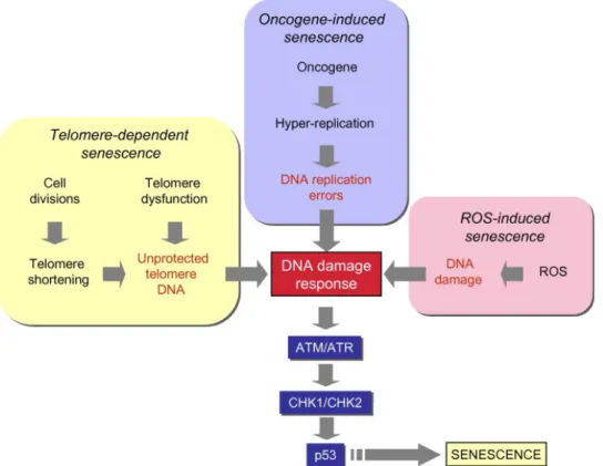
Cellular senescence is a permanent arrest of the cell cycle in response to diverse stress conditions such as dysfunctional telomeres, DNA damage, strong mitotic signals and disrupted chromatin (figure 1.4). Senescence, a strong anti-cancer mechanism is regulated by p53-retinoblastoma pathway and involves the activation of p21Cip1, p16Ink4a and p15Ink4b cyclin-dependent kinase (CDK) inhibitors [128]. Frequent inactivation of TP53 and CDKN2A genes, together with frequent upregulation of telomerase reverse transcriptase (TERT) strongly suggest that HCC tumor cells may bypass senescence to become immortalized [13]. Nevertheless, the detection of senescent cells in nearly 50% of HCC tumors provides evidence for the capacity of HCC cells to undergo senescence arrest under appropriate conditions [129]. Immortal HCC cell lines can spontaneously generate progeny that undergo replicative senescence [130]. Murine HCC tumors generated by the expression of a mutant Ras gene in p53-deficient hepatoblasts can be cleared by a massive senescence response upon reactivation of p53 expression [131-132]; and c-myc oncogene inactivation in murine HCCs results in senescence-mediated tumor regression
Fig. 1.4 DNA damage and p53 activation play a central role in different senescence pathways DNA damage (often in the form of double-strand breaks) activate upstream kinases (ATM and ATR) leading to p53 phosphorylation by CHK1 and CHK2 kinases. Phosphorylated p53 is released from MDM, and stabilized in order to induce senescence arrest or apoptosis (not shown here). (Modified from [13]).
1.8 Histone modifications
Major histone modifications include DNA methylation, covalent modifications of histones and repositioning of histones. Among these three, we started to understand the role of post-translational modifications of histones recently compared to DNA methylation.
Chromatin consists of repeating units of nucleosomes and the nucleosome consists of 146bp DNA wrapped to an octamer of four histone proteins (H3, H4, H2A and H2B). Basically, the protruding N-terminal ends of histone proteins are covalently modified by eight distinct types of mechanisms. Acetylations, methylation of lysines and arginines, phosphorylation, ubiquitylation, sumoylation, ADP ribosylation and proline isomeration are the eight major histone modifications and dictate the complexicity of regulation and biological roles. These covalent modifications work together to dictate and regulate the functioning of genome and change the local structure of
as well as diseases like cancer. To sum up, we can call these modifications as ‘histone codes’, a complicated code written by histone modifying enzymes and read by histone binding proteins. The complexity of code comes not only from different numbers of histone modifications reaching as high as 70 but also degree of modification (methylation at lysines or arginines may be one of three different forms: mono-, di-, or trimethyl for lysines and mono- or di- (asymmetric or symmetric) for arginines [133] and cross-talk between these codes [134]. The function of each code is difficult to understand and only recently we started to understand the biological functions. Modifications like lysine acetylation may affect the chromatin structure by neutralizing the basic negative charge of lysines and causing a permissive chromatin conformation for transcription to start. Phosphorylation is another way of changing the charge of chromatin thus leading to important consequences like mitosis and apoptosis [135-136]. Lysine methylation, in contrast to lysine acetylation has a different meaning depending on which lysine residues being methylated. This is because of the fact that histone methyl transferases have substrate specificity compared to histone acetyltransferases. The complexes in which enzymes are found [137], proteins associating with the enzymes of residue to modify [138], and the degree of methylation (mono-, di-, or tri-) [139] are the factors for residue specificity and thus biological roles. For instance, trimethylation of Histone 3 lysine 4 (H3K4Me3) on gene promoters means transcriptional activation, trimethylation of H3 lysine 36 (H3K36Me3) on gene coding region and histone 3 lysine 79 trimethylation (H3K79Me3) means transcriptional activation [140-141], whereas trimethylation of histone 3 lysine 9 (H3K9Me3) and lysine27 (H3K27Me3) on gene promoters means transcriptional repression [134]. After these codes are written by histone modifying enzymes, these codes are read by proteins that are recruited to these specific modifications and that have binding domains suitable for these histone marks. For example, methylation is recognized by chromo-like domains of the Royal family (chromo, tudor, MBT) and nonrelated PHD domains, acetylation is recognized by bromodomains, and phosphorylation is recognized by a domain within 14-3-3 proteins (figure 1.5).
Figure 1.5 Recruitment of proteins to histones (modified from [134])
The dynamic and reversible modifications of each histone residue are regulated by specific enzymes. Histone acetyltransferases (HATs) add acetyl groups and histone deacetylases (HDACs) removes these and histone methyltransferases (HMTs) transfers methyl groups and histone demethylases (HDMs) reverses this [142-143]. The list of each type of enzymes is listed in table 1.4.
Table 1.4 Histone modifying enzymes and residues (modified from [134])
Enzymes that Modify
Histones Residues Modified
Acetyltransferase Lysine Methyltransferase
HAT1 H4 (K5, K12) SUV39H1 H3K9 CBP/P300 H3 (K14, K18) H4 (K5, K8) H2A (K5) H2B (K12, K15) SUV39H2 H3K9 PCAF/GCN5 H3 (K9, K14, K18) G9a H3K9 TIP60 H4 (K5, K8, K12, K16) H3 K14 ESET/SETDB1 H3K9 HB01 (ScESA1, SpMST1) H4 (K5, K8, K12) EuHMTase/GLP H3K9 ScSAS3 H3 (K14, K23) CLL8 H3K9 ScSAS2 (SpMST2) H4 K16 SpClr4 H3K9
SirT2 (ScSir2) H4 K16 MLL3 H3K4 Lysine Demethylases MLL4 H3K4 LSD1/BHC110 H3K4 MLL5 H3K4 JHDM1a H3K36 SET1A H3K4 JHDM1b H3K36 SET1B H3K4 JHDM2a H3K9 ASH1 H3K4 JHDM2b H3K9 Sc/Sp SET1 H3K4 JMJD2A/JHDM3A H3K9, H3K36 SET2 (Sc/Sp SET2) H3K36 JMJD2B H3K9 NSD1 H3K36 JMJD2C/GASC1 H3K9, H3K36 SYMD2 H3K36 JMJD2D H3K9 DOT1 H3K79 JMJD6 H3R2, H4R3 H3K9 H3K79
Arginine Methlytransferases Residues Modified Pr-SET 7/8 H4K20
CARM1 H3(R2,R17,R26) SUV4 20H1 H4K20
PRMT4 H4R3 SUV420H2 H4K20
PRMT5 H3R8,H4R3 PRMT5 H4K20
Serine/Threonine Kinases Residues Modified EZH2 H3K27
Haspin H3T3 RIZ1 H3K9
MSK1 H3S28 Ubiquitilases
MSK2 H3S28 Bmi/Ring1A H2AK119
CKII H4S1 RNF20/RNF40 H2BK120
Mst1 H2BS14
1.8.1 Histone modifications in HCC and other cancers
High-throughput sequencing technologies (Chip-seq) have enabled to elucidate the roles and characterization of histone modifications and aberrations in enzymes in diseases and tumorigenesis as well. Fraga et al, showed that global loss of acetylated H4-lysine 16 (H4K16Ac) and trimethylated H4-lysine 20 is a hallmark of human cancer including breast and liver cancer (H4K20Me3) [133]. HDACs are often overexpressed in various types of cancers including prostate and gastric tumors including liver cancer and there are drugs targeting HDACs under clinical trials [144-146]. Abnormal HAT activities are also observed in cancers, viral oncogene proteins such as E1A target CBP/P300 and disrupt their interaction with PCAF [147]. In
alterations in methylation of H3K9 and H3K7 in cancer are also reported [149-150]. EZH2 is a histone methyl transferase acting on H3K27 residue is overexpressed in breast, prostate and liver cancers [149, 151]. Another histone methyltransferase acting on H3K9, G9a is associated with liver cancer [152]. MLL gene, acting on H3K4 is frequently makes fusion proteins with Hox genes and involves in leukemia [153]. Histone demethylases are also upregulated in prostate cancer and involved in tumorigenesis [142].
1.8.2 H3K4 methylation
Trithroax (Trx-G) group proteins are involved in H3K4 methylation and evolutionary conserved from flies to mammals. They are involved in epigenetic activation of homeodomain genes, whereas Polycomb repressor group (PcG) proteins mediate their silencing. These two antagonistic groups of proteins control important aspects of differentiation and proliferation [154].
1.8.2.1 MLL and SMYD3 – H3K4 methyltransferases
The mixed lineage leukemia (MLL) protein, SET domain containing H3K4 methyltransferase and activates of Hox gene expression during development. MLL and Hox genes frequently make fusion proteins, and involves in Leukemia [153]. SET- and MYND-domain-containing protein 3 (SMYD3), methyltransferase of H3K4, is found to be upregulated in colorectal and hepatocellular carcinoma cell lines and enhances cell growth and promotes transformation, whereas its inhibition represses cell growth in cancer cell lines [155].
1.8.3 H3K9 Methylation
Histone H3 lysine 9 (H3K9) methylation is recognized by HP1 and involves in heterochromatin formation and correlates with gene silencing in a variety of organisms. SUV39H1 and SUV39H2, mammalian homologues of Drosophila SU (VAR)3–9, are SET domain containing Histone methyltransferases acting on H3K9 residues.
![Figure 1.3 Schematic view of β β-catenin protein domains, principal interacting proteins and monoclonal β β antibody epitopes [Yuzugullu et al, unpublished data]](https://thumb-eu.123doks.com/thumbv2/9libnet/5765134.116751/33.892.124.775.464.775/schematic-principal-interacting-proteins-monoclonal-antibody-yuzugullu-unpublished.webp)
![Table 1.2 Kinase distribution by major groups in human and model systems (modified from [105])](https://thumb-eu.123doks.com/thumbv2/9libnet/5765134.116751/35.892.98.807.940.1096/table-kinase-distribution-major-groups-human-systems-modified.webp)
![Table 1.3. Phosphatase distribution by major groups in human and model systems(modified from [106-110])](https://thumb-eu.123doks.com/thumbv2/9libnet/5765134.116751/37.892.102.794.156.467/table-phosphatase-distribution-major-groups-human-systems-modified.webp)